
1. Introduction
A hematoma of the pinna, clinically recognized as an auricular hematoma, is an acute accumulation of blood located between the cartilage of the external ear and its overlying perichondrium. This condition presents as a sudden, tense, and painful swelling on the outer ear, requiring prompt medical intervention to evacuate the trapped blood.
The external ear relies on a precise anatomical relationship between its rigid cartilaginous framework and the thin vascular membrane supplying it with nutrients. When mechanical trauma disrupts this relationship, bleeding occurs within the tight subperichondrial space. The pooled blood acts as a physical barrier, isolating the cartilage from its sole source of blood flow and oxygen.
Failure to address this vascular separation results in irreversible cartilage necrosis and subsequent fibrotic scarring. This progressive structural degradation leads to a permanent deformity commonly known as cauliflower ear. Timely procedural drainage, combined with specialized compression dressings, remains the definitive strategy for preserving the aesthetic and functional integrity of the auricle.
2. Anatomy of the Auricle
Understanding the pathogenesis of this condition requires examining the structural architecture of the external ear. The pinna is composed predominantly of a single, continuous sheet of elastic cartilage. This cartilaginous framework is uniquely avascular, containing no internal blood vessels to sustain its cellular metabolism.
Instead, the cartilage derives all essential oxygen and nutrients through passive diffusion from the perichondrium. The perichondrium is a dense, richly vascularized connective tissue membrane that firmly adheres to the surface of the cartilage. Overlying the perichondrium is a very thin layer of subcutaneous tissue and skin, which provides minimal mechanical padding.
Because the anterior surface of the ear features skin that is tightly bound directly to the perichondrium without an intervening protective fat layer, this region is exceptionally vulnerable to sheer forces. Any significant physical impact can easily tear the delicate blood vessels bridging the perichondrial and subcutaneous layers.
3. Mechanism of Shearing Trauma
The primary catalyst for an auricular hematoma is a specific type of mechanical force applied to the external ear. While direct, blunt perpendicular impacts can cause bleeding, tangential or shearing forces are the most common instigators. A shearing force drags the skin and perichondrium abruptly across the underlying stationary cartilage.
This rapid, opposing movement generates profound mechanical stress, effectively ripping the perichondrium away from the cartilaginous framework. The sudden separation tears the fragile perforating blood vessels that travel through the perichondrial layer.
Once these vessels are severed, whole blood forcefully leaks into the newly created artificial space between the perichondrium and the cartilage. Because the overlying skin is tight and unyielding, the accumulating blood rapidly generates substantial internal pressure, forming a distinct, fluctuant mass on the anterior surface of the ear.
4. Pathophysiology of Cartilage Ischemia
The accumulation of blood within the subperichondrial space initiates a rapid sequence of destructive biological events. The most immediate threat is acute ischemia. The hematoma acts as an impenetrable physical barrier, completely halting the passive diffusion of oxygen and nutrients from the perichondrium to the chondrocytes, the specialized cells that maintain the cartilage.
Without a constant supply of nutrients, chondrocytes begin to undergo necrosis within hours of the initial injury. As the cartilage cells die, the structural collagen matrix they support begins to degrade and collapse. The cartilage loses its inherent elasticity and structural rigidity.
Simultaneously, the pooled blood triggers a robust localized inflammatory response. The immune system dispatches white blood cells to break down the clotted blood, releasing inflammatory cytokines and proteolytic enzymes into the confined space. These enzymes further accelerate the enzymatic degradation of the starving cartilage framework.
5. Development of Cauliflower Ear
If the hematoma is left untreated or inadequately drained, the body attempts to repair the damaged tissue through a process of disorganized fibrosis and neocartilage formation. As the clotted blood eventually organizes and fibroses, the separated perichondrium is stimulated to produce new, abnormal cartilage.
This neocartilage is deposited haphazardly within the subperichondrial space, filling the area previously occupied by the blood clot. The original, necrotic cartilage framework is concurrently replaced by dense, contracted scar tissue.
The culmination of this aberrant healing process is a thick, nodular, and rigid deformity that obscures the delicate, natural contours of the pinna. This permanent disfigurement, clinically termed cauliflower ear, is exceptionally difficult to correct surgically once the fibrotic tissue has fully matured and calcified.
6. Clinical Presentation and Symptoms
A patient presenting with an auricular hematoma reports the rapid development of localized swelling following a traumatic incident. The swelling typically appears within minutes to a few hours post-injury. The mass is usually located on the anterior, concave surface of the pinna, frequently obscuring the natural folds of the scaphoid fossa or the conchal bowl.
Upon physical inspection, the clinician observes a tense, fluctuant, and often deeply ecchymotic swelling. The overlying skin may appear red, purplish, or pale due to the significant underlying pressure. The mass feels rubbery and warm to the touch.
Pain is a prominent symptom. Patients describe a constant, severe throbbing ache that is significantly exacerbated by any direct contact or manipulation of the ear. Depending on the size of the hematoma, the swelling may physically occlude the external auditory meatus, leading to a temporary, conductive reduction in hearing acuity.
7. Risk Factors and Sports Injuries
The demographic most frequently affected by auricular hematomas consists of individuals participating in close-contact sports. Activities that involve repetitive friction, grappling, or direct strikes to the head carry a pronounced risk of sheer trauma to the external ear.
Wrestling, mixed martial arts, judo, and rugby are the disciplines most notoriously associated with this condition. In these sports, the athlete’s ear is continually pressed and dragged against the opponent’s body or the floor mat, creating the exact mechanical forces required to separate the perichondrium.
Boxing and full-contact martial arts also present substantial risk due to the velocity of direct strikes. The consistent utilization of properly fitted, rigid protective headgear is the only definitive preventative measure capable of shielding the vulnerable pinna from these targeted mechanical forces.
8. Differential Diagnosis
While the clinical diagnosis of an auricular hematoma is generally straightforward, clinicians must differentiate it from other pathologies affecting the external ear. Auricular pseudocyst is a primary consideration. This condition presents as a painless, fluid-filled swelling, typically occurring without a clear history of acute trauma, and contains sterile, yellowish fluid rather than whole blood.
Relapsing polychondritis is an autoimmune condition that causes episodic, severe inflammation of the ear cartilage. Unlike a hematoma, polychondritis presents with widespread, fiery redness and tenderness affecting the entire cartilaginous portion of the ear, while characteristically sparing the soft, non-cartilaginous earlobe.
A severe localized skin infection, such as erysipelas or perichondritis, can also cause notable swelling and pain. However, infectious processes are typically accompanied by systemic symptoms such as fever, profound warmth, and an advancing border of erythema, distinguishing them from the localized, fluctuant blood pool of a hematoma.
9. When to Seek Medical Attention
Immediate medical evaluation is mandated whenever a discrete, painful swelling develops on the ear following physical trauma. The critical window for optimal intervention is narrow. To prevent irreversible cartilage necrosis, the hematoma must be completely evacuated within the first several days following the injury.
Delaying treatment allows the blood clot to organize and adhere firmly to the surrounding tissues, rendering simple drainage procedures ineffective. Once the blood begins to fibrose and the perichondrium starts generating neocartilage, the window for preserving the normal anatomical contour closes rapidly.
Patients who experience a recurrence of swelling after an initial drainage procedure must return for prompt re-evaluation, as continuous blood accumulation will inflict the same ischemic damage as the initial injury.
10. Diagnostic Evaluation
The diagnosis relies heavily on clinical history and targeted physical examination. The physician ascertains the mechanism of injury, the timeline of the swelling, and any previous history of similar trauma or bleeding disorders.
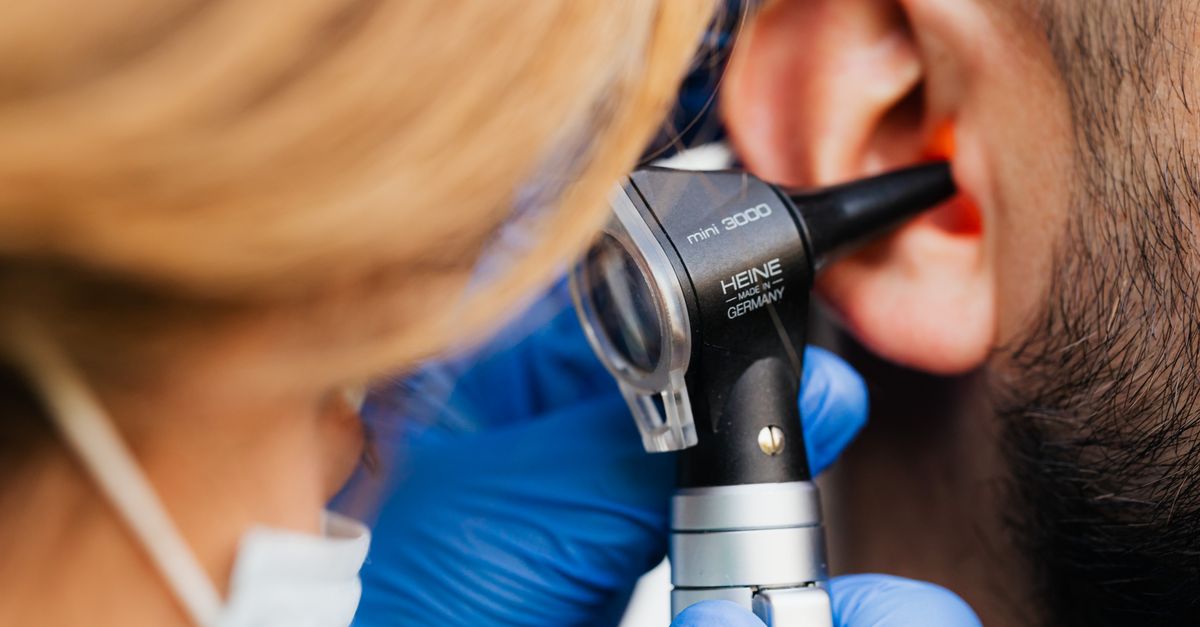
During the physical examination, the clinician carefully palpates the auricle to determine the extent and fluctuance of the mass. A thorough assessment of the external auditory canal and the tympanic membrane using an otoscope is necessary to rule out concurrent traumatic injuries, such as a cutaneous hematoma within the canal or a ruptured eardrum resulting from changes in ambient pressure during the impact.
Radiological imaging is rarely indicated for a simple auricular hematoma unless the mechanism of injury suggests a concurrent fracture of the temporal bone or underlying intracranial trauma.
11. Needle Aspiration
For small, acute hematomas presenting within the first twenty-four hours, needle aspiration may be considered. Following the administration of a local anesthetic block to numb the external ear, the physician inserts a large-bore needle directly into the fluctuant mass.
The trapped liquid blood is aspirated into a syringe, immediately decompressing the subperichondrial space. While needle aspiration is less invasive and rapid, it carries a substantially higher rate of recurrence compared to surgical incision. The small puncture wound seals quickly, and the damaged blood vessels often resume bleeding, refilling the cavity.
Consequently, needle aspiration is generally reserved for very small collections of fluid or as a temporary temporizing measure when definitive surgical incision is not immediately available.
12. Incision and Drainage
The definitive and most reliable treatment for an auricular hematoma is a formal incision and drainage procedure. Under local anesthesia, the clinician makes a small, strategic incision through the skin and perichondrium, typically tracing the natural curvature of the scaphoid or conchal folds to minimize visible scarring.
The incision allows for the complete evacuation of both liquid blood and any organized, gelatinous clots that have formed. The physician thoroughly irrigates the subperichondrial pocket with sterile saline to ensure no residual debris remains to impede healing or harbor bacterial growth.
If the hematoma has been present for several days and neocartilage has already begun to form, the surgeon may need to carefully excise this abnormal tissue to allow the perichondrium to lie flat against the native cartilage framework once again.
13. Bolster Dressings and Compression
Evacuating the blood is only the first step in successful management; preventing the fluid from re-accumulating is equally critical. Because the potential space between the perichondrium and the cartilage has been stretched open, blood will inevitably pool again unless the tissues are physically forced back together.
To achieve this, the physician applies a specialized compression bolster dressing immediately following the drainage procedure. This typically involves placing sterile dental rolls, customized silicone splints, or molded thermoplastic materials against both the anterior and posterior surfaces of the ear.
These bolsters are firmly sutured in place, squeezing the ear tightly between them. This constant mechanical compression closes the dead space, halts any ongoing capillary bleeding, and forces the perichondrium to re-adhere firmly to the underlying cartilage. The bolster is usually left in place for seven to ten days.
14. Antibiotic Prophylaxis and Follow-up
The ear cartilage has a notoriously poor blood supply, rendering it exceptionally susceptible to bacterial infection once the overlying skin barrier is breached during the drainage procedure. Suppurative perichondritis is a devastating complication that can rapidly destroy the entire cartilaginous framework.
To mitigate this risk, patients are routinely prescribed a prophylactic course of oral antibiotics covering common skin flora and specific pathogens like Pseudomonas aeruginosa. The patient must keep the dressing clean and dry and avoid all contact sports until the bolster is removed and the incision is fully healed.
Close clinical follow-up is necessary. The physician must evaluate the ear after the removal of the compression dressing to ensure that no recurrent fluid has accumulated and to monitor for any delayed signs of infection or cartilage necrosis.
15. Summary of Procedural Interventions
The management of an auricular hematoma focuses on prompt decompression and sustained tissue approximation. The table below outlines the primary interventional strategies.
| Intervention Type | Clinical Indication | Primary Mechanism |
|---|---|---|
| Needle Aspiration | Small, very acute lesions (< 24 hours) | Rapid fluid removal; high recurrence rate |
| Incision and Drainage | Standard definitive treatment | Complete clot evacuation, irrigation of pocket |
| Bolster Compression | Mandatory following any drainage | Eliminates dead space, prevents blood re-accumulation |
| Antibiotic Therapy | Routine post-procedural prophylaxis | Prevents suppurative perichondritis and cartilage loss |
Strict adherence to post-procedural compression and abstaining from traumatic activities are essential components of preventing permanent anatomical disfigurement.
16. Frequently Asked Questions (FAQ)
1. Can an auricular hematoma heal properly without being drained?
No. If the blood is not physically evacuated, the body will replace the clot with dense scar tissue and abnormal cartilage. This leads to the permanent, bumpy deformity known as cauliflower ear.
2. Does the draining procedure hurt?
The physician will use an injectable local anesthetic to completely numb the ear before making any incisions or inserting needles. While the initial numbing injection causes a brief sting, the actual drainage procedure should be entirely painless.
3. Why is buttons or padding sewn onto the ear after the procedure?
The padding, known as a bolster, squeezes the skin tightly against the cartilage. Without this constant pressure, the empty pocket where the blood was located will quickly fill up with new blood, requiring the entire procedure to be repeated.
4. When can an athlete return to wrestling or martial arts after treatment?
Athletes must strictly avoid any contact sports while the bolster is in place and until the incision is completely healed, which typically takes several weeks. Returning too early guarantees a recurrent bleed and further cartilage damage.
5. Will a cauliflower ear affect a person’s ability to hear?
The deformity itself primarily affects the outer appearance of the ear. However, if the swelling or resulting scar tissue is substantial enough to physically block the ear canal opening, it can cause a noticeable reduction in hearing acuity.
17. Bibliography
Disclaimer: The content is for informational purposes only and does not replace medical advice. Always consult your doctor for personalized treatment.
