1. Introduction
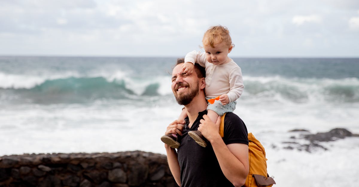
The hip joint aches after carrying a toddler unilaterally because this asymmetric posture forces the pelvis into a lateral tilt, placing severe mechanical load on the gluteus medius muscle, the trochanteric bursa, and the sacroiliac joint. To balance the weight of the child, the caregiver instinctively juts one hip outward, which stretches the stabilizing tendons and compresses the local articular structures. Over time, this uneven weight distribution triggers localized tissue inflammation and profound muscular fatigue.
Carrying a child is a daily necessity for caregivers, but as the toddler grows heavier, the biomechanical strain on the adult skeleton increases exponentially. The human pelvis is designed to distribute weight evenly across both lower extremities. When a thirty-pound child is continually hoisted onto a single hip, the center of gravity shifts entirely, forcing the body to adopt compensatory postures to prevent falling over.
Understanding the specific anatomical structures affected by this uneven load provides crucial clarity. The resulting ache is an active neurological signal of mechanical overuse and soft tissue irritation. By analyzing the anatomy of the hip and modifying daily carrying mechanics, caregivers can relieve the acute discomfort and prevent chronic joint degradation.
2. Biomechanics of the Unilateral Hold
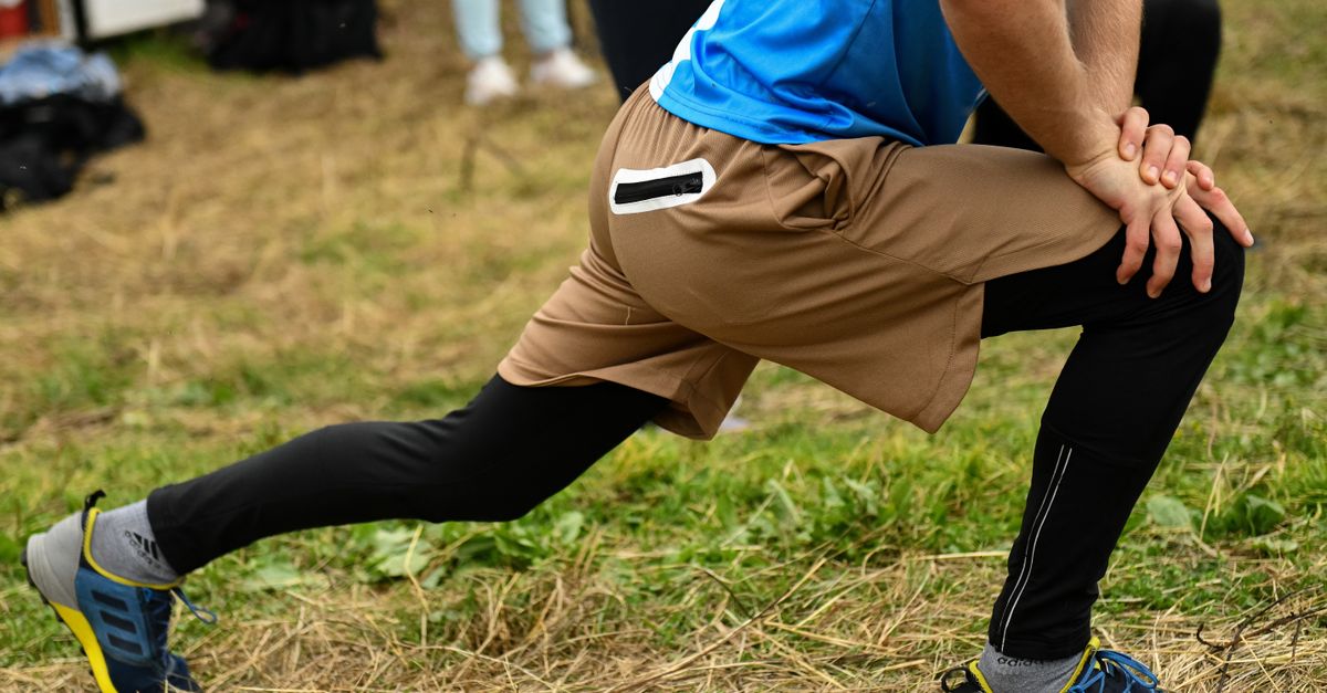
When a person holds a child on one side, they perform a specific postural shift called a lateral pelvic tilt. To create a supportive bony shelf for the toddler to rest upon, the caregiver drops the opposite side of the pelvis and thrusts the weight-bearing hip outward.
This posture significantly alters the natural alignment of the lower kinetic chain. The femur on the weight-bearing side is forced into adduction, meaning it angles inward toward the midline of the body. This angle places the lateral structures of the hip under immense, continuous stretch.
Simultaneously, the lumbar spine must curve away from the child to keep the torso upright. This lateral spinal flexion compresses the intervertebral discs on one side while overstretching the paraspinal muscles on the other, compounding the mechanical stress far beyond the hip joint itself.
3. The Gluteus Medius and Lateral Stabilizers
The primary muscle responsible for stabilizing the pelvis during single-leg stance or asymmetrical loading is the gluteus medius. Located on the outer surface of the pelvis, this fan-shaped muscle prevents the opposite side of the pelvis from dropping when walking or lifting.
Carrying a toddler on the hip forces the gluteus medius into a state of relentless isometric contraction. The muscle must work continuously to hold the pelvis steady against the gravitational pull of the child’s weight.
Because isometric contractions restrict localized blood flow, the gluteus medius rapidly depletes its cellular energy reserves. The muscle shifts into anaerobic metabolism, accumulating lactic acid. This metabolic waste chemically irritates the sensory nerves, creating a deep, burning ache in the outer hip.
4. Trochanteric Bursitis Explained
The greater trochanter is the bony prominence on the outer edge of the femur. To reduce friction between this bone and the overlying gluteal tendons, the body utilizes a small, fluid-filled sac called a bursa.
When the hip is thrust outward repeatedly, the tight gluteus medius tendon snaps forcefully back and forth across the trochanteric bursa. This continuous friction damages the delicate synovial lining of the sac, triggering an inflammatory cascade.
The bursa swells with excess fluid, a condition diagnosed clinically as trochanteric bursitis. The swollen bursa is exquisitely tender. Caregivers often experience a sharp, localized pain on the outside of the hip that worsens when lying on the affected side at night or when climbing stairs.
5. Sacroiliac Joint Dysfunction
The sacroiliac joint connects the sacrum at the base of the spine to the ilium bones of the pelvis. This joint is designed for stability and shock absorption, possessing very little natural movement.
Asymmetrical loading places significant shear force across the sacroiliac joint on the weight-bearing side. The ligaments that secure this joint are stretched unevenly. Over months of repetitive toddler carrying, these ligaments can become strained and micro-torn.
Sacroiliac joint dysfunction presents as a deep, dull ache in the lower back that often radiates into the buttocks and down the back of the thigh. The pain frequently sharpens when the caregiver attempts to stand up from a seated position or shifts weight suddenly.
6. Lumbar Spine Compensation
The hip and the lower spine operate as a functionally integrated unit. When the pelvis tilts laterally to support the child, the lumbar spine must compensate to keep the eyes level and the head centered over the feet.
This compensation involves the quadratus lumborum, a deep muscle in the lower back. On the side opposite the child, this muscle contracts fiercely to pull the spine upright. This sustained tension frequently causes the muscle to spasm.
A spasming quadratus lumborum refers pain directly down into the hip crest and the gluteal region. Often, what the caregiver perceives as a primary hip problem is actually referred pain originating from exhausted spinal stabilizers.
7. Muscle Fatigue and Lactic Acid
Skeletal muscles rely on a steady supply of oxygenated blood to function efficiently. The continuous static tension required to brace the body against a squirming toddler physically squeezes the local capillary beds shut.
Without fresh blood flow, the metabolic waste products generated by muscle contraction cannot be cleared. Lactic acid pools within the tissue, lowering the local pH and creating a toxic cellular environment that inhibits further muscular function.
This localized toxicity is responsible for the heavy, dead sensation in the leg and hip. The muscle fibers become stiff and unyielding, resulting in a profound ache that lingers for hours after the toddler is placed on the floor.
8. Joint Cartilage Compression
The hip is a ball-and-socket joint designed to bear the body’s weight evenly. The head of the femur and the acetabulum of the pelvis are lined with smooth articular cartilage that provides frictionless movement.
Carrying a heavy load exclusively on one side alters the contact mechanics within the joint. The compressive force is concentrated onto a smaller surface area of the cartilage, rather than being distributed evenly across the entire socket.
Over time, this focused pressure squeezes the lubricating synovial fluid out of the cartilage matrix. The dry, unlubricated cartilage surfaces grind together, which can accelerate cellular wear and produce a deep, toothache-like pain directly inside the groin or deep within the joint capsule.
9. Ligament Strain in the Pelvis
The stability of the pelvic ring relies on a robust network of ligaments. The iliofemoral ligament, the strongest ligament in the human body, wraps over the front of the hip joint to prevent hyperextension.
When a caregiver juts their hip forward and outward, this ligament is stretched to its absolute maximum capacity. Sustaining this stretch for prolonged periods causes the collagen fibers within the ligament to begin fraying on a microscopic level.
A strained iliofemoral ligament causes a diffuse ache across the front of the hip and groin. The joint may feel unstable or wobbly, prompting the surrounding muscles to tighten further in a defensive guarding mechanism.
10. Leg Length Discrepancy Illusion
Chronic asymmetrical carrying can induce a functional leg length discrepancy. The muscles on the weight-bearing side become permanently tight and shortened, while the muscles on the opposite side become overstretched and weak.
This severe muscular imbalance physically pulls the pelvis out of alignment, causing one side of the pelvis to rest higher than the other even when the caregiver is standing without the child.
Because the pelvis is tilted, one leg functionally behaves as if it were shorter than the other. This alters the entire gait cycle, causing the caregiver to limp slightly and introducing secondary mechanical strain to the knees and ankles.
11. Identifying Pathological Joint Pain
It is clinically vital to distinguish between a mechanical strain caused by poor posture and structural joint pathology. Muscular aches and bursitis typically present with tenderness on the outside of the hip and resolve with rest and stretching.
Conversely, pain that is felt deep within the groin, accompanied by a catching or clicking sensation during movement, may indicate a labral tear. The acetabular labrum is a ring of cartilage that seals the hip joint, and it can tear under severe shear stress.
If the hip pain is accompanied by a sudden inability to bear weight, unexplained swelling, or pain that consistently wakes the individual from deep sleep, an orthopedic evaluation is necessary to rule out advanced joint degeneration or occult stress fractures.
12. Ergonomic Carrying Alternatives
The definitive intervention for carrying-induced hip pain is altering the biomechanical load. Caregivers must consciously stop resting the child on the jutting hip. The child should be held centered on the front of the body or squarely on the back.
Utilizing a high-quality, structured ergonomic baby carrier transfers the weight of the child away from the arms and isolated hip, distributing the load evenly across the caregiver’s shoulders, back, and both sides of the pelvis.
If the child must be carried in arms, the caregiver should hold the child centrally against the chest, wrapping both arms around the toddler’s back. This posture keeps the spine neutral and ensures both legs share the compressive load equally.
13. Physical Therapy and Strengthening
Resolving the muscular imbalances requires targeted physical therapy. The primary focus must be on strengthening the gluteus medius and the core stabilizers to ensure the pelvis remains level under load.
Exercises such as side-lying leg raises and clamshells isolate and condition the lateral hip rotators. Strengthening these specific muscles provides a robust dynamic brace for the hip joint, absorbing the mechanical force before it stresses the bursae or ligaments.
Additionally, releasing the chronically tight muscles through massage or foam rolling restores normal tissue elasticity. A physical therapist can guide the patient through specific stretches for the quadratus lumborum and the hip flexors to correct the functional pelvic tilt.
14. Rest and Inflammatory Control
When the hip joint is actively aching and inflamed, the body requires an environment conducive to cellular repair. Absolute avoidance of the asymmetrical carrying posture is mandatory to stop the cycle of micro-trauma.
Applying cold packs to the lateral hip for fifteen minutes multiple times a day induces localized vasoconstriction. This limits the blood flow to the swollen bursa, rapidly reducing acute inflammation and numbing the irritated sensory nerves.
Over-the-counter nonsteroidal anti-inflammatory drugs can assist in dampening the systemic inflammatory cascade. However, medication should be utilized to facilitate pain-free physical therapy exercises rather than to mask the pain while continuing the damaging carrying habits.
15. When to Consult an Orthopedist
If the hip ache persists for more than four weeks despite strict ergonomic modifications and rest, professional medical intervention is required. Chronic inflammation can lead to the calcification of the trochanteric bursa or severe tendinopathy.
An orthopedic specialist can perform diagnostic ultrasound or magnetic resonance imaging to accurately visualize the soft tissue structures. These imaging modalities differentiate between a simple fluid-filled bursa and a completely torn gluteal tendon.
In cases of severe, recalcitrant bursitis, the physician may administer a targeted corticosteroid injection directly into the bursa to extinguish the chronic inflammation, allowing the caregiver to engage in rehabilitation and return to daily activities pain-free.
| Biomechanical Error | Physiological Consequence |
|---|---|
| Lateral Pelvic Tilt (Jutting Hip) | Isolates mechanical load on the gluteus medius, causing fatigue. |
| Asymmetrical Spinal Flexion | Strains the quadratus lumborum, referring pain to the hip. |
| Tendon Snapping over Bone | Irritates the trochanteric bursa, leading to severe localized swelling. |
| Uneven Leg Loading | Creates shear force across the sacroiliac joint, straining ligaments. |
16. Frequently Asked Questions (FAQ)
1. Can carrying a toddler cause permanent arthritis in my hip?
While poor posture accelerates joint wear, carrying a toddler for a few years is unlikely to cause permanent osteoarthritis on its own. It primarily causes soft tissue inflammation, like bursitis and tendinitis, which are entirely reversible.
2. Why does my hip hurt worse when I try to sleep on that side?
Sleeping on the affected side applies direct mechanical compression to the inflamed trochanteric bursa. This pressure squeezes the swollen tissue against the bone, causing a sharp spike in pain that often disrupts sleep.
3. Should I start carrying my child on the opposite hip to balance it out?
No, switching sides will simply transfer the same damaging biomechanics to the healthy hip, potentially leaving you with bilateral pain. You must change the technique entirely and carry the child centrally.
4. Is it normal for the pain to shoot down the side of my leg to my knee?
Yes, pain radiating down the lateral aspect of the thigh is a classic symptom of trochanteric bursitis and a tight iliotibial band. The inflamed tissues share nerve pathways, causing the brain to perceive pain further down the leg.
5. Will wearing a pelvic support belt help my hip pain?
A pelvic support belt, such as an SI joint belt, can provide external compression that stabilizes a hypermobile pelvis. It is a helpful temporary tool to reduce pain while you actively strengthen your gluteal and core muscles.
17. Bibliography
- Read relevant clinical studies on PubMed
- Search for academic articles on Google Scholar
- Read relevant clinical studies on PubMed
- Search for academic articles on Google Scholar
Disclaimer: The content is for informational purposes only and does not replace medical advice. Always consult your doctor for personalized treatment.