
1. Introduction to Hypoxemia
Hypoxemia is a critical physiological state defined by an abnormally low concentration of oxygen in the arterial blood. Oxygen is the fundamental catalyst required for cellular respiration and energy production. When the partial pressure of oxygen in the blood falls below normal physiological thresholds, the delivery of this vital gas to the tissues is severely compromised.
This condition must be clearly distinguished from hypoxia. While hypoxemia refers strictly to low oxygen levels within the blood vessels, hypoxia refers to the consequent state where the body’s peripheral tissues and vital organs are actively starved of adequate oxygen to sustain normal metabolic function.
Hypoxemia is not a standalone disease, but rather the primary physiological warning sign of severe underlying respiratory, cardiovascular, or environmental pathology. Identifying the specific mechanism driving the oxygen deficit is crucial. Rapid clinical intervention is mandated to reverse the hypoxemia, as prolonged tissue hypoxia inexorably leads to cellular death, irreversible neurological damage, and multi-organ system failure.
2. The Physiology of Oxygen Transport
To comprehend how hypoxemia develops, the intricate physiological process of oxygen transport must be detailed. The journey begins with ventilation, where the mechanical action of the diaphragm draws ambient air into the lungs, allowing oxygen to reach the millions of tiny air sacs called alveoli.
Surrounding these alveoli is a dense network of pulmonary capillaries. Oxygen crosses the ultra-thin alveolar-capillary membrane via passive diffusion, moving from an area of high concentration in the lungs to an area of lower concentration in the returning venous blood.
Once in the bloodstream, approximately two percent of the oxygen dissolves directly into the blood plasma. The remaining ninety-eight percent binds tightly to hemoglobin molecules housed within the red blood cells. The heart then forcefully pumps this oxygen-saturated blood throughout the systemic circulation, delivering it precisely where cellular demand dictates.
3. Mechanism One: Ventilation-Perfusion Mismatch
The most prevalent physiological mechanism responsible for hypoxemia in clinical practice is a ventilation-perfusion mismatch. In a healthy lung, the amount of air reaching the alveoli (ventilation) is perfectly matched with the amount of blood flowing through the adjacent capillaries (perfusion), ensuring maximum oxygen uptake.
A mismatch occurs when this delicate balance is disrupted. For instance, in conditions like severe asthma, chronic obstructive pulmonary disease, or pneumonia, sections of the lung are physically blocked by mucus, severe bronchospasm, or inflammatory fluid. Consequently, blood flows through these areas without being exposed to fresh oxygen, creating wasted perfusion.
Alternatively, in cases of pulmonary embolism, a massive blood clot blocks perfusion to a specific lung segment. Even though the area receives perfect ventilation, no blood flows past to pick up the oxygen, creating wasted ventilation. Both scenarios drastically lower the overall oxygen concentration in the arterial blood leaving the lungs.
4. Mechanism Two: Right-to-Left Shunting
A right-to-left shunt is a severe and dangerous mechanism of hypoxemia where deoxygenated venous blood entirely bypasses the ventilated areas of the lungs and mixes directly into the oxygenated arterial blood stream. This mixing profoundly dilutes the overall oxygen concentration.
Anatomical shunts occur due to structural cardiac defects, such as a ventricular septal defect combined with high right-sided heart pressures, typical in congenital heart diseases. In these cases, blue venous blood from the right ventricle flows straight through a hole into the left ventricle, skipping the lungs completely.
Physiological shunting occurs in profound lung diseases like acute respiratory distress syndrome. Here, the alveoli are entirely filled with inflammatory fluid or completely collapsed. Blood flows through the pulmonary capillaries, but absolutely zero gas exchange can occur. Crucially, hypoxemia caused by a severe right-to-left shunt does not improve significantly even when the patient is given 100 percent pure supplemental oxygen.
5. Mechanism Three: Alveolar Hypoventilation
Alveolar hypoventilation occurs when the physical act of breathing is insufficient to refresh the air within the alveoli adequately. The lungs themselves may be perfectly healthy and structurally sound, but the mechanical pump driving respiration is failing.
Because fresh oxygen is not drawn in fast enough, and carbon dioxide is not expelled efficiently, the concentration of oxygen within the alveoli plummets, while carbon dioxide levels rise dangerously. This invariably leads to a parallel drop in arterial blood oxygen levels.
This failure of the respiratory pump is frequently caused by severe depression of the central nervous system respiratory center due to drug overdoses, specifically opioids or heavy sedatives. It is also the primary mechanism of hypoxemia in severe neuromuscular disorders, such as ALS or Guillain-Barré syndrome, where the diaphragm and intercostal muscles are too paralyzed to expand the chest wall.
6. Mechanism Four: Diffusion Impairment
Diffusion impairment restricts the physical movement of oxygen molecules across the alveolar-capillary membrane. The membrane naturally functions as a microscopic, permeable barrier. For efficient gas exchange, this membrane must remain exceptionally thin.
In interstitial lung diseases, such as idiopathic pulmonary fibrosis or severe asbestosis, chronic inflammation replaces the delicate membrane with dense, rigid scar tissue. The physical distance the oxygen must travel between the air sac and the blood vessel is substantially thickened.
At rest, the blood passes through the lungs slowly enough to still pick up adequate oxygen despite the thickened barrier. However, during exercise or physical exertion, the heart pumps blood much faster. The blood rushes past the thickened membrane too quickly for the oxygen to fully diffuse, resulting in profound, exercise-induced hypoxemia.
7. Mechanism Five: Low Inspired Oxygen Tension
The simplest physical cause of hypoxemia is environmental. If the absolute concentration of oxygen in the inhaled air is low, the blood cannot reach normal saturation levels, regardless of how healthy the lungs or the cardiovascular system are.
This phenomenon is almost exclusively encountered at high altitudes. While the percentage of oxygen in the air remains constant at 21 percent, the overall atmospheric pressure drops significantly as altitude increases. This decrease in pressure lowers the partial pressure of oxygen driving the gas into the bloodstream.
Mountain climbers ascending rapidly without supplemental oxygen or passengers in an aircraft experiencing a sudden loss of cabin pressure will immediately develop severe hypoxemia strictly due to this environmental drop in oxygen tension.
8. Pulmonary Conditions Leading to Hypoxemia
The vast majority of hypoxemic events are directly tied to acute or chronic pulmonary pathologies. Pneumonia, a severe bacterial or viral infection, fills the alveolar spaces with pus and inflammatory exudate, physically blocking oxygen diffusion and creating a severe ventilation-perfusion mismatch.
Chronic obstructive pulmonary disease physically destroys the alveolar walls, reducing the total surface area available for gas exchange and trapping stagnant air within the lungs. During acute exacerbations, the combination of thick mucus production and severe bronchospasm triggers acute, life-threatening drops in arterial oxygen.
Acute respiratory distress syndrome represents a catastrophic inflammatory failure of the entire lung structure, usually triggered by severe sepsis or massive trauma. The lungs become incredibly stiff and universally flooded with fluid, creating massive physiological shunting and profound hypoxemia that is notoriously difficult to manage even with advanced life support.
9. Cardiovascular Causes and Heart Failure
The cardiovascular system plays a pivotal role in oxygen delivery, and its failure directly induces hypoxemia. Congestive heart failure, particularly severe left ventricular failure, is a primary culprit.
When the left ventricle fails to pump blood forward effectively, the pressure backs up into the pulmonary circulation. This immense pressure forces blood plasma to leak out of the pulmonary capillaries directly into the alveoli, a condition known as cardiogenic pulmonary edema. As the air sacs flood with fluid, oxygen transfer abruptly halts, leading to sudden and severe hypoxemia.
Recognizing the cardiovascular origin is vital. The symptom of severe breathlessness and sudden oxygen drops often mirrors presentations that require rapid assessment, much like investigating the root causes of sudden chest pain, where cardiac ischemia and heart failure are primary concerns.
10. Clinical Symptoms and Cyanosis
The clinical presentation of hypoxemia reflects the body’s desperate physiological attempts to compensate for the lack of oxygen and the neurological fallout of tissue starvation. The universal early symptom is severe dyspnea, a distressing sensation of breathlessness or air hunger, often accompanied by tachypnea, a rapid and shallow breathing pattern.
The sympathetic nervous system activates immediately, resulting in significant tachycardia as the heart attempts to circulate the limited oxygen faster. The patient often appears intensely anxious, restless, and profusely sweaty.
Cyanosis is the classic, unmistakable physical sign of profound hypoxemia. It is characterized by a distinct bluish or purplish discoloration of the skin, particularly visible in the lips, tongue, and nail beds. This color change occurs when high concentrations of deoxygenated hemoglobin circulate in the superficial blood vessels, visually confirming that the blood lacks adequate oxygen saturation.
11. Neurological and Systemic Effects of Hypoxia
The brain demands an immense, continuous supply of oxygen to function. It is exquisitely sensitive to hypoxemia. Early mild hypoxemia impairs judgment, coordination, and short-term memory. As the oxygen levels drop further, the patient becomes profoundly confused, lethargic, and disoriented.
If the arterial oxygen saturation falls to critical levels, the brain simply shuts down, leading to rapid loss of consciousness, seizures, and eventual hypoxic brain death within a matter of minutes.
Systemically, severe hypoxemia forces the organs to switch to anaerobic metabolism, rapidly generating lactic acid and plunging the body into severe metabolic acidosis. This acidic environment impairs cardiac contractility and triggers widespread cellular failure, culminating in irreversible multi-organ dysfunction.
12. Diagnostic Tools and Arterial Blood Gases
The most accurate and definitive diagnostic tool for measuring hypoxemia is the arterial blood gas test. Blood is drawn directly from an artery, typically the radial artery in the wrist, and analyzed immediately.
This test provides the precise partial pressure of oxygen, the partial pressure of carbon dioxide, and the strict acid-base balance (pH) of the blood. It definitively identifies the severity of the hypoxemia and helps pinpoint the underlying mechanism, such as distinguishing hypoventilation from a severe shunt.
To clarify clinical assessment tools, the table below outlines the primary diagnostic modalities used in evaluating hypoxemia.
| Diagnostic Modality | Primary Measurement | Clinical Application |
|---|---|---|
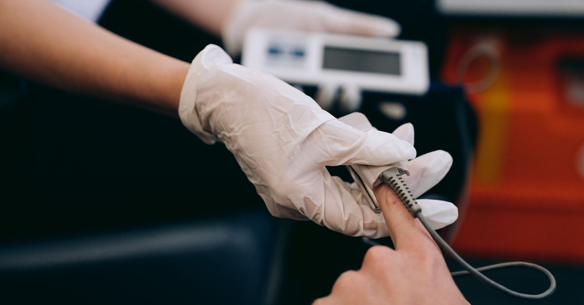
| Pulse Oximetry | Percentage of hemoglobin saturated with oxygen. | Rapid, continuous, non-invasive bedside monitoring. |
| Arterial Blood Gas | Precise arterial partial pressures of O2 and CO2. | Definitive diagnosis of severity and acid-base status. |
| Chest Radiograph (X-ray) | Visualization of lung structure and fluid. | Identifying pneumonia, edema, or collapsed lung areas. |
13. Oxygen Therapy and Delivery Systems
The immediate first-line treatment for any hypoxemic episode is the administration of supplemental oxygen. The goal is to rapidly increase the fraction of inspired oxygen, forcefully pushing more oxygen molecules across the alveolar-capillary membrane into the blood.
For mild hypoxemia, low-flow systems like a standard nasal cannula provide modest increases in oxygen concentration. For more severe deficits, high-flow systems or non-rebreather masks are utilized. A non-rebreather mask features an attached reservoir bag filled with pure oxygen and one-way valves, allowing the delivery of near 100 percent oxygen to the patient.
Oxygen is treated as a critical pharmacological intervention. The flow rate and delivery device are carefully titrated to achieve a safe target saturation level, avoiding the toxicity associated with prolonged exposure to excessively high oxygen concentrations.
14. Non-Invasive and Mechanical Ventilation
When supplemental oxygen alone fails to reverse the hypoxemia, or if the patient becomes too exhausted to maintain the muscular effort of breathing, advanced ventilatory support is required.
Non-invasive positive pressure ventilation, utilizing tight-fitting facial masks, delivers pressurized air and oxygen directly into the lungs. This continuous positive pressure splints the airways open, forcibly pushing fluid out of flooded alveoli, which is particularly effective in severe heart failure or chronic lung disease exacerbations.
If non-invasive measures fail, endotracheal intubation and invasive mechanical ventilation are instituted. A machine entirely takes over the work of breathing, allowing precise control of oxygen delivery, lung expansion pressures, and respiratory rate until the underlying lung injury begins to heal.
15. Treating the Underlying Pathology
Reversing the hypoxemia via oxygen and ventilation is a critical, life-saving bridge, but true medical resolution demands definitively treating the root cause. Without addressing the underlying pathology, the hypoxemia will immediately return once support is withdrawn.
If bacterial pneumonia is the cause, aggressive broad-spectrum intravenous antibiotics are immediately administered to clear the infectious exudate from the alveoli. If a massive pulmonary embolism is blocking perfusion, powerful thrombolytic drugs, known as clot busters, are utilized to chemically dissolve the blockage and restore blood flow.
In cases of cardiogenic pulmonary edema, potent intravenous diuretics are rapidly pushed to force the kidneys to excrete massive amounts of fluid, pulling the edema out of the lungs and allowing oxygen to reach the bloodstream once again.
16. Long-Term Management and Prognosis
For patients surviving acute, catastrophic hypoxemic events, prolonged respiratory rehabilitation is frequently required to regain lung capacity and muscular strength. The prognosis depends entirely on the reversibility of the initial lung or heart injury.
Individuals with chronic lung diseases, such as severe emphysema or pulmonary fibrosis, often develop persistent, chronic hypoxemia. These patients require long-term home oxygen therapy. Utilizing continuous supplemental oxygen through a portable concentrator profoundly improves their quality of life, reduces the dangerous strain placed on the right side of the heart, and significantly extends life expectancy.
Diligent follow-up with a pulmonologist is strictly required to adjust oxygen flow rates dynamically, monitor for the development of pulmonary hypertension, and optimize inhaled pharmacological therapies.
17. Frequently Asked Questions (FAQ)
1. What is the difference between oxygen saturation and partial pressure?
Oxygen saturation, measured by a finger clip, indicates the percentage of your hemoglobin that is carrying oxygen. Partial pressure, measured by an arterial blood draw, indicates the actual physical pressure of the oxygen gas dissolved in your blood plasma.
2. Why do my lips turn blue when I cannot breathe?
This is known as cyanosis. Hemoglobin carrying oxygen is bright red, making healthy skin look pink. When oxygen drops severely, the hemoglobin turns dark blue or purple, which shows through thin skin areas like the lips and fingernails.
3. Is breathing pure oxygen dangerous?
In a medical emergency, high doses of pure oxygen are life-saving. However, breathing 100 percent oxygen for prolonged periods (several days) can produce toxic free radicals that actively damage the lung tissue, so doctors carefully lower the dose as soon as it is safe to do so.
4. Can panic attacks cause hypoxemia?
No. Panic attacks cause hyperventilation (breathing too fast), which actually increases oxygen and lowers carbon dioxide, leading to dizziness and tingling. True hypoxemia involves a physical failure of oxygen entering the bloodstream.
5. What is considered a dangerously low oxygen level on a pulse oximeter?
Normal levels are between 95 and 100 percent. A reading consistently below 90 percent is a clinical warning sign of significant hypoxemia requiring immediate medical evaluation.
18. Bibliography
Disclaimer: The content is for informational purposes only and does not replace medical advice. Always consult your doctor for personalized treatment.

