
1. Introduction
An incipient cataract represents the earliest stage of clouding in the natural crystalline lens of the eye. The bottom line up front is that while an incipient cataract causes minimal to no immediate visual impairment, it signals the beginning of structural protein changes within the lens that will gradually progress over time, requiring routine monitoring rather than immediate surgical intervention. This stage is primarily a clinical finding discovered during a comprehensive eye examination.
The term “incipient” implies a beginning or an initial stage. At this point, the opacities are localized and have not yet coalesced to block a significant portion of light entering the eye. Understanding the mechanisms of how these early changes form allows patients to comprehend their symptoms, adopt preventative lifestyle measures, and prepare for the eventual progression of the condition.
2. Anatomy of the Crystalline Lens
To understand how a cataract forms, it is essential to review the anatomy of the natural lens. The lens is a transparent, biconvex structure located directly behind the iris and the pupil. Its primary function is to focus incoming light rays precisely onto the retina to create a sharp, clear image.
The lens consists of three main parts. The outermost layer is the elastic capsule. Beneath the capsule is the cortex, which is composed of soft, newly formed lens fibers. At the center lies the nucleus, the hardest and oldest part of the lens. The transparency of the lens is maintained by a precise arrangement of lens proteins and strict regulation of its water content.
3. Pathophysiology of Early Cataracts
As the human body ages, the natural physiological processes within the lens begin to degrade. The most significant changes occur at the biochemical level. The delicate crystallin proteins that make up the lens fibers slowly undergo oxidation and begin to misfold and clump together.
Simultaneously, the metabolic pump systems that regulate fluid within the lens become less efficient. This leads to an accumulation of fluid, causing the lens fibers to swell and separate slightly. These microscopic areas of fluid accumulation and protein aggregation disrupt the smooth transmission of light, scattering it and creating the first visual signs of a cataract.
4. Classification of Incipient Opacities
Incipient cataracts are not uniform; they can develop in different regions of the lens, and their location dictates the nature of the early symptoms. The classification is primarily based on anatomy. While these early opacities are small, the eye care professional will meticulously document their location and density.
The development may begin in the periphery of the lens, moving slowly inward like the spokes of a wheel, or it may begin deep within the central core. Identifying the specific type helps the clinician predict the timeline of visual decline and anticipate the specific visual complaints the patient might develop.
5. Cortical vs. Nuclear Changes
The two most common patterns seen in the incipient stage are cortical and nuclear changes. An incipient cortical cataract occurs in the outer cortex of the lens. It often begins as small, whitish, wedge-shaped opacities or fluid clefts at the outer edge of the lens, eventually progressing toward the center.
Conversely, an incipient nuclear sclerotic cataract affects the central nucleus. In the early stages, the center of the lens begins to harden and take on a subtle yellowish hue due to the deposition of urochrome pigments. This central hardening slowly changes the refractive power of the eye, often altering the patient’s eyeglass prescription before creating significant blur.
6. Primary Causes and Risk Factors
The vast majority of incipient cataracts are senile cataracts, meaning they are a direct result of the natural aging process. Advancing age is the single most significant risk factor, with biochemical changes becoming clinically detectable in most individuals over the age of sixty.
However, several other factors can accelerate this process. Prolonged, unprotected exposure to ultraviolet radiation from the sun significantly increases oxidative stress within the lens. Metabolic conditions, most notably diabetes mellitus, alter glucose metabolism in the eye, leading to rapid hydration changes in the lens cortex. Additionally, chronic use of corticosteroid medications and a history of smoking are established catalysts for early cataract formation.
7. Visual Symptoms in the Early Stage
Because an incipient cataract is small and often peripheral, many patients are entirely asymptomatic. When symptoms do emerge, they are typically subtle and insidious. A patient might notice a slight, overall dimming of vision, as if they are looking through a very faintly smudged piece of glass.
Colors may begin to lose their vibrancy, appearing slightly washed out or yellowed, particularly blues and purples. Patients often find themselves requiring brighter light to read fine print or complete close-up tasks, even with their updated reading glasses.
8. Glare and Contrast Sensitivity Issues
One of the hallmark early symptoms of an incipient cortical cataract is increased sensitivity to glare. Because the fluid clefts in the lens cortex scatter light irregularly, oncoming headlights during night driving can create intense starbursts or halos.
Furthermore, contrast sensitivity often declines before visual acuity (the ability to read letters on an eye chart) is affected. The patient may struggle to distinguish objects from their background in low-light environments, such as identifying the edges of stairs in a dimly lit hallway.
9. Diagnostic Eye Examination
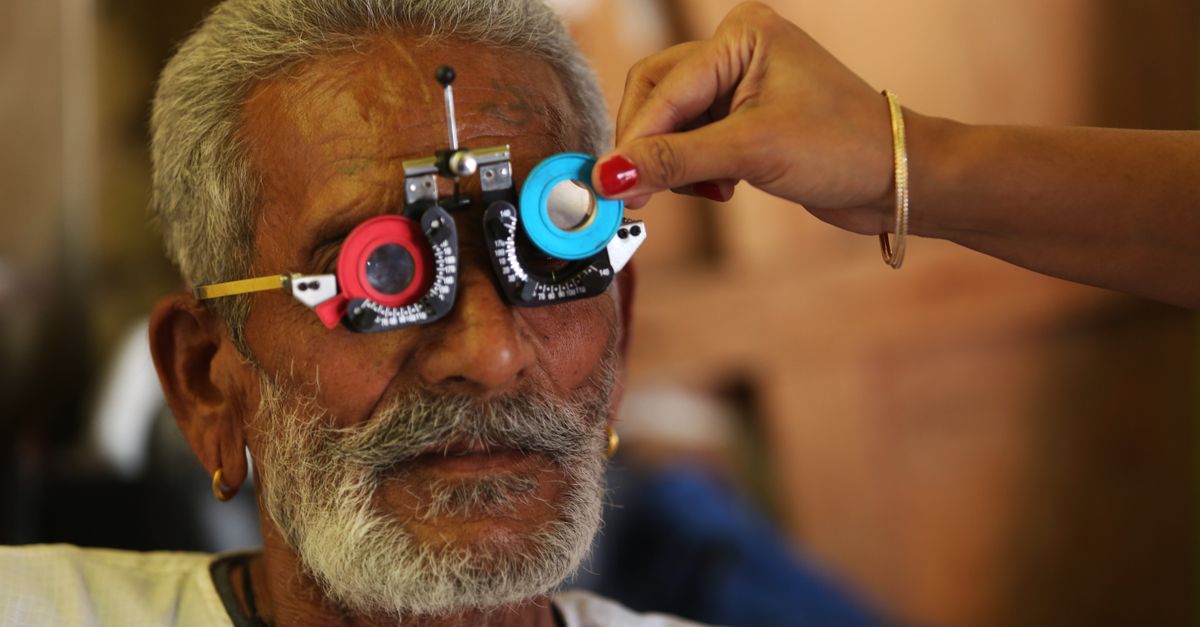
The diagnosis of an incipient cataract is straightforward and painless, performed during a comprehensive, dilated eye examination. The clinician will evaluate visual acuity under various lighting conditions and perform a careful refraction to determine if the lens changes have altered the patient’s need for glasses.
Dilation of the pupil is mandatory. Specialized eyedrops are used to enlarge the pupil, providing the clinician with a wide, unobstructed view of the entire crystalline lens, from the center to the far periphery, to accurately assess the extent of the opacification. It shares diagnostic similarities with evaluating other lens anomalies, such as those seen in an aging eye condition.
10. Role of Slit-Lamp Biomicroscopy
The primary diagnostic instrument is the slit-lamp biomicroscope. This device provides highly magnified, stereoscopic, and intensely illuminated views of the anterior structures of the eye. By directing a narrow beam of light through the lens at an angle, the clinician obtains an optical cross-section of the tissue.
Through the slit lamp, the clinician can clearly visualize the distinct layers of the lens, identifying the exact depth, density, and pattern of the protein aggregates and fluid vacuoles that define the incipient stage.
| Stage of Cataract | Anatomical Findings | Visual Symptoms |
|---|---|---|
| Incipient | Mild fluid clefts, early yellowing, peripheral opacities | Minimal blur, mild glare at night, frequent prescription changes |
| Immature | Opacities extend centrally, lens is mostly cloudy | Significant blurred vision, loss of color vibrancy, heavy glare |
| Mature | Lens is entirely opaque and dense | Severe vision loss, inability to read or drive safely |
11. Differential Diagnosis of Blurred Vision
When a patient presents with early visual complaints, the clinician must rule out other causes of vision degradation. Presbyopia, the natural loss of focusing ability for near vision, occurs in the same age demographic and can mimic the need for brighter reading light.
Macular degeneration affects the central retina and causes blurred or distorted central vision, but unlike a cataract, it does not cause a generalized haze or severe glare. Glaucoma, in its early stages, primarily affects peripheral vision. A comprehensive exam ensures that the visual symptoms are correctly attributed to the lens changes and not a more sight-threatening pathology.
12. Progression Timeline
The progression from an incipient cataract to an immature or mature cataract is highly variable. In some individuals, the opacities remain stable or progress very slowly over a decade or more. In others, particularly those with poorly controlled diabetes or those on high-dose steroids, the progression can occur rapidly over months.
Because of this unpredictability, routine monitoring is the standard of care. There is no biological necessity to remove a cataract simply because it has begun to form; intervention is based entirely on how the progression impacts the patient’s daily functional vision.
13. Non-Surgical Management and Vision Correction
During the incipient stage, surgery is generally not indicated. Management focuses on optimizing the patient’s current vision through conservative measures. As the lens changes shape and density, it induces refractive errors, often increasing myopia (nearsightedness).
Simply updating the patient’s prescription for eyeglasses or contact lenses can frequently restore crisp, clear vision for several years. Adding anti-reflective coatings to eyeglass lenses can significantly reduce the annoying glare from oncoming headlights and digital screens.
14. Lifestyle Modifications and Prevention
While the aging process cannot be halted, certain lifestyle modifications can slow the oxidative stress that accelerates cataract progression. Consistent use of sunglasses that block one hundred percent of UVA and UVB radiation is critical whenever the patient is outdoors.
A diet rich in antioxidants, particularly vitamins C and E, lutein, and zeaxanthin, provides the cellular building blocks necessary to defend the lens proteins against oxidative damage. Smoking cessation and strict management of systemic conditions like diabetes are vital to preserving lens clarity for as long as possible.
15. Monitoring and Indicators for Surgery
The clinical team will monitor the incipient cataract with annual or bi-annual examinations. The indicator for surgical intervention is subjective and depends heavily on the patient’s lifestyle requirements.
When conservative measures like new glasses no longer provide satisfactory vision, and the visual decline begins to interfere with safe driving, reading, or the enjoyment of hobbies, the cataract has progressed beyond the incipient stage. At this point, the clinician will recommend phacoemulsification surgery to remove the cloudy lens and replace it with a clear artificial intraocular implant.
16. Frequently Asked Questions (FAQ)
1. Does an incipient cataract mean I am going blind?
No. An incipient cataract simply means that the natural lens in your eye is showing the very first signs of aging and clouding. It is a slow, progressive condition that is completely treatable with standard outpatient surgery once it begins to affect your daily life.
2. Do I need surgery right away for an incipient cataract?
Surgery is rarely recommended during the incipient stage. Because the visual symptoms are very mild or non-existent, the risks of surgery outweigh the benefits at this point. Your doctor will monitor the changes over time.
3. Can I reverse a cataract with eye drops or a special diet?
No. Once the proteins in the lens have clumped together and clouded, the process cannot be reversed with medications, eye drops, or dietary changes. However, a healthy diet and UV protection can slow down further progression.
4. Why does my vision seem fine during the day but blurry at night?
Early cataracts, especially those in the outer layers of the lens, cause incoming light to scatter. During the day, your pupil is small, allowing light to pass through the clearer center of the lens. At night, your pupil dilates, exposing the clouded edges, which scatters light and causes glare from headlights.
5. How frequently should my eyes be checked if I have an incipient cataract?
Your eye care professional will generally recommend a comprehensive eye examination every one to two years to monitor the slow changes in the lens and update your eyeglass prescription to keep your vision as sharp as possible.
17. Bibliography
Disclaimer: The content is for informational purposes only and does not replace medical advice. Always consult your doctor for personalized treatment.
