
1. Introduction
An infection caused by the Mycobacterium avium-intracellulare group represents a challenging opportunistic illness involving environmental, nontuberculous mycobacteria that primarily affects the lungs or disseminates systemically in immunocompromised hosts. The bottom line up front is that while these environmental bacteria are ubiquitous and harmless to healthy individuals, they cause chronic, destructive pulmonary disease in patients with underlying lung damage, or life-threatening systemic infections in severely immunodeficient individuals, requiring a prolonged, multi-drug antimicrobial regimen anchored by a macrolide to eradicate the slow-growing bacilli and prevent structural tissue damage.
The Mycobacterium avium-intracellulare group, frequently referred to in clinical practice as the Mycobacterium avium complex or MAC, consists of several closely related, slow-growing mycobacterial species. Unlike classic tuberculosis, these infections are not considered contagious between humans. Understanding the environmental nature of the pathogen and the specific vulnerabilities of the human host is crucial for diagnosing the insidious onset of symptoms and deploying appropriate, long-term pharmacological interventions.
2. Microbiology of Nontuberculous Mycobacteria
Mycobacterium avium and Mycobacterium intracellulare are the two dominant species within this complex. They are classified as nontuberculous mycobacteria, distinguishing them from the organism that causes tuberculosis. Morphologically, they are aerobic, non-motile bacilli characterized by a remarkably thick, lipid-rich cell wall.
This waxy cell wall is the defining biological feature of the organism. It renders the bacteria highly resistant to standard environmental stressors, many common disinfectants, and most conventional classes of antibiotics, such as penicillins and cephalosporins. In the laboratory, this cell wall prevents standard Gram staining from working properly; instead, the bacteria retain a specific dye even when washed with acid alcohol, earning them the designation of “acid-fast bacilli.”
3. Environmental Sources and Transmission
The bacteria comprising the Mycobacterium avium complex are ubiquitous in the natural and constructed environment. They thrive in fresh and saltwater, soil, and dust. Notably, they have adapted exceptionally well to engineered water systems, frequently colonizing municipal water supplies, showerheads, hot tubs, and indoor plumbing pipes.
Transmission occurs entirely through environmental exposure. Humans acquire the infection primarily by inhaling aerosolized water droplets containing the bacteria, such as while showering or using a humidifier. Ingestion of contaminated water or soil is an alternative route, particularly relevant for disseminated infections. There is no documented human-to-human transmission, meaning infected patients do not need to be isolated from family members or the public.
4. Host Susceptibility Factors
Because the bacteria are ubiquitous, almost all humans are exposed to them regularly, yet only a fraction develop an infection. A robust, healthy immune system and normal lung architecture easily clear the inhaled bacilli. Disease occurs only when specific host vulnerabilities exist.
The two primary phenotypes of susceptible patients are those with structural lung disease and those with profound cellular immunosuppression. Patients with pre-existing damage to their lung tissues, such as those suffering from chronic obstructive pulmonary disease, prior tuberculosis scarring, or cystic fibrosis, lack the mechanical ability to clear mucus and bacteria from their airways, allowing the mycobacteria to establish a chronic foothold.
5. Pulmonary Disease Presentation
Pulmonary involvement is the most frequent clinical manifestation. The disease progresses insidiously, often mimicking other chronic respiratory conditions, leading to significant delays in accurate diagnosis.
Patients typically present with a chronic, lingering cough that may produce purulent sputum or, occasionally, hemoptysis (coughing up blood). Systemic symptoms often accompany the respiratory decline, including profound fatigue, low-grade fevers, unintentional weight loss, and night sweats. Because these symptoms overlap with chronic bronchitis or emphysema, a high index of clinical suspicion is required when a patient’s respiratory status fails to improve on standard therapies.
6. Disseminated Disease in Advanced Immunosuppression
In patients with severe cellular immunodeficiency, particularly those with advanced HIV and CD4 cell counts below fifty cells per microliter, the bacteria behave as aggressive, opportunistic pathogens. Rather than remaining confined to the lungs, the bacteria enter the bloodstream and disseminate throughout the body.
Disseminated disease involves the widespread infiltration of the bone marrow, liver, spleen, and lymphatic system. Patients present with persistent fevers, drenching night sweats, massive weight loss, and severe anemia. Before the advent of potent antiretroviral therapy, disseminated MAC was a leading cause of mortality in patients with AIDS, emphasizing the critical role of cellular immunity in containing these environmental organisms. Evaluating these extensive symptoms is standard in comprehensive opportunistic infections management.
7. Pathophysiology of Granuloma Formation
When the mycobacteria enter the lungs of a vulnerable host, local macrophages attempt to engulf and destroy them. However, the thick, waxy cell wall of the bacteria allows them to survive and multiply directly inside the macrophages.
The immune system responds by recruiting more white blood cells to the area, attempting to wall off the infection by forming dense, nodular structures called granulomas. While granulomas help contain the bacteria, the chronic, localized inflammation eventually leads to tissue necrosis and structural damage to the lung architecture, contributing to the development of bronchiectasis and cavitary lesions.
8. Radiographic Imaging and High-Resolution CT
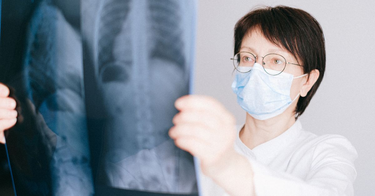
Diagnostic imaging plays a pivotal role in assessing the extent of pulmonary damage. While a standard chest X-ray can show non-specific infiltrates or cavities, High-Resolution Computed Tomography of the chest is the gold standard imaging modality.
The scan typically reveals one of two distinct patterns. The fibrocavitary form primarily affects older males with a history of smoking, displaying large, destructive cavities in the upper lobes of the lungs. The nodular bronchiectatic form frequently affects older females without a smoking history, showing clustered nodules and widened, scarred airways (bronchiectasis), predominantly in the middle lobe and lingula.
9. Diagnostic Criteria and Culturing
Diagnosing a pulmonary infection requires meeting a strict combination of clinical, radiographic, and microbiological criteria. Because the bacteria can temporarily colonize the airways without causing active disease, a single positive sputum sample is insufficient for a diagnosis.
The guidelines require the patient to have appropriate respiratory symptoms and matching CT scan abnormalities, combined with microbiological proof. This proof involves obtaining at least two separate positive sputum cultures on different days, or one positive culture obtained directly from the deep airways via bronchoscopy.
| Diagnostic Category | Required Criteria for Pulmonary Infection |
|---|---|
| Clinical | Chronic cough, fatigue, weight loss, appropriate exclusion of other diseases. |
| Radiographic | Nodular or cavitary opacities on chest X-ray, or bronchiectasis on High-Resolution CT. |
| Microbiological | Two positive sputum cultures, or one positive bronchoscopy culture/biopsy. |
10. The Role of Molecular Diagnostics
Traditional culturing of these bacteria on specialized media is necessary but exceptionally slow; it can take two to six weeks for the bacilli to grow visibly in a laboratory.
To expedite treatment, clinical laboratories heavily utilize molecular diagnostics, specifically polymerase chain reaction tests and nucleic acid probes. These tests identify the specific genetic material of the bacteria directly from the sputum sample within a few days. They rapidly differentiate the avium-intracellulare group from classic tuberculosis, which is a critical step in determining the necessary public health protocols and initial antibiotic regimens.
11. Multidrug Antimicrobial Therapy
Eradicating these slow-growing mycobacteria requires a complex, multi-drug pharmacological strategy. Using a single antibiotic inevitably leads to rapid bacterial resistance. The standard treatment regimen involves at least three different antimicrobial agents administered simultaneously.
The absolute cornerstone of therapy is a newer-generation macrolide antibiotic, either azithromycin or clarithromycin. The macrolide is typically combined with ethambutol and a rifamycin, such as rifampin or rifabutin. This three-drug regimen attacks the bacteria at multiple stages of their slow replication cycle, gradually reducing the bacterial burden within the lungs or bloodstream.
12. Managing Drug Toxicity and Interactions
The prolonged nature of the multi-drug therapy introduces significant challenges regarding medication side effects and drug interactions. The clinical team must monitor the patient closely throughout the entire treatment course.
Macrolides can cause gastrointestinal distress and, rarely, cardiac arrhythmias. Ethambutol carries a specific risk of optic neuritis, requiring patients to undergo baseline and routine visual acuity and color vision testing. Rifampin is a potent inducer of liver enzymes, heavily altering the metabolism of numerous other prescription medications, including oral contraceptives and certain cardiovascular drugs, requiring careful pharmacist review.
13. Treatment Duration and Monitoring
Treating an active infection is a marathon, not a sprint. Because the bacteria grow so slowly and hide within the lung tissue and macrophages, a short course of antibiotics is entirely ineffective.
Clinical guidelines mandate that the multi-drug therapy continue for a minimum of twelve months after the patient provides their first negative sputum culture. In practice, this often means the patient will take three different antibiotics daily or three times a week for eighteen to twenty-four months. Sputum cultures are checked every one to two months to monitor the bacteriological response to therapy.
14. Surgical Resection Considerations
In specific clinical scenarios, pharmacological therapy alone is insufficient to cure the disease. If the infection is highly localized to a single, large cavity in the lung, the antibiotics may not be able to penetrate the thick, avascular scar tissue surrounding the cavity.
If a patient continues to produce positive sputum cultures despite many months of optimal multi-drug therapy, and they possess adequate lung function, a thoracic surgeon may surgically remove the heavily diseased lobe of the lung. This surgical resection physically eliminates the bacterial reservoir, significantly improving the chances of a medical cure.
15. Prophylaxis in Immunocompromised Patients
For patients with advanced HIV, primary prevention is highly effective. If a patient’s CD4 cell count drops below fifty cells per microliter, clinical guidelines recommend initiating prophylactic therapy with azithromycin to prevent the environmental bacteria from establishing a disseminated infection.
This prophylaxis is continued until antiretroviral therapy successfully restores the patient’s immune system, raising the CD4 count above the safe threshold. The widespread use of modern HIV medications has drastically reduced the incidence of disseminated disease in the developed world.
16. Frequently Asked Questions (FAQ)
1. Is an infection with MAC the same as tuberculosis?
No. While the bacteria belong to the same overarching family, they are completely different diseases. Tuberculosis is highly contagious and spreads from person to person. MAC is an environmental bacterium and is not contagious; you cannot catch it from or give it to another person.
2. Where do these bacteria come from?
The bacteria live naturally in the environment, particularly in soil and water. They are commonly found in municipal tap water, showerheads, hot tubs, and garden soil. Everyone breathes them in, but they only cause disease in vulnerable individuals.
3. Why does the treatment take over a year to complete?
The bacteria have a very thick, waxy outer wall and multiply incredibly slowly compared to normal bacteria. It takes many months of constant exposure to multiple antibiotics to completely destroy every single slow-growing bacterium hidden in the lung tissue.
4. Why do I need to take three different antibiotics at once?
If you only take one or two medications, these specific bacteria will quickly mutate and become immune to the drugs. Taking three different antibiotics attacks the bacteria in three different ways, preventing them from developing resistance.
5. Do I need to stop showering to prevent reinfection?
You do not need to stop showering, but certain precautions can help if you are highly susceptible. Clinicians often recommend ensuring your hot water heater is turned up to a safe but higher temperature to limit bacterial growth, cleaning showerheads regularly, and ensuring good bathroom ventilation.
17. Bibliography
Disclaimer: The content is for informational purposes only and does not replace medical advice. Always consult your doctor for personalized treatment.
