
1. Introduction
Normal periapical tissue is the highly specialized complex of anatomical structures located immediately surrounding the apex, or the very tip, of a tooth root. This dynamic biological zone serves as the critical transition point between the internal environment of the tooth and the surrounding jawbone. It is a bustling intersection of nerves, blood vessels, connective tissues, and immune cells, all working synchronously to anchor the tooth and maintain vitality.
The health of the periapical region is an absolute prerequisite for a functional, pain-free dentition. Because the dental pulp inside the tooth communicates directly with the systemic circulation through this narrow apical gateway, the periapical tissue acts as a vigilant biological guardian. It provides essential nourishment to the internal tooth structures while simultaneously maintaining a strict defensive perimeter against microbial invasion.
Understanding the anatomy and physiological mechanics of normal periapical tissue provides essential insight into endodontic health. When this tissue remains uninflamed and structurally intact, the tooth can withstand the immense mechanical forces of daily mastication. Recognizing the clinical and radiographic hallmarks of a healthy apex allows dental professionals to detect the earliest signs of internal tooth disease before extensive bone damage occurs.
2. Anatomy of the Tooth Root and Apex
The root of a human tooth is the anatomical segment anchored firmly beneath the gumline, hidden entirely within the jawbone. The very terminal end of this root is called the apex. While the crown of the tooth is covered by hard, impenetrable enamel, the outer surface of the root is covered by a slightly softer, bone-like material known as cementum.
The apex is not a solid, closed point. It contains one or more microscopic openings known as apical foramina. These minute channels act as the primary structural gateways connecting the hollow internal pulp chamber of the tooth directly to the tissues surrounding the root tip.
Normal periapical tissue conforms perfectly to the contours of this root apex. The structural architecture in this region must remain highly organized to support the blood vessels passing through the small foramina, ensuring that they are not crushed or sheared by the physical movements of the tooth during normal biting and chewing.
3. Components of the Periapical Region
The term periapical tissue does not describe a single structure but rather a functional triad of tissues surrounding the root tip. This triad includes the apical portion of the periodontal ligament, the surrounding alveolar bone, and the apical cementum covering the root tip itself.
Each component performs a distinct yet heavily integrated function. The cementum provides an attachment surface. The periodontal ligament acts as a highly specialized biological shock absorber and sensory network. The alveolar bone provides the rigid, structural housing that supports the entire dental complex.
When normal periapical tissue is assessed clinically, all three of these components must demonstrate specific signs of health. The presence of dense bone, a uniform ligament space, and intact cementum indicates that the internal pulp is healthy and that no inflammatory process has breached the apex.
4. The Periodontal Ligament at the Apex
The periodontal ligament is a dense, fibrous connective tissue network that spans the narrow gap between the tooth root and the bony socket. At the apex of the tooth, this ligament forms a complex, web-like cushion around the root tip.
The primary function of the apical periodontal ligament is mechanical support. It consists of thick bundles of collagen fibers that act like a physiological hammock, suspending the tooth within the bone. When pressure is applied to the tooth crown, these apical fibers stretch, absorbing and distributing the physical load evenly into the surrounding bone to prevent fracturing.
Normal periapical tissue requires this ligament to remain pliable and well-hydrated. In a healthy state, the width of the periodontal ligament space around the apex remains exceptionally narrow and uniform, measuring roughly a fraction of a millimeter on a dental radiograph.
5. Alveolar Bone Surrounding the Apex
The alveolar bone is the specialized portion of the jawbone that forms the sockets housing the teeth. The bone immediately adjacent to the periapical region is known as the lamina dura, which appears as a distinct, dense white line on dental X-rays.
This bone is remarkably dynamic, constantly remodeling itself in response to the mechanical forces transmitted through the periodontal ligament. Normal periapical tissue implies that the alveolar bone completely encases the root tip without any areas of resorption or localized destruction.
If bacteria from a severely decayed tooth manage to travel down the root canal and exit through the apex, the immune system will trigger a massive inflammatory response in this bone. This inflammation causes specialized cells to dissolve the bone tissue rapidly, creating a dark, hollow lesion on an X-ray, which signals the immediate loss of normal periapical architecture.
6. Cementum and Apical Sealing
Cementum is the mineralized connective tissue covering the outer surface of the tooth root. At the periapical region, cellular cementum is particularly thick and active. This tissue provides a sturdy anchor point for the collagen fibers of the periodontal ligament to insert themselves.
A unique feature of apical cementum is its continuous deposition throughout a person’s life. As the biting surface of the tooth slowly wears down from decades of chewing, new cementum is actively deposited at the apex. This continuous buildup subtly pushes the tooth upward, ensuring it remains in active contact with the opposing teeth.
Furthermore, if the tooth suffers minor trauma or irritation, the cementum at the apex attempts to seal off the microscopic accessory canals. This biological sealing mechanism is a vital component of normal periapical tissue defense, minimizing the pathways through which bacterial toxins can enter the surrounding bone.
7. Blood Supply and Vascularization
The vitality of the tooth depends entirely on a robust vascular supply navigating through the periapical region. Tiny arterioles branch off from the main alveolar arteries running through the jawbone. These arterioles enter the periapical tissue, branch into microscopic capillary networks, and funnel directly through the apical foramen into the tooth pulp.
Simultaneously, venules drain deoxygenated blood and metabolic waste products out of the pulp, exiting through the same tight apical opening. Normal periapical tissue must maintain an intricate spatial arrangement to prevent these vessels from being compressed.
Because the apical foramen is an unyielding, hard opening, any significant inflammation or swelling inside the tooth pulp will rapidly compress these blood vessels against the rigid walls of the root. This cuts off the blood supply, leading to the rapid death of the internal tooth tissue, underscoring the delicate vascular balance at the apex.
8. Neural Pathways and Sensory Function
The periapical tissue is densely innervated by branches of the trigeminal nerve. Sensory nerve bundles travel alongside the blood vessels, passing through the periapical space and entering the root canal to provide sensation to the internal pulp.
Additionally, the apical periodontal ligament itself is heavily populated with highly specialized proprioceptive nerve endings. These sensors are incredibly sensitive to pressure and spatial orientation. When a person bites down, these receptors instantly measure the exact amount of mechanical force being applied to the tooth.
This sensory feedback is crucial for normal dental function. It signals the brain to adjust the strength of the jaw muscles, preventing a person from biting down with destructive force on a hard object like a seed or a bone fragment, protecting both the tooth and the underlying jawbone from catastrophic fracture.
9. The Role of the Apical Foramen
The apical foramen is the defining structural feature of the periapical region. It is the primary anatomical gateway connecting the systemic body to the isolated environment of the dental pulp. The diameter of this opening changes over a lifespan.
In a newly erupted tooth, the apical foramen is wide open, resembling a blunderbuss, allowing massive blood flow necessary to complete root formation. Once the root is fully developed, normal periapical tissue continues to lay down cementum, gradually narrowing the opening to a microscopic pinpoint.
This constricted anatomy in mature teeth is highly protective, limiting the volume of toxins that can escape an infected tooth. However, this same narrow constriction creates a significant challenge during endodontic treatment, demanding exceptional precision from the clinician attempting to clean and seal the canal to the exact depth of the apex.
10. Immune Defense in the Periapical Space
The periapical tissue acts as a heavily fortified immune checkpoint. Even in a perfectly normal, healthy state, a small number of specialized immune cells, including macrophages and lymphocytes, constantly patrol the connective tissue surrounding the apex.
Their primary mission is continuous biological surveillance. Because the oral cavity is teeming with bacteria, microscopic toxins occasionally diffuse down the root canal towards the apex. The local immune cells immediately recognize and neutralize these transient threats without initiating a destructive inflammatory cascade.
Normal periapical tissue function represents a state of perfect immunological homeostasis. The defensive cells manage minor irritants efficiently, maintaining the structural integrity of the bone and ligament without causing pain or pathological tissue destruction.
11. Radiographic Appearance of Normal Tissue
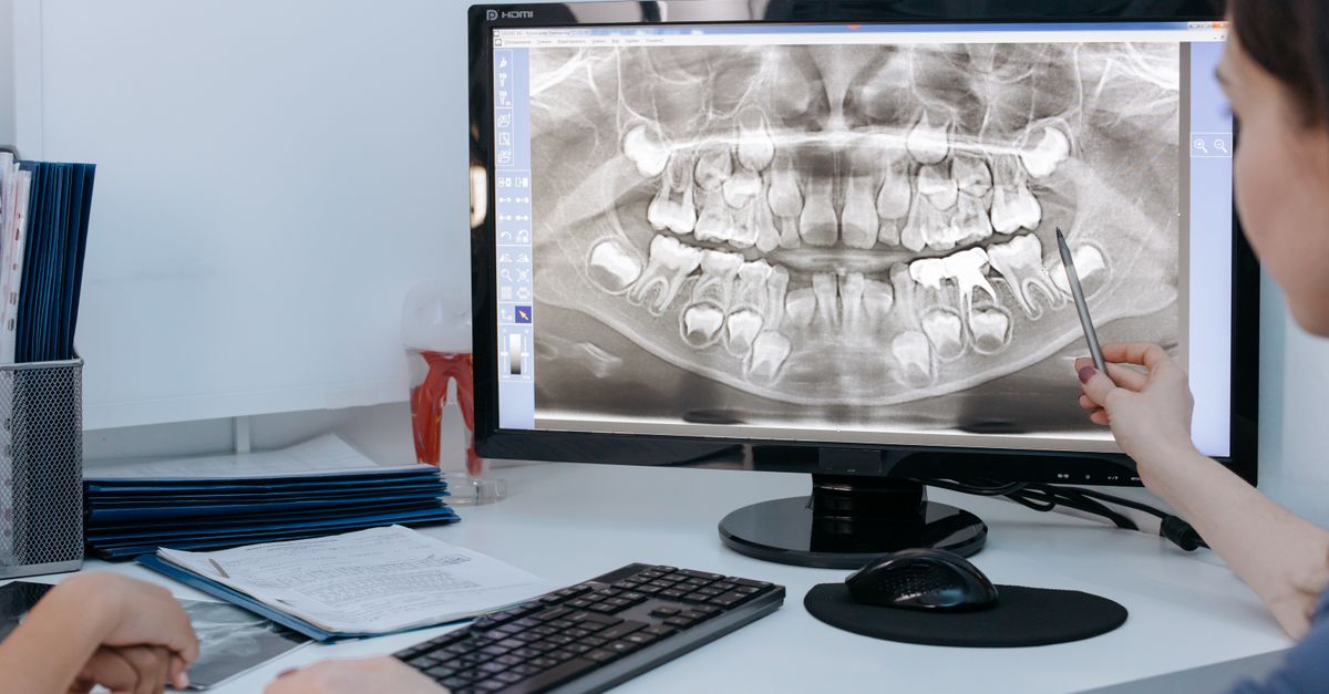
Because the periapical region is buried deep within the jawbone, clinicians rely heavily on dental radiographs to evaluate its health. Recognizing the strict visual parameters of normal periapical tissue is the foundation of endodontic diagnosis.
On a healthy X-ray, the tooth root appears as a solid white structure. The periodontal ligament space appears as a very thin, uniform, continuous dark line hugging the entire contour of the root apex. Immediately outside this dark line, the lamina dura appears as a crisp, continuous white border of dense bone.
The bone surrounding the lamina dura should display a normal trabecular pattern, resembling a tightly woven, uniform sponge. An absence of any dark, expanding shadows or widening of the ligament space at the root tip confirms that the periapical tissue is biologically stable and uninfected.
12. Response to Biomechanical Stress
Normal periapical tissue is incredibly resilient and adapts to changes in functional demands. If a tooth is subjected to unusually heavy biting forces—such as from chronic teeth grinding or a high dental filling—the periapical tissue undergoes specific physiological adaptations.
The apical periodontal ligament will widen slightly to accommodate the increased shock absorption requirement. The surrounding alveolar bone will become noticeably denser, thickening the lamina dura to provide stronger support against the excessive mechanical load.
These adaptations, while clinically visible on an X-ray, are entirely normal physiological responses, not indicators of infection. However, if the mechanical stress becomes too extreme, the adaptive capacity fails, resulting in severe toothache and localized tissue trauma at the apex.
13. Preserving Periapical Health
The health of the periapical tissue is entirely dependent on the health of the internal dental pulp. Therefore, preserving normal periapical tissue function requires preventing deep bacterial decay and severe dental trauma.
Meticulous daily oral hygiene, including proper brushing and flossing, is necessary to prevent the formation of dental caries. When a cavity forms, it must be treated promptly by a dentist. If decay is allowed to progress unchecked, bacteria will eventually penetrate the deep pulp chamber.
Once the pulp becomes infected, the bacteria will inevitably migrate down the root canal and exit through the apical foramen, transforming the healthy periapical tissue into a highly destructive inflammatory lesion, known as an apical abscess, which actively destroys the jawbone.
14. When to Consult an Endodontist
You should seek professional dental evaluation immediately if you experience deep, throbbing pain that originates within the jawbone, particularly if the pain prevents you from sleeping or is significantly worsened by biting down or applying pressure to a specific tooth.
Schedule an emergency appointment with an endodontist or general dentist if you develop a visible, pimple-like swelling on the gums near the base of a tooth root, or if you experience severe facial swelling. These symptoms indicate that bacteria have breached the apex and caused a massive periapical infection that is spreading into the surrounding tissues.
Furthermore, if a tooth suddenly becomes highly sensitive to hot or cold temperatures and the pain lingers long after the stimulus is removed, professional clinical and radiographic assessment of the periapical tissue is urgently required to determine if a root canal procedure is necessary to save the tooth.
15. Frequently Asked Questions (FAQ)
1. Where exactly is the periapical tissue located?
The prefix “peri” means around, and “apical” refers to the apex. Therefore, periapical tissue refers specifically to the bone, ligament, and connective tissue located immediately around the very tip of the tooth root, buried deep under your gums.
2. Can an X-ray show if my periapical tissue is normal?
Yes. Dental X-rays are the primary tool dentists use to check this area. If the tissue is normal, the X-ray will show healthy, solid bone hugging the root tip. If there is an infection, a distinct dark circle or shadow will appear around the tip of the root.
3. What causes a periapical infection?
A periapical infection almost always originates from inside the tooth. When a deep cavity or severe trauma kills the nerve inside the tooth, bacteria multiply inside the hollow root and eventually spill out through the tip into the surrounding bone.
4. How does a root canal treat periapical tissue?
A root canal cleans the bacteria and dead nerve tissue out from the inside of the tooth. By removing the source of the bacteria and sealing the root tip, the immune system can then heal the damaged periapical tissue and regenerate the bone around the apex.
5. Does it hurt when periapical tissue adapts to heavy chewing?
A mild, temporary ache or soreness can occur if the tooth is subjected to sudden, heavy stress, like a new filling that is slightly too high. The tissue becomes bruised. Once the heavy pressure is corrected by a dentist, the normal tissue recovers quickly and the pain disappears.
16. Bibliography
Disclaimer: The content is for informational purposes only and does not replace medical advice. Always consult your doctor for personalized treatment.
