1. Introduction
Pain on penetration, clinically classified as dyspareunia, is a complex medical condition characterized by persistent or recurrent genital discomfort occurring just before, during, or after sexual intercourse. The primary causes range from localized mucosal inflammation and hormonal deficiencies to deep pelvic tissue pathology. Initial management requires a meticulous clinical evaluation to identify the specific anatomical source of the discomfort.
This condition affects a significant portion of the female population and can lead to profound distress, impacting both sexual function and overall quality of life. The sensation may present as a sharp superficial burning or a deep pelvic ache, depending entirely on the underlying etiology. Addressing this pain requires a systematic medical approach rather than dismissing it as a purely psychological issue.
Accurate diagnosis is paramount. By delineating whether the pain occurs at the vaginal entrance or deep within the pelvis, medical professionals can formulate a targeted therapeutic strategy. Successful intervention frequently involves a multidisciplinary approach encompassing gynecological treatment, physical therapy, and appropriate pain management.
2. Anatomy of the Female Pelvic Region
The female reproductive tract and its surrounding structures form a highly innervated and vascularized network. The introitus, or vaginal opening, is lined with sensitive mucosal tissue and contains the Bartholin glands, which provide necessary lubrication. The integrity of this mucosal lining relies heavily on adequate systemic estrogen levels.
Surrounding the vaginal canal is a complex hammock of pelvic floor muscles, primarily the levator ani group. These muscles provide structural support for the pelvic organs and must relax synchronously to allow for comfortable penetration. Involuntary spasms in this muscular layer directly impede penetration and generate severe mechanical pain.
Deeper within the pelvis lie the cervix, uterus, fallopian tubes, and ovaries. These internal structures are suspended by ligaments that can be subjected to tension or pathological adhesion formation. Deep penetration impacts these organs, meaning any inflammatory or structural anomaly in the upper pelvis will be registered as deep-seated pain.
3. Pathophysiology of Penetration Pain
The mechanism of pain during penetration is broadly divided into two pathophysiological categories: superficial and deep. Superficial pain involves the neural pathways of the vulva and vaginal introitus. Inflammation in this area sensitizes the local peripheral nerves, leading to an exaggerated pain response to normal tactile stimuli, a phenomenon known as allodynia.
Deep penetration pain originates from the visceral organs of the pelvis. When the cervix or vaginal fornices are impacted, mechanical pressure is transmitted to the uterus and surrounding ligaments. If these structures are inflamed, tethered by scar tissue, or abnormally enlarged, the mechanical force triggers deep visceral nociceptors.
A secondary pathophysiological mechanism involves the pelvic floor musculature. Chronic pain, regardless of its original source, often leads to a reflex guarding response. The pelvic floor muscles become hypertonic and ischemic. This localized muscle tension not only exacerbates the initial pain but can become an independent source of chronic discomfort.
4. Superficial Dyspareunia and Introital Causes
Superficial dyspareunia is defined by pain localized to the vulva or vaginal entrance upon initial penetration. One of the most prevalent causes is inadequate lubrication, which creates mechanical friction and micro-abrasions in the delicate mucosal lining. This lack of lubrication may result from insufficient arousal, medication side effects, or hormonal shifts.
Vulvodynia is a chronic pain condition of the vulva without an identifiable infectious or structural cause. Patients typically describe a severe burning or stinging sensation upon any localized pressure. A specific subset, provoked vestibulodynia, involves acute pain strictly localized to the vulvar vestibule during attempted penetration.
Infectious agents are also common culprits. Fungal infections, such as candidiasis, or bacterial vaginosis cause acute mucosal inflammation. The resulting tissue edema and erythema make any frictional contact highly painful until the underlying microbial imbalance is medically eradicated.
5. Deep Dyspareunia and Pelvic Pathology
Deep dyspareunia occurs when the male partner or a penetrative object impacts the deeper structures of the vaginal canal or cervix. Endometriosis is a leading cause of this specific pain pattern. In this condition, endometrial-like tissue implants on the pelvic organs and ligaments, creating dense, painful scar tissue that restricts organ mobility during intercourse.
Pelvic inflammatory disease, usually resulting from an ascending bacterial infection, leaves the pelvic organs chronically inflamed and tender. The resulting adhesions tether the uterus and ovaries to the bowel or pelvic sidewall. Any mechanical movement of the cervix during penetration stretches these adhesions, causing sharp internal pain.
Uterine fibroids and ovarian cysts can also mechanically obstruct or alter the anatomical alignment of the pelvis. A retroverted uterus, while often a normal anatomical variant, can orient the cervix in a manner that makes it more susceptible to direct collision during intercourse, leading to localized aching.
| Category of Pain | Common Clinical Causes |
|---|---|
| Superficial Dyspareunia | Vulvovaginal atrophy, vulvodynia, localized infections. |
| Deep Dyspareunia | Endometriosis, pelvic inflammatory disease, ovarian cysts. |
| Muscular Pain | Pelvic floor hypertonicity, vaginismus. |
| Dermatological Pain | Lichen sclerosus, contact dermatitis. |
6. The Role of Hormonal Changes
Estrogen plays a critical role in maintaining the structural integrity, elasticity, and moisture of the vulvovaginal tissues. During menopause, postpartum periods, or specific oncological treatments, systemic estrogen levels drop significantly. This hormonal deprivation leads to a condition known as genitourinary syndrome of menopause.
As estrogen levels decline, the vaginal mucosa becomes progressively thinner, drier, and less elastic. The localized blood flow diminishes, and the natural acidic pH of the vagina shifts, increasing susceptibility to irritation. Attempted penetration under these conditions causes micro-tears in the fragile tissue, resulting in acute, stinging pain.
Hormonal contraceptives can occasionally contribute to superficial pain. Certain low-dose oral contraceptives may alter the localized hormonal environment of the vulvar vestibule, rendering the mucosal tissues excessively dry and highly sensitive to mechanical friction.
7. Dermatological and Mucosal Conditions
Chronic dermatological conditions affecting the vulvar region represent a specialized source of penetration pain. Lichen sclerosus is an autoimmune-mediated inflammatory disorder that causes the vulvar skin to become thin, white, and exceptionally fragile. Over time, it can lead to anatomical scarring and narrowing of the vaginal opening.
Lichen planus is another inflammatory condition that can affect mucosal surfaces, including the vagina. It often presents with erosive lesions that are intensely painful. When the vaginal canal is involved, the chronic inflammation can lead to the formation of synechiae, which are internal scar tissue bands that physically obstruct penetration.
Contact dermatitis is an acute allergic or irritant response to external agents. Soaps, synthetic underwear, latex condoms, or spermicides can trigger a severe inflammatory reaction in the vulvar tissues. The resulting swelling and blistering make sexual activity impossible until the offending agent is removed and the skin barrier heals.
8. Pelvic Floor Muscle Dysfunction
The pelvic floor muscles are intimately involved in the mechanics of sexual intercourse. Vaginismus is a specific functional disorder characterized by the involuntary, reflexive spasm of the musculature surrounding the outer third of the vagina. This intense muscle contraction acts as a physical barrier, making penetration extremely painful or entirely impossible.
Hypertonic pelvic floor dysfunction occurs when the muscles remain in a constant state of elevated tension, often secondary to chronic pelvic pain from other sources. These shortened, fatigued muscles develop painful trigger points. Pressure applied to these trigger points during intercourse elicits sharp, radiating pain.
Evaluating muscle tone is an essential part of the clinical assessment. It requires specialized training to differentiate between primary muscular disorders and muscle spasms that are occurring reflexively to guard against a deep inflammatory process like urinary bladder pain or endometriosis.
9. Psychological and Psychosomatic Factors
The perception of pain is highly influenced by psychological states and neurobiological feedback loops. Anxiety regarding sexual intercourse, particularly following a history of painful experiences, activates the sympathetic nervous system. This autonomic arousal directly triggers pelvic floor muscle tension and reduces localized lubrication.
A history of physical or sexual trauma can leave profound somatic imprints. In such cases, the brain’s threat-detection circuitry is hypersensitized, interpreting the physical act of penetration as a danger. This leads to immediate, severe muscle guarding and a heightened perception of pain through central sensitization.
Addressing the psychological components of dyspareunia does not imply the pain is imaginary. The pain signals are anatomically real, but their amplification requires a comprehensive treatment strategy that may include cognitive behavioral therapy to recalibrate the nervous system’s response to tactile stimuli.
10. Clinical Evaluation and Gynecological Examination
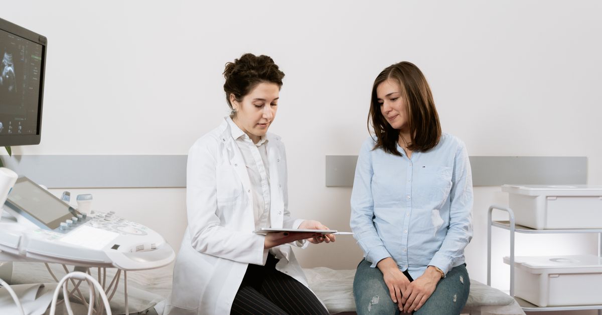
A thorough clinical evaluation begins with a detailed, empathetic medical history. The physician must ascertain the exact timing, location, and character of the pain. Distinguishing whether the pain is primary occurring since the first attempt at intercourse or secondary developing after a period of pain-free function is diagnostically crucial.
The physical examination is conducted with the utmost care, prioritizing patient comfort. A visual inspection assesses the vulva for dermatological changes, erythema, or anatomical narrowing. The Q-tip test is frequently employed to map localized hyperesthesia in the vulvar vestibule by applying light pressure to specific glandular regions.
A gentle digital examination evaluates the tone and tenderness of the pelvic floor muscles. The clinician then palpates the internal pelvic organs, assessing the mobility of the uterus and identifying any nodularity along the uterosacral ligaments that might suggest deep infiltrating endometriosis.
11. Diagnostic Imaging and Laboratory Tests
When deep dyspareunia is reported, transvaginal ultrasonography is the primary imaging modality. It provides high-resolution visualization of the uterus, endometrium, and ovaries. This ultrasound can easily detect structural anomalies such as ovarian cysts, uterine fibroids, or signs of adenomyosis.
In complex cases where deep endometriosis is suspected, magnetic resonance imaging provides a more comprehensive anatomical map of the pelvic basin. MRI is highly sensitive for identifying deep fibrotic plaques tethering the bowel to the posterior vaginal fornix, which are a major source of deep penetration pain.
Laboratory testing typically includes targeted vaginal swabs. These swabs assess the localized flora to rule out subclinical bacterial, fungal, or trichomonas infections. A urinalysis may also be ordered to ensure that adjacent structures are not harboring a chronic infectious process.
12. Differential Diagnosis
Distinguishing the exact cause of dyspareunia requires careful differentiation from overlapping pelvic pain syndromes. Interstitial cystitis, a chronic inflammatory condition of the bladder, frequently causes referred pain to the anterior vaginal wall. Mechanical pressure on the anterior wall during intercourse exacerbates bladder discomfort.
Gastrointestinal disorders, particularly irritable bowel syndrome or chronic constipation, can cause generalized pelvic floor dysfunction. An impacted rectum places pressure on the posterior vaginal wall, narrowing the vaginal canal and causing deep aching pain during penetrative acts.
Neuropathic conditions must also be considered. Pudendal neuralgia involves the compression or irritation of the primary nerve supplying the perineum. This presents as severe, electric, shooting pain that is often exacerbated by sitting and can be triggered by the physical pressure of intercourse.
13. When to See a Doctor
Medical intervention is necessary when pain during penetration is persistent, causes significant emotional distress, or leads to the avoidance of intimacy. Occasional mild discomfort due to transient lack of arousal is normal, but sharp, deep, or consistently reproducible pain indicates an underlying structural or inflammatory issue.
Immediate clinical evaluation is imperative if the pain is accompanied by abnormal vaginal bleeding, foul-smelling discharge, or systemic signs such as fever and chills. These symptoms strongly suggest an acute infectious process, such as pelvic inflammatory disease, which requires urgent antibiotic therapy to prevent irreversible reproductive damage.
Patients should also seek care if they experience a sudden, severe onset of deep pelvic pain during intercourse. This can indicate the rupture of an ovarian cyst or the torsion of an adnexal mass, both of which are acute medical situations requiring immediate assessment.
14. Pharmacological Treatments and Hormonal Therapy
The pharmacological approach directly targets the identified underlying etiology. For genitourinary syndrome of menopause, localized estrogen therapy is the gold standard. Administered as a vaginal cream, tablet, or ring, low-dose estrogen rejuvenates the mucosal lining, restores elasticity, and dramatically reduces superficial pain without significant systemic absorption.
Infectious etiologies are treated with specific antimicrobial or antifungal regimens. For chronic inflammatory conditions like lichen sclerosus, high-potency topical corticosteroids are prescribed to suppress the localized autoimmune response, halt tissue scarring, and relieve burning sensations.
For patients with central sensitization or localized nerve pain, neuromodulating medications such as topical gabapentin or oral amitriptyline may be utilized. These medications alter the transmission of pain signals along the peripheral nerves, raising the pain threshold and providing significant symptomatic relief.
15. Pelvic Floor Physical Therapy
Pelvic floor physical therapy is a cornerstone of treatment for dyspareunia, particularly when muscular hypertonicity or vaginismus is present. Specialized therapists use manual techniques to release trigger points within the levator ani muscles, restoring normal resting muscle length and improving local blood flow.
Vaginal dilator therapy is frequently incorporated into the rehabilitation program. Using a series of progressively sized medical dilators, the patient works to gently stretch the vaginal opening and desensitize the local neural pathways. This systematic approach gradually retrains the nervous system to accept penetration without triggering a reflexive muscle spasm.
Biofeedback is another highly effective therapeutic tool. Small sensors are utilized to provide real-time visual or auditory feedback regarding pelvic muscle tension. This allows the patient to consciously identify and release involuntary muscle guarding, granting them active control over their pelvic floor dynamics.
16. Minimally Invasive and Surgical Options
Surgical intervention is strictly reserved for cases where structural pathology cannot be managed conservatively. For severe endometriosis causing deep dyspareunia, laparoscopic excision of the endometrial implants and scar tissue is highly effective. Removing these dense adhesions restores organ mobility and significantly reduces mechanical pain.
In cases of localized provoked vestibulodynia that entirely resist conservative management, a vestibulectomy may be considered. This surgical procedure involves the precise excision of the hyper-sensitive mucosal tissue at the vaginal entrance. Following recovery, the newly advanced tissue typically demonstrates a normal pain threshold.
The surgical management of uterine fibroids or large ovarian cysts via myomectomy or cystectomy also resolves deep mechanical pain. By restoring the normal anatomical volume and alignment of the pelvic organs, the mechanical collisions that trigger visceral nociceptors during intercourse are eliminated.
17. Frequently Asked Questions (FAQ)
1. Is pain during initial penetration always caused by a lack of lubrication?
No. While insufficient lubrication is a frequent cause of superficial friction, immediate pain upon entry can also indicate provoked vestibulodynia, localized mucosal infections, or reflex muscle spasms associated with vaginismus.
2. Can taking birth control pills cause painful intercourse?
In some cases, yes. Certain low-dose oral contraceptives can suppress natural androgen and estrogen production enough to cause localized vaginal dryness and mucosal thinning, leading to pain during friction.
3. Will pelvic floor exercises cure my pain?
If your pain is caused by hypertonic muscle spasms or vaginismus, targeted pelvic floor down-training and relaxation techniques are highly effective. However, standard strengthening exercises like Kegels can actually worsen pain caused by muscle tightness.
4. Why does the pain sometimes continue for hours after intercourse?
Post-coital aching is typical of deep pelvic conditions like endometriosis or severe pelvic floor muscle spasms. The mechanical irritation triggers an inflammatory response and sustained muscular cramping that persists long after the physical stimulus is removed.
5. How long does localized estrogen take to relieve menopausal vaginal pain?
Patients typically notice a significant improvement in mucosal moisture and a reduction in superficial pain within two to four weeks of initiating consistent localized estrogen therapy, with maximum tissue rejuvenation occurring around twelve weeks.
18. Bibliography
Disclaimer: The content is for informational purposes only and does not replace medical advice. Always consult your doctor for personalized treatment.