
1. Introduction
A parotid gland mass refers to an abnormal growth or lump occurring within the parotid gland, which is the largest of the major salivary glands. Located on either side of the face, just in front of the ears and extending down to the jawline, the parotid glands are responsible for producing a significant portion of the saliva that aids in digestion and maintains oral health.
The discovery of a mass in the cheek or jaw area naturally raises clinical concern. However, in the realm of head and neck pathology, the parotid gland follows a reassuring statistical rule: approximately eighty percent of all tumors arising in the parotid gland are entirely benign. These are typically slow-growing, painless nodules that pose no immediate threat to life, though they require careful medical evaluation and eventual surgical removal.
The remaining twenty percent of parotid masses represent malignant cancers, which can be locally aggressive and require comprehensive oncological treatment. The most complex aspect of managing any parotid mass, whether benign or malignant, is its intimate anatomical relationship with the facial nerve. This critical nerve runs directly through the center of the gland, making diagnostic biopsies and surgical interventions highly delicate procedures.
2. Anatomy of the Parotid Region and the Facial Nerve
To understand the clinical presentation and surgical challenges of a parotid mass, one must understand the unique anatomy of the region. The parotid gland is a wedge-shaped structure. It is not divided by a true anatomical membrane, but surgeons conceptually divide it into a “superficial lobe” and a “deep lobe.”
This division is defined entirely by the path of the facial nerve, the seventh cranial nerve. The facial nerve exits the base of the skull, enters the parotid gland, and immediately branches out like the fingers of a hand. These nerve branches control all the muscles of facial expression, allowing a person to smile, frown, and close their eyes tightly.
The portion of the gland sitting above these nerve branches is the superficial lobe, where the vast majority of parotid masses develop. If a mass develops in the deep lobe, beneath the nerve, it can sometimes grow inward toward the throat, presenting as a bulge in the tonsil area rather than a visible lump on the cheek.
3. Pleomorphic Adenoma: The Most Common Benign Tumor
The pleomorphic adenoma, also known as a benign mixed tumor, is overwhelmingly the most common mass found in the parotid gland. It accounts for roughly sixty to seventy percent of all parotid neoplasms. The term “pleomorphic” refers to its diverse cellular makeup, consisting of both epithelial gland cells and connective tissue elements like cartilage.
Clinically, a pleomorphic adenoma presents as a firm, smooth, painless, and slow-growing lump just in front of or below the earlobe. It can take years for the patient to notice the mass. They are benign and do not spread to lymph nodes or distant organs.
Despite being benign, pleomorphic adenomas must be surgically removed. If left alone for many years, they have a small but significant potential to undergo a malignant transformation, turning into an aggressive cancer known as carcinoma ex pleomorphic adenoma. Furthermore, if the tumor is simply “shelled out” or ruptured during surgery, microscopic cells will spill into the surgical bed, leading to multiple recurrent tumors years later.
4. Warthin Tumor
The Warthin tumor, or papillary cystadenoma lymphomatosum, is the second most common benign tumor of the parotid gland. It has a very distinct clinical profile. It occurs almost exclusively in older adults and has a very strong association with a history of tobacco smoking.
Unlike most other salivary gland tumors, Warthin tumors are frequently bilateral, meaning a patient may develop a mass in both the left and right parotid glands either simultaneously or sequentially. They are also known to be multicentric, with several distinct nodules forming within the same gland.
Pathologically, a Warthin tumor is a cystic mass filled with thick brown fluid and surrounded by dense lymphatic tissue. They are completely benign, carry zero risk of malignant transformation, and grow very slowly. Surgical removal is the standard treatment to confirm the diagnosis and eliminate the mass.
5. Malignant Parotid Tumors
While less common, malignancies do occur in the parotid gland. The most frequent primary malignant tumor is mucoepidermoid carcinoma. The biological behavior of this cancer varies widely; low-grade variants are slow-growing and highly curable with surgery alone, while high-grade variants are very aggressive and prone to metastasis.
Adenoid cystic carcinoma is another significant salivary malignancy. It is notorious for its tendency to exhibit perineural invasion, meaning the cancer cells actively seek out and crawl along the branches of the facial nerve or other regional nerves, causing pain and facilitating spread far beyond the visible boundaries of the main tumor.
Other malignancies include acinic cell carcinoma and salivary duct carcinoma. Furthermore, because there are lymph nodes located within the parotid gland itself, the gland can be a site for metastatic spread from aggressive skin cancers, such as squamous cell carcinoma or melanoma, originating on the scalp or upper face.
6. Infectious and Inflammatory Causes
Not all parotid masses are true neoplasms. Inflammatory conditions can cause unilateral or bilateral swelling of the glands. Acute suppurative parotitis is a severe bacterial infection of the gland, presenting with rapid swelling, severe pain, redness, and the expression of pus into the mouth from the parotid duct. It requires acute antibiotic therapy and hydration.
Sialolithiasis, the formation of a salivary stone, can physically block the drainage duct of the parotid gland. This causes the gland to swell rapidly and painfully when the patient eats, as the saliva produced cannot escape. The swelling typically subsides slowly after the meal.
Viral infections can also cause parotid enlargement. The classic example is the mumps virus, which typically causes painful, bilateral parotid swelling in children, though this has become rare due to modern vaccination protocols.
7. Autoimmune and Systemic Conditions
Chronic, painless, bilateral enlargement of the parotid glands can be a manifestation of a systemic autoimmune disease, most notably Sjogren’s syndrome. In this condition, the body’s immune system erroneously attacks its own moisture-producing glands.
Patients with Sjogren’s syndrome typically present with enlarged parotid glands accompanied by profound dry mouth and dry eyes. The chronic inflammation destroys the functional salivary tissue, replacing it with lymphatic infiltrates.
Diagnosis involves specialized blood tests for specific autoimmune antibodies and sometimes a minor biopsy of the lip to examine the small salivary glands. Management focuses on rheumatological therapies to suppress the immune system and supportive care to manage the severe dryness, rather than surgical removal of the parotid glands.
8. Identifying Red Flag Symptoms
When evaluating a parotid mass, the physician vigilantly looks for clinical signs that strongly suggest a malignant process rather than a benign adenoma. The most critical red flag is the presence of facial nerve weakness or paralysis.
A benign tumor pushes the nerve aside as it grows, leaving its function intact. A malignant tumor invades and destroys the nerve fibers. If a patient with a parotid mass experiences an inability to raise their eyebrow, close their eye completely, or smile evenly on the affected side, the mass is considered malignant until proven otherwise.
Other red flags include rapid growth over a few weeks, severe localized pain, the mass becoming rock-hard and fixed to the skin or underlying bone, or the presence of enlarged, firm lymph nodes in the neck, indicating regional metastatic spread.
9. The Diagnostic Physical Examination
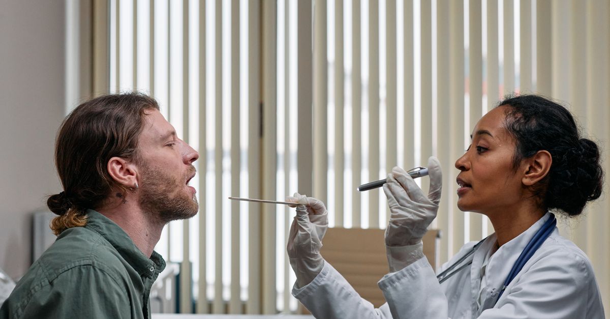
The clinical evaluation of a parotid mass requires a meticulous physical examination of the head and neck. The physician visually inspects the symmetry of the face and neck. They systematically palpate the mass to assess its size, consistency, and mobility. A soft, mobile mass is reassuring, while a hard, fixed mass is concerning.
The physician will thoroughly examine the inside of the mouth. They inspect the opening of the parotid duct (Stensen’s duct) on the inside of the cheek to see if clear saliva or abnormal pus is flowing. They will also palpate the tonsil area to ensure a deep lobe tumor is not pushing inward.
A comprehensive neurological examination of all branches of the facial nerve is mandatory and carefully documented, establishing a baseline of nerve function prior to any needle biopsy or surgical intervention.
10. Advanced Imaging Modalities
Radiological imaging is essential for surgical planning. While ultrasound is an excellent, non-invasive tool to confirm if a mass is solid or a fluid-filled cyst, Magnetic Resonance Imaging is the gold standard for evaluating parotid tumors.
An MRI provides exquisite soft-tissue contrast. It accurately maps the exact size of the tumor and its precise location within the superficial or deep lobe. Crucially, the MRI helps the surgeon visualize the predicted plane of the facial nerve in relation to the tumor, which dictates the surgical approach.
If a malignancy is suspected based on clinical signs or initial imaging, a computed tomography scan of the neck and chest may be ordered to check for the spread of cancer to the cervical lymph nodes or the lungs, similar to the staging process used for a neck mass suspected of being squamous cell carcinoma.
11. Fine Needle Aspiration Biopsy
To determine the cellular nature of the mass before surgery, a fine needle aspiration biopsy is routinely performed. This is a safe, quick procedure performed in the clinic, often under ultrasound guidance to ensure accuracy.
A very thin needle is inserted into the mass to extract a small sample of cells. The cells are examined by a cytopathologist. This technique is highly accurate for differentiating between benign lesions like a pleomorphic adenoma or a Warthin tumor, and malignant carcinomas.
Importantly, an open surgical biopsy—cutting directly into the mass on the cheek—is strictly contraindicated for parotid tumors. Doing so violates the tumor capsule, potentially spreading cancer cells or microscopic benign tumor cells throughout the surgical field, making future curative surgery impossible.
12. Data Summary: Differentiating Parotid Masses
The following table outlines the key clinical and pathological differences between the most common benign and malignant parotid masses.
| Characteristic | Pleomorphic Adenoma (Benign) | Malignant Carcinoma (e.g., Mucoepidermoid) |
|---|---|---|
| Growth Rate | Very slow, often present for years. | Variable, can be rapid. |
| Pain and Nerve Function | Painless, normal facial movement. | May cause pain, numbness, or facial muscle weakness. |
| Physical Texture | Firm, smooth, mobile. | Hard, irregular, often fixed to skin or bone. |
| Treatment Focus | Surgical removal with a margin of healthy tissue. | Aggressive surgery, possible nerve sacrifice, post-op radiation. |
This clinical distinction drives the entire preoperative counseling and surgical planning process.
13. Surgical Principles: The Parotidectomy
The definitive treatment for almost all solid parotid masses is surgery, specifically a parotidectomy. The fundamental principle of parotid surgery is the identification and meticulous preservation of the facial nerve.
For a tumor in the superficial lobe, a superficial parotidectomy is performed. The surgeon makes an incision hidden in the creases in front of the ear and down into the neck. They identify the main trunk of the facial nerve as it exits the skull and carefully dissect every tiny branch away from the glandular tissue, removing the tumor surrounded by a cuff of healthy normal gland.
If the tumor involves the deep lobe, a total parotidectomy is required. The surgeon must elevate the delicate facial nerve network completely out of the way to reach and remove the underlying deep tumor tissue. Throughout the procedure, a continuous electronic nerve monitor is used to ensure the nerve fibers are not damaged.
14. Managing the Facial Nerve in Malignancy
If a patient is diagnosed with a malignant parotid tumor, the surgical strategy changes. If the cancer is growing near the facial nerve but not actively invading it, the surgeon will make every effort to peel the tumor off the nerve to preserve facial movement.
However, if the cancer has directly invaded and grown into the nerve fibers, the affected section of the facial nerve must be surgically sacrificed (cut and removed) to ensure all cancer is eradicated. Leaving cancer cells behind on the nerve guarantees a fatal recurrence.
When a nerve must be sacrificed, the surgeon will immediately attempt nerve reconstruction. They may harvest a small sensory nerve from the patient’s leg or neck and graft it into the face to bridge the gap, helping to restore facial tone and movement over the subsequent year.
15. Complications and Post-Operative Care
Parotid surgery carries unique specific risks. The most common immediate complication is transient facial weakness. Due to the stretching and manipulation of the nerve during surgery, the nerve may temporarily stop working, leading to a temporary facial droop that usually recovers fully over several weeks or months.
A late complication is Frey’s syndrome. After the gland is removed, the tiny nerves that previously commanded the gland to produce saliva can mistakenly attach to the sweat glands in the overlying cheek skin. Consequently, when the patient eats, they experience sweating and flushing on the side of their face instead of salivating.
For malignant tumors, surgery is frequently followed by a course of radiation therapy directed at the parotid bed and neck. This adjuvant treatment destroys any remaining microscopic cancer cells and significantly reduces the risk of the tumor returning.
16. Frequently Asked Questions FAQ
1. If I have a lump on my cheek, is it definitely a tumor?
Not necessarily. It could be a swollen lymph node, a blocked saliva duct, or an infection. However, any painless, firm lump that does not go away after a few weeks needs to be evaluated by an Ear, Nose, and Throat specialist.
2. Is a pleomorphic adenoma cancer?
No, a pleomorphic adenoma is a benign tumor. It does not spread to other parts of the body. However, it must be removed because it will continue to grow, and if left in your cheek for decades, it has a small chance of turning into cancer.
3. Will I be paralyzed after parotid surgery?
Permanent facial paralysis is very rare when surgery is performed for a benign tumor by an experienced surgeon. You may have a temporary weakness or a crooked smile for a few weeks due to the nerve being stretched during the operation, but this usually heals completely.
4. How is the biopsy done if you can’t cut into the cheek?
Doctors use a Fine Needle Aspiration. They use a very tiny needle, similar to drawing blood, to suck a few cells out of the lump. This provides a safe diagnosis without cutting the tumor open and risking the spread of cells.
5. Do I need my parotid gland to produce saliva?
You have two large parotid glands, two submandibular glands under your jaw, and hundreds of minor saliva glands in your mouth. Removing one parotid gland will not leave you with a dry mouth; the other glands easily compensate for the loss.
17. Bibliography
Disclaimer: The content is for informational purposes only and does not replace medical advice. Always consult your doctor for personalized treatment.
