
1. Introduction
A peripheral corneal epithelial staining pattern is a critical diagnostic finding in clinical ophthalmology, utilized to assess the structural integrity of the ocular surface. The cornea, the transparent, avascular dome serving as the eye’s primary refractive element, is protected by a continuous layer of stratified squamous epithelium. When this delicate epithelial barrier is compromised by mechanical friction, desiccation, chemical toxicity, or active inflammation, specialized vital dyes applied to the eye will pool within the microscopic epithelial defects.
Evaluating the specific location and pattern of this dye uptake allows the ophthalmologist to trace the cellular damage back to its exact pathological source. Staining that is localized strictly to the periphery of the cornea—the zone adjacent to the limbus where the clear cornea meets the white sclera—points toward a highly specific set of etiologies, distinguishing it from central or diffuse corneal disease. Accurate interpretation of these peripheral staining patterns is paramount for deploying targeted immunomodulatory treatments, adjusting contact lens parameters, and preventing irreversible vision loss due to corneal ulceration or scarring.
2. Anatomy of the Corneal Epithelium
The corneal epithelium is a highly organized, regenerating layer of non-keratinized cells, five to seven layers thick. The outermost superficial cells are firmly locked together by tight junctions, establishing an impermeable barrier against tear fluid, external pathogens, and environmental debris. These superficial cells possess microscopic projections called microvilli, which anchor the protective tear film directly to the ocular surface.
The peripheral cornea is anatomically distinct from the central cornea. It lies immediately adjacent to the limbus, the vascularized border zone harboring the corneal epithelial stem cells responsible for continuous tissue regeneration. Because the peripheral cornea is in direct anatomical proximity to the immune-rich blood vessels of the conjunctiva and sclera, it is uniquely susceptible to localized, immune-mediated inflammatory attacks that the avascular central cornea typically escapes.
3. The Role of Vital Dyes in Diagnosis
Clinical assessment of the corneal epithelium relies on the application of vital dyes, primarily sodium fluorescein, rose bengal, and lissamine green. Sodium fluorescein is a water-soluble dye that does not penetrate intact, healthy epithelial cells. However, when the tight junctions between cells are broken, or cells are entirely missing (creating a microscopic pothole), the fluorescein dye seeps into the underlying stroma and pools in the defect. When illuminated with a cobalt blue light, the pooled dye fluoresces brilliantly, revealing the exact area of cellular damage.
Rose bengal and lissamine green dyes function differently. They actively stain dead or devitalized epithelial cells, as well as areas where the protective mucin layer of the tear film is absent. By utilizing these dyes in tandem, the clinician gains a comprehensive, color-coded map of the ocular surface, identifying not only areas of active cellular loss but also the regions of chronic cellular stress surrounding the periphery.
4. Staphylococcal Marginal Keratitis
A classic presentation of severe peripheral corneal epithelial staining is Staphylococcal marginal keratitis. This is not a direct bacterial infection of the cornea, but rather an intense, localized hypersensitivity reaction to the exotoxins produced by Staphylococcus aureus bacteria colonizing the patient’s eyelid margins (blepharitis).
The bacterial antigens accumulate in the tear film and pool in the peripheral sulcus, exactly where the eyelid rests against the cornea. The immune system launches a robust inflammatory attack against these pooled antigens, leading to the formation of small, sterile infiltrates and epithelial breakdown in the peripheral cornea. The staining pattern classically appears as a distinct crescent or small punctate dots located at the 2 o’clock, 4 o’clock, 8 o’clock, or 10 o’clock positions, separated from the limbus by a clear zone of healthy tissue.
5. Exposure Keratopathy and Lagophthalmos
When the eyelids fail to close completely during a blink or while sleeping, a condition clinically termed lagophthalmos, the exposed section of the cornea undergoes severe desiccation. The tear film rapidly evaporates, causing the epithelial cells to dry out, slough off, and die.
The resulting staining pattern is highly specific. Because the eyelids typically rest slightly apart horizontally during lagophthalmos, the epithelial damage appears as a distinct band of dense, confluent staining across the inferior peripheral cornea, exactly mapping the anatomical region left exposed to the ambient air. Managing this requires aggressive lubrication with thick ophthalmic ointments at night and, in severe cases, taping the eyelids shut or performing surgical correction of the eyelid position.
6. Contact Lens-Induced Mechanical Toxicity
Contact lens wearers frequently present with distinct peripheral corneal staining patterns resulting from mechanical friction or hypoxia. A contact lens that fits too tightly or has a steep edge profile will physically dig into the peripheral epithelium during every blink. The constant mechanical abrasion shears off the superficial epithelial cells, resulting in a thin, continuous arc of fluorescein staining that perfectly traces the outer edge of the contact lens.
Furthermore, if the contact lens material prevents adequate oxygen transmission to the peripheral cornea, the epithelial cells undergo hypoxic stress and subsequent death, leading to diffuse punctate staining. Resolving this pattern requires the optometrist or ophthalmologist to immediately refit the patient into a lens with a higher oxygen permeability profile or a more appropriate base curve to eliminate the abrasive mechanical edge.
7. Solution-Induced Corneal Staining (SICS)
Patients utilizing multipurpose chemical disinfecting solutions for their contact lenses may develop Solution-Induced Corneal Staining. This occurs when the preservatives or biocides within the cleaning solution bind to the matrix of the contact lens and are slowly released onto the cornea throughout the day, causing a direct, toxic chemical reaction that kills the delicate epithelial cells.
The staining pattern associated with SICS typically presents as diffuse, fine, punctate dots scattered across the cornea, frequently concentrated in an annular (ring-like) pattern in the peripheral zones where the lens rests most heavily. The clinical resolution is remarkably simple: discontinuing the chemical multipurpose solution and transitioning the patient to a preservative-free, hydrogen peroxide-based lens cleaning system.
8. Dry Eye Syndrome and Tear Film Instability
Dry eye syndrome, specifically evaporative dry eye caused by meibomian gland dysfunction, frequently manifests with characteristic peripheral staining. The meibomian glands, located in the eyelids, secrete the lipid (oil) layer of the tear film, which prevents the underlying aqueous tears from evaporating.
When these glands are blocked, the tears evaporate rapidly. The inferior peripheral cornea is usually the first to suffer from this desiccation due to gravitational pooling of the inadequate tears and incomplete blinking dynamics. The staining appears as fine, scattered stippling along the lower third of the cornea, indicating widespread cellular distress from chronic friction and hyperosmolarity.
9. Structural Data: Staining Patterns and Etiologies
Differentiating the exact location and shape of the peripheral stain guides the immediate clinical diagnosis.
| Staining Pattern & Location | Primary Clinical Etiology | Mechanism of Cellular Damage |
|---|---|---|
| Inferior Band (lower third) | Lagophthalmos / Exposure Keratopathy. | Desiccation due to incomplete eyelid closure. |
| Crescent-shaped near the limbus | Staphylococcal Marginal Keratitis. | Immune hypersensitivity to bacterial exotoxins. |
| Arcuate line tracking the lens edge | Contact Lens Mechanical Abrasion. | Physical shearing of the epithelium by a tight lens. |
| Annular (Ring-like) punctate dots | Solution-Induced Toxicity (SICS). | Chemical destruction of cells by lens preservatives. |
10. Autoimmune and Connective Tissue Disorders
Severe, progressive peripheral staining can be the initial ocular manifestation of systemic autoimmune and connective tissue diseases, such as Rheumatoid Arthritis, Lupus, or Sjögren’s syndrome. In these conditions, circulating autoantibodies attack the vascular network supplying the limbus.
This localized vasculitis cuts off the nutrient supply to the peripheral cornea, causing a highly destructive condition known as Peripheral Ulcerative Keratitis. The staining reveals a deep, crescent-shaped ulceration that rapidly melts the corneal stroma. This is an absolute ophthalmological emergency. Localized eye drops are insufficient; the patient requires urgent systemic immunosuppressive therapy to halt the autoimmune attack before the cornea perforates.
11. Diagnostic Evaluation via Slit Lamp Biomicroscopy
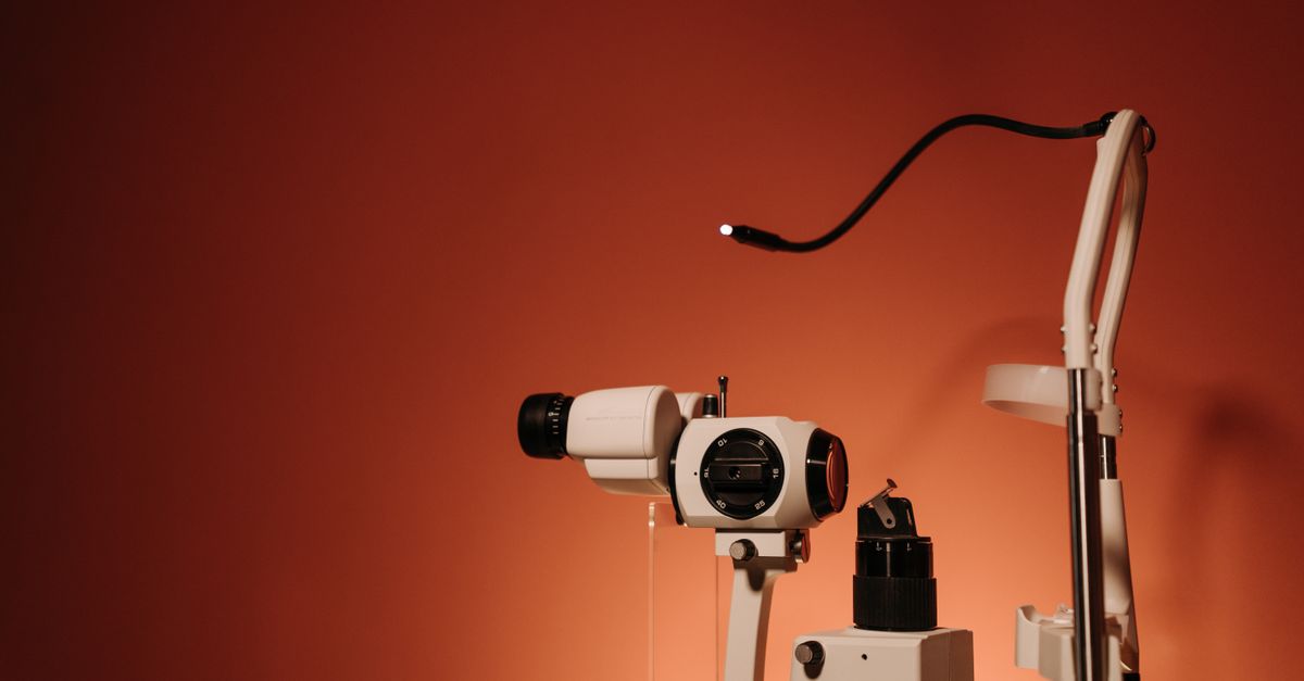
The definitive tool for evaluating peripheral corneal staining is the slit lamp biomicroscope. This instrument provides the clinician with intense, highly focused illumination and high-magnification stereoscopic viewing. The physician instills a tiny volume of fluorescein dye into the lower conjunctival fornix and asks the patient to blink, spreading the dye across the ocular surface.
Using a cobalt blue filter to excite the fluorescein, and a Wratten yellow filter to enhance the contrast, the clinician systematically scans the entire perimeter of the cornea. They carefully assess the depth, density, and precise anatomical distribution of the staining, correlating these microscopic visual findings directly with the patient’s subjective symptoms of foreign body sensation, photophobia, or burning.
12. Targeted Lubrication and Barrier Repair
For peripheral staining caused by exposure, dry eyes, or mild mechanical friction, the foundational treatment is the aggressive restoration of the tear film barrier. Clinicians prescribe highly viscous, preservative-free artificial tears to be used frequently throughout the day. Preservative-free formulations are mandatory, as the chemicals in standard bottled eye drops (such as benzalkonium chloride) will cause further toxic staining if used more than four times daily.
For nighttime use, dense ophthalmic ointments containing mineral oil and white petrolatum are applied. These ointments physically coat the damaged peripheral epithelium, preventing further desiccation while the patient sleeps and providing a secure biological environment for the basal cells to rapidly replicate and replace the stained, missing superficial cells.
13. Pharmacological Immunomodulation
When the peripheral staining is driven by an underlying inflammatory or immune process, such as Staphylococcal hypersensitivity or early autoimmune melt, aggressive pharmacological intervention is required. Topical corticosteroids are utilized to rapidly suppress the localized immune cascade, halting the destruction of the epithelial and stromal layers.
Because prolonged steroid use carries risks of elevating intraocular pressure or inducing cataracts, clinicians frequently transition patients to long-term, steroid-sparing topical immunomodulators, such as cyclosporine or lifitegrast. These medications specifically target and inhibit the inflammatory T-cells present on the ocular surface, promoting the generation of healthy, functional tears and the sustained resolution of the epithelial staining.
14. Eyelid Hygiene and Environmental Management
Resolving peripheral staining frequently requires addressing the anatomical structures immediately adjacent to the cornea. If the staining is caused by blepharitis and bacterial exotoxins, the patient must initiate a strict daily regimen of eyelid hygiene. This involves the application of warm compresses to melt thickened glandular oils, followed by gentle mechanical scrubbing of the eyelid margins with specialized lid cleansers or hypochlorous acid sprays.
Furthermore, environmental modifications are advised. Patients are instructed to redirect air conditioning or heating vents away from their face, utilize indoor humidifiers, and employ moisture-chamber goggles while sleeping to actively prevent the evaporative stress that leads to severe peripheral exposure staining.
15. When to Seek Urgent Ophthalmological Care
While mild, punctate peripheral staining associated with dry eyes is a chronic, manageable condition, certain clinical presentations require immediate, emergency evaluation by an ophthalmologist. If the patient experiences a sudden, severe increase in ocular pain, pronounced redness in the white of the eye, or a dramatic reduction in visual acuity, immediate assessment is mandatory.
Furthermore, if a contact lens wearer notices a dense, white spot on their cornea corresponding to the area of pain, they must remove the lens immediately and seek urgent care. This combination strongly suggests that the peripheral epithelial defect has become infected, rapidly evolving into a sight-threatening microbial corneal ulcer that requires immediate, intensive fortified antibiotic therapy to prevent permanent scarring or perforation.
16. Frequently Asked Questions (FAQ)
1. What exactly does it mean when the doctor says my eye is staining?
Staining means the doctor put a specialized yellow or green dye on your eye, and the dye filled into microscopic scratches, dry patches, or missing cells on the surface of your cornea. It is a visual map showing the doctor exactly where your eye is damaged.
2. Why did my contact lenses cause a ring of staining around my eye?
If your contact lens fits too tightly or does not let enough oxygen through, it acts like a tight rubber band, scraping the delicate cells off the outer edge of your cornea every time you blink, leading to a visible ring of damage.
3. Is corneal staining permanent damage?
Usually, no. The cells on the surface of the cornea (the epithelium) are incredible at healing. Once the doctor identifies the cause—such as a toxic cleaning solution or severe dryness—and removes it, the cells typically regenerate and heal the staining completely within a few days.
4. Can sleeping with my eyes slightly open cause this?
Yes. If your eyelids do not close entirely while you sleep, the exposed band of your eye dries out completely, causing the cells to die and flake off. The doctor will see a distinct band of staining across the bottom of your eye.
5. What is the treatment for peripheral corneal staining?
The treatment depends entirely on the cause. It ranges from using thick, preservative-free lubricating drops to heal dry spots, changing your contact lens prescription, to using prescription steroid drops to calm down an allergic reaction on the edge of the cornea.
17. Bibliography
Disclaimer: The content is for informational purposes only and does not replace medical advice. Always consult your doctor for personalized treatment.

