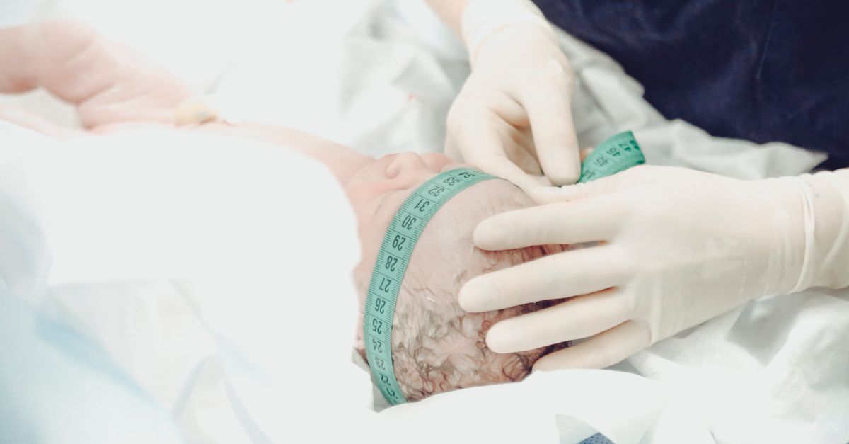
1. Introduction to Post-Term Infancy
A post-term infant is medically defined as a neonate born after the completion of forty-two weeks of gestation. While human gestation is optimally programmed to culminate around forty weeks, extending the intrauterine environment significantly beyond this threshold introduces a complex array of physiological risks. The primary challenge in post-term pregnancies lies in the biological limitation of the placenta, an organ inherently designed for a finite lifespan.
As the pregnancy stretches past forty-two weeks, the infant is subjected to an increasingly hostile environment marked by dwindling nutritional supply and compromised oxygen exchange. Caring for a post-term infant requires high clinical vigilance, as these neonates frequently present with distinct metabolic derangements, profound respiratory vulnerabilities, and a characteristic physical appearance indicating intrauterine starvation. Accurate assessment and immediate stabilization in the delivery room are critical to preventing long-term neurological sequelae.
2. Defining the 42-Week Threshold
The classification of a post-term infant relies entirely on accurate gestational dating, typically established through precise first-trimester crown-rump length ultrasound measurements. It is vital to distinguish between a late-term infant, born between forty-one weeks and forty-one weeks and six days, and a true post-term infant, born at exactly forty-two weeks or later.
The forty-two-week mark is highly significant in clinical obstetrics and neonatology. Statistical data demonstrates a sharp, non-linear increase in perinatal morbidity and mortality once this threshold is crossed. Consequently, modern medical protocols universally advocate for labor induction before a pregnancy becomes definitively post-term, making the true post-term infant an increasingly rare, high-risk presentation requiring specialized pediatric care.
3. Placental Senescence and Uteroplacental Insufficiency
The central pathophysiological driver of post-term complications is placental senescence. Beyond forty weeks, the placenta begins to age rapidly, developing micro-infarctions, areas of calcification, and fibrotic tissue changes. This morphological deterioration progressively impairs the efficiency of the uteroplacental circulation.
This state of uteroplacental insufficiency deprives the fetus of vital oxygen and nutrients. To survive this chronic hypoxic stress, the fetus reflexively centralizes its blood flow, prioritizing perfusion to the brain, heart, and adrenal glands, while sacrificing blood supply to the peripheral tissues, gastrointestinal tract, and kidneys. This redistribution mechanism ultimately leads to a severe decrease in amniotic fluid, known as oligohydramnios.
4. Dysmaturity Syndrome: Physical Hallmarks
When a post-term fetus is subjected to prolonged placental insufficiency, it develops a specific clinical presentation known as dysmaturity syndrome. Because the placenta can no longer support growth, the fetus must metabolize its own subcutaneous fat and muscle glycogen stores to meet basic energy requirements.
Upon delivery, the dysmature infant appears strikingly thin and malnourished, lacking the typical plumpness of a term newborn. The skin is remarkably dry, cracked, and heavily peeling, resembling parchment paper, due to the total absence of vernix caseosa. These infants often have an abundance of scalp hair, unusually long fingernails, and a wide-eyed, hyper-alert facial expression indicative of chronic in utero stress.
5. Macrosomia and Birth Trauma Risks
Conversely, if the placenta maintains adequate function beyond forty-two weeks, the fetus will continue to grow, leading to fetal macrosomia, defined as a birth weight exceeding 4,000 or 4,500 grams. The macrosomic post-term infant faces entirely different, yet equally severe, mechanical risks during delivery.
The excessive size of the fetal shoulders in relation to the maternal pelvis significantly increases the risk of shoulder dystocia. This obstetric emergency can result in severe neonatal birth trauma, including clavicular fractures and brachial plexus nerve injuries. Furthermore, the prolonged, difficult extraction places the macrosomic infant at a high risk for acute intrapartum asphyxia.
6. Meconium Aspiration Syndrome
One of the most dangerous respiratory complications for the post-term infant is meconium aspiration syndrome. Chronic hypoxia triggers a vagal response in the fetus, causing the relaxation of the anal sphincter and the passage of meconium, the infant’s first stool, into the amniotic fluid. Because amniotic fluid volume is already low, the meconium becomes thick and highly concentrated.
If the distressed infant gasps in utero or takes a deep breath during delivery, this toxic, tar-like substance is drawn deep into the lungs. Meconium physically blocks the bronchioles, inactivates pulmonary surfactant, and incites a severe chemical pneumonitis, leading to profound neonatal respiratory distress and persistent pulmonary hypertension.
7. Neonatal Hypoglycemia Management
The post-term infant, particularly one exhibiting signs of dysmaturity, is at an exceptionally high risk for profound neonatal hypoglycemia. The depletion of subcutaneous fat and hepatic glycogen stores prior to birth leaves the infant without any energy reserves to sustain normal blood glucose levels once the umbilical cord is cut.
Because the neonatal brain relies exclusively on a continuous supply of glucose, preventing hypoglycemia is a paramount clinical priority. The care team must initiate early and frequent feedings, accompanied by strict, protocol-driven blood glucose monitoring. In cases of severe or refractory hypoglycemia, intravenous dextrose administration is required to prevent seizures and irreversible neurological injury.
8. Polycythemia and Hyperviscosity
In response to the chronic low-oxygen environment of the aging placenta, the fetal kidneys increase the production of erythropoietin, stimulating the bone marrow to produce an excess of red blood cells. While this polycythemia attempts to maximize oxygen delivery before birth, it becomes a severe liability postpartum.
The excessively high red blood cell count makes the infant’s blood highly viscous, or thick. This hyperviscosity impairs normal blood flow through the tiny capillary beds of the brain, lungs, and intestines, increasing the risk of microthrombosis, respiratory distress, and necrotizing enterocolitis. These infants often appear deeply ruddy or plethoric and require diligent monitoring of their hematocrit levels.
9. Thermoregulation Challenges in the Neonate
Maintaining a stable core body temperature is a significant challenge for the dysmature post-term infant. The loss of insulating subcutaneous fat, combined with a depletion of specialized brown fat utilized for non-shivering thermogenesis, severely impairs the infant’s ability to generate and retain heat.
Hypothermia in the neonate creates a dangerous metabolic cascade. It rapidly accelerates the depletion of already low glucose stores and increases cellular oxygen consumption, further straining a potentially compromised respiratory system. Immediate placement under a radiant warmer or prolonged skin-to-skin contact is strictly required to maintain euthermia.
10. Delivery Room Resuscitation Protocols
The delivery of a post-term infant necessitates the presence of a specialized neonatal resuscitation team prepared for complex interventions. The immediate focus is assessing the infant’s respiratory effort, heart rate, and tone.
If the infant is born through thick meconium and is non-vigorous (exhibiting poor muscle tone, inadequate breathing, or a heart rate below 100 beats per minute), current resuscitation guidelines dictate rapid clearing of the airway and the immediate initiation of positive pressure ventilation. Swift and efficient restoration of oxygenation is the critical determinant of long-term neurological outcome.
11. Neonatal Intensive Care Unit Observation
Infants demonstrating signs of respiratory distress, severe hypoglycemia, or neurological depression are rapidly transferred to the neonatal intensive care unit (NICU) for continuous, specialized monitoring and therapeutic intervention.
| Clinical Complication | Physiological Etiology | NICU Intervention |
|---|---|---|
| Hypoglycemia | Exhaustion of glycogen and fat reserves. | Continuous IV dextrose, frequent blood monitoring. |
| Meconium Aspiration | Inhalation of fetal stool due to hypoxia. | Mechanical ventilation, surfactant therapy. |
| Polycythemia | Excess RBC production from chronic hypoxia. | Intravenous hydration, possible partial exchange transfusion. |
12. Neurological Monitoring and Assessment
Because post-term infants frequently endure subtle, chronic oxygen deprivation during the final weeks of pregnancy, the clinical team must perform meticulous, ongoing neurological assessments. The clinician observes for abnormal tone, exaggerated reflexes, irritability, or lethargy, which may indicate hypoxic-ischemic encephalopathy.
In high-risk neonates, cerebral function monitoring using continuous electroencephalography (EEG) is utilized to detect subclinical seizure activity. Early identification of neurological injury allows the medical team to implement neuroprotective strategies, such as therapeutic hypothermia, which must be initiated within hours of birth to be effective.
13. Nutritional Support and Feeding Protocols
Feeding the post-term infant requires a delicate clinical balance. While the infant desperately needs calories to reverse the state of intrauterine starvation and correct hypoglycemia, their gastrointestinal tract may have suffered ischemic injury due to blood flow redistribution before birth.
Enteral feeds are introduced cautiously, prioritizing maternal breast milk for its optimal digestibility and protective immunological factors. The clinical team monitors the infant closely for signs of feeding intolerance, abdominal distension, or bloody stools, which could indicate the early onset of severe intestinal complications.
14. Frequently Asked Questions FAQ
1. What does a baby look like if born past 42 weeks?
A post-term baby often looks long and thin because they have lost body fat in the womb. Their skin is typically very dry, peeling, and wrinkled. They may have long fingernails, lots of hair, and an unusually alert, wide-eyed expression.
2. Why is meconium in the fluid dangerous?
If the baby becomes stressed in the womb, they may pass their first stool (meconium) into the fluid. If they breathe this thick, sticky substance into their lungs, it blocks the airways and causes severe inflammation, making it very difficult for the baby to breathe after birth.
3. Why do post-term babies have problems with their blood sugar?
Because the placenta stopped providing adequate nutrients at the end of the pregnancy, the baby had to use up all their stored energy and fat just to survive in the womb. Consequently, they are born with very low energy reserves, leading to a sudden drop in blood sugar.
4. Are post-term babies always bigger than normal babies?
No. If the placenta kept working well, the baby will be very large (macrosomic). However, if the placenta failed, the baby stops growing and begins to lose weight, resulting in a thin, undernourished appearance.
5. Will a post-term baby have long-term health issues?
Most post-term infants recover fully with proper neonatal care and quickly catch up in weight. However, babies who suffered severe oxygen deprivation or inhaled massive amounts of meconium may require long-term pediatric monitoring for developmental or respiratory issues.
15. Bibliography
Disclaimer: The content is for informational purposes only and does not replace medical advice. Always consult your doctor for personalized treatment.