
1. Introduction to Fetal Postmaturity
Fetal postmaturity is a high-risk obstetric condition that occurs when a pregnancy advances significantly beyond the physiological forty-week mark, specifically extending past forty-two weeks of gestation. While the intrauterine environment provides perfect life support throughout normal gestation, maintaining a pregnancy beyond this natural timeline introduces escalating risks to fetal health. The fundamental issue lies in the placenta, a temporary organ not designed to sustain a growing fetus indefinitely.
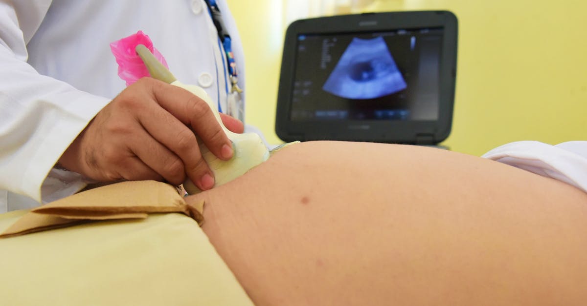
Managing a prolonged pregnancy requires vigilant obstetric surveillance to balance the goal of a natural, spontaneous labor against the increasing dangers of placental failure. Modern clinical guidelines emphasize early detection of fetal compromise and proactive intervention. Through comprehensive ultrasound evaluations, non-stress testing, and well-timed labor induction, obstetricians can safely navigate the complexities of postmaturity and ensure optimal outcomes for both mother and child.
2. Defining Prolonged vs. Post-term Gestation
Precision in gestational dating is the cornerstone of managing advanced pregnancies. A pregnancy is considered full-term between thirty-nine and forty weeks. The period between forty-one weeks and forty-one weeks and six days is classified as late-term. True postmaturity, or a post-term pregnancy, is strictly defined as an exact gestational age of forty-two weeks or greater.
Accurate dating must be established through first-trimester ultrasound measurements of the fetal crown-rump length, as calculating the due date based solely on the last menstrual period is notoriously inaccurate. Ensuring precise chronological dating prevents unnecessary, premature medical interventions while preventing the dangerous oversight of a truly postmature fetus.
3. The Etiology of Prolonged Pregnancy
The exact biological trigger that initiates spontaneous human labor remains one of the great mysteries of obstetrics. Consequently, the specific etiology behind a prolonged pregnancy is often elusive. It is understood that labor requires a complex, synchronized cascade of maternal and fetal hormonal signals, primarily involving the release of oxytocin, prostaglandins, and placental corticotropin-releasing hormone.
When these intricate endocrine pathways fail to activate sequentially, the cervix remains firm, the uterus stays quiescent, and the pregnancy simply continues. Nulliparity, advanced maternal age, maternal obesity, and a prior history of a post-term pregnancy are recognized statistical risk factors that increase the likelihood of delayed spontaneous labor onset.
4. Pathophysiology of Placental Senescence
The central clinical concern of fetal postmaturity is placental senescence. The placenta achieves its maximum efficiency near the thirty-seventh week of gestation. Beyond forty weeks, the organ undergoes a predictable pattern of morphological and functional decline. The microscopic villi, responsible for nutrient and gas exchange, begin to calcify and suffer structural fibrosis.
This deterioration leads to a gradual reduction in uteroplacental blood flow. The placenta becomes increasingly inefficient at transferring oxygen to the fetus and clearing carbon dioxide. The resulting state of chronic hypoxia forces the fetus to adapt metabolically, altering its blood flow to prioritize the brain and heart while sacrificing peripheral organ perfusion.
5. Oligohydramnios and Cord Compression
As the postmature fetus adapts to placental insufficiency, it constricts blood flow to its kidneys. A reduction in renal perfusion directly causes a profound decrease in fetal urine production. Because fetal urine is the primary component of amniotic fluid in the third trimester, its reduction results in severe oligohydramnios, a dangerous lack of amniotic fluid.
Amniotic fluid acts as a crucial mechanical buffer, protecting the umbilical cord from compression against the uterine walls. In an oligohydramnic environment, the umbilical cord is highly vulnerable to being pinched between the fetal body and the uterus, particularly during the intense muscular contractions of labor. Repeated cord compression causes acute fetal hypoxia and sudden, dangerous decelerations in the fetal heart rate.
6. The Paradox of Macrosomia and Growth Restriction
The postmature fetus can present with one of two distinct growth trajectories, depending entirely on the functional state of the aging placenta. If the placenta maintains adequate perfusion, the fetus will continue to grow, leading to fetal macrosomia. Macrosomic infants face an increased risk of obstructed labor and shoulder dystocia, a severe obstetric emergency where the fetal shoulders become impacted behind the maternal pubic bone.
Conversely, if the placenta undergoes rapid senescence, the fetus is subjected to intrauterine starvation. This leads to the classic postmaturity syndrome, characterized by intrauterine growth restriction, loss of subcutaneous fat, and profound metabolic instability. Clinicians must identify which growth trajectory the fetus is following to plan the safest mode of delivery.
7. Risks of Intrauterine Demise
The most devastating complication associated with unmonitored fetal postmaturity is sudden intrauterine fetal demise. Statistical data unequivocally demonstrates that the rate of stillbirth rises progressively after the forty-first week of gestation. This increased mortality is directly correlated with the unpredictable nature of sudden placental failure and umbilical cord accidents in a depleted fluid environment.
To mitigate this severe risk, standard obstetric protocols mandate the initiation of intensive antenatal surveillance as soon as a pregnancy reaches the late-term window. The goal is to identify any subtle signs of fetal distress and expedite delivery before irreversible hypoxic injury or fetal death occurs.
8. Antenatal Surveillance Techniques
Antenatal testing is designed to evaluate the physiological well-being of the fetus in real-time. The primary tool utilized is the non-stress test, a non-invasive procedure that continuously records the fetal heart rate over a twenty to forty-minute period.
The clinician assesses the tracing for spontaneous accelerations in the heart rate, which indicate a well-oxygenated, neurologically intact fetus. If the tracing lacks accelerations, it suggests that the fetus may be conserving energy due to underlying hypoxia, prompting further, more comprehensive diagnostic evaluations.
9. Ultrasound and the Biophysical Profile
When the non-stress test yields non-reassuring results, a biophysical profile is immediately performed using high-resolution ultrasound. This comprehensive assessment evaluates four specific sonographic markers: fetal breathing movements, gross body movements, fetal muscle tone, and the total volume of amniotic fluid.
Each component is assigned a score, reflecting the acute and chronic neurological status of the fetus. A low biophysical profile score indicates severe fetal compromise and a highly hostile intrauterine environment, serving as an absolute indication for immediate delivery, typically via cesarean section.
10. Guidelines for Labor Induction
Based on compelling clinical evidence demonstrating that active management reduces the risk of perinatal mortality, international obstetric guidelines recommend offering induction of labor for all pregnancies reaching forty-one weeks. Prolonging the pregnancy to forty-two weeks without medical indication is universally discouraged.
The decision to induce requires a careful assessment of the maternal cervix. The Bishop score is a clinical tool used to quantify the readiness of the cervix for labor. A high score indicates a soft, dilating cervix favorable for induction, while a low score indicates a firm, closed cervix that requires extensive ripening before labor can be initiated.
11. Pharmacological Cervical Ripening
When the cervix is unfavorable, pharmacological agents are deployed to initiate the physical changes necessary for dilation. Prostaglandin analogs are administered either orally or directly into the vaginal fornix. These biochemical agents dissolve the collagen network within the cervical tissue, causing it to soften and efface, mimicking the natural enzymatic processes of spontaneous labor.
While highly effective, prostaglandins must be monitored closely, as they can occasionally cause uterine tachysystole, a condition where the uterus contracts too frequently. Excessive contractions impede placental blood flow, exacerbating fetal distress in an already vulnerable postmature fetus.
12. Mechanical Methods of Induction
Mechanical cervical ripening offers an effective alternative to pharmacological agents, particularly for patients with a previous cesarean section who are at a higher risk of uterine rupture. The most common mechanical method involves the insertion of a Foley balloon catheter through the cervical canal.
Once in place, the balloon is inflated with saline, creating sustained mechanical pressure against the internal cervical os. This pressure gently forces the cervix to dilate and stimulates the localized, natural release of maternal prostaglandins without causing hyperstimulation of the uterine muscle.
13. Intrapartum Fetal Monitoring
Once labor is successfully induced, the postmature fetus is subjected to the intense mechanical and hypoxic stress of uterine contractions. Because the fetus has limited physiological reserves, continuous electronic fetal monitoring throughout the entire duration of labor is an absolute clinical requirement.
| Fetal Heart Rate Pattern | Underlying Pathology | Clinical Action |
|---|---|---|
| Variable Decelerations | Umbilical cord compression due to low fluid. | Maternal repositioning, oxygen administration, amnioinfusion. |
| Late Decelerations | Uteroplacental insufficiency, fetal hypoxia. | Discontinue induction agents, prepare for operative delivery. |
| Decreased Variability | Fetal sleep cycle or severe neurological depression. | Fetal scalp stimulation, escalate monitoring. |
14. Cesarean Delivery Considerations
Despite meticulous induction protocols, a significant percentage of postmature pregnancies require delivery via cesarean section. If the fetal heart rate monitoring reveals persistent late decelerations or profound bradycardia, it signifies that the aging placenta cannot sustain the fetus through the stress of labor. An emergency cesarean section is performed to rapidly extricate the hypoxic fetus.
Additionally, if the fetus has grown macrosomic due to prolonged gestation, an elective cesarean delivery may be recommended to prevent severe maternal perineal trauma and traumatic neonatal birth injuries, ensuring the safest possible outcome for both patients.
15. Frequently Asked Questions FAQ
1. What is the difference between a late-term and a post-term pregnancy?
A late-term pregnancy is between 41 weeks and 41 weeks and 6 days. A true post-term, or postmature pregnancy, strictly reaches or exceeds exactly 42 weeks of gestation.
2. Why is amniotic fluid levels so important at the end of pregnancy?
Amniotic fluid protects the umbilical cord. If the aging placenta causes fluid levels to drop, the umbilical cord can be easily squeezed during contractions, cutting off the oxygen supply to the baby.
3. Why do doctors recommend inducing labor at 41 weeks?
Medical data clearly shows that the risk of stillbirth and severe complications increases significantly as the pregnancy approaches 42 weeks due to the unpredictable decline of placental function. Induction safely prevents these risks.
4. Does a postmature fetus always stop growing?
No. If the placenta remains healthy, the fetus will continue to grow, leading to a very large baby that may be difficult to deliver vaginally. If the placenta fails, the fetus stops growing and begins to lose weight.
5. How do doctors check if a late-term baby is still healthy inside the womb?
Doctors use ultrasound to perform a biophysical profile, which checks the baby’s movements, muscle tone, breathing motions, and amniotic fluid volume. They also perform non-stress tests to monitor the fetal heart rate.
16. Bibliography
Disclaimer: The content is for informational purposes only and does not replace medical advice. Always consult your doctor for personalized treatment.
