
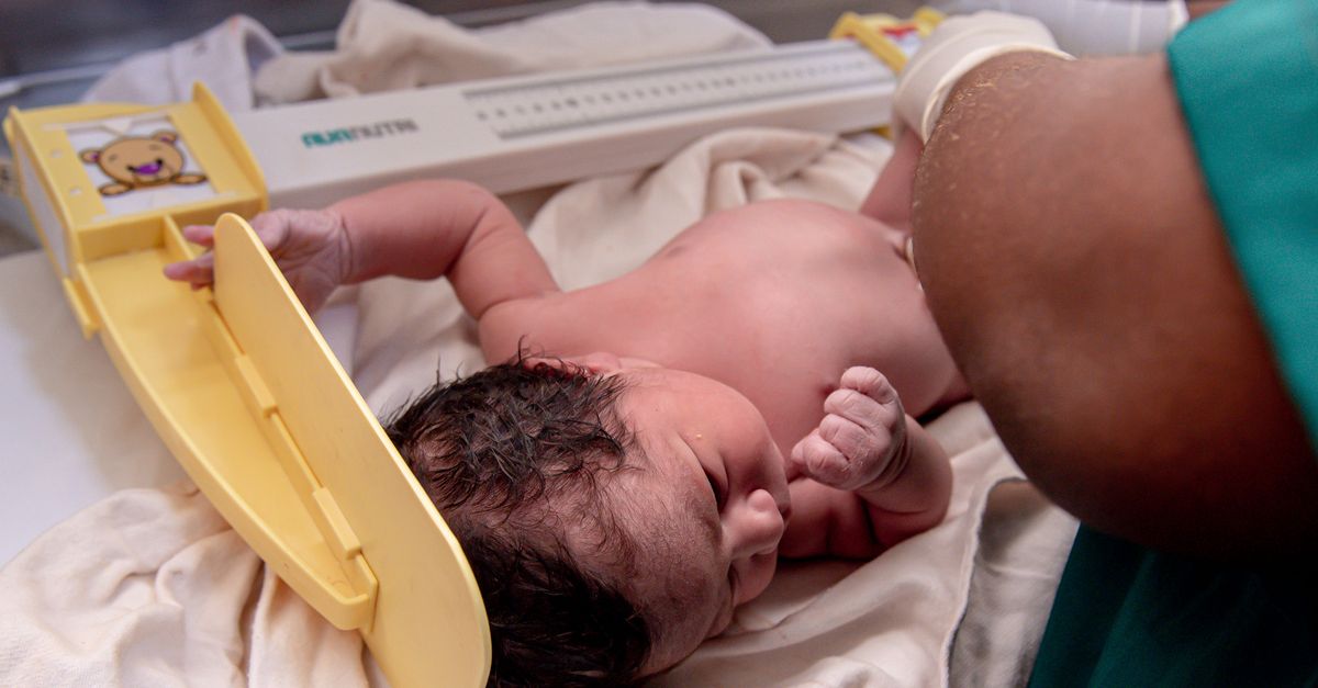
1. Introduction to Premature Birth
Premature birth, clinically defined as the delivery of a live infant before the completion of thirty-seven weeks of gestation, is one of the most critical challenges in modern obstetrics and neonatology. The intrauterine environment is meticulously designed to support a highly specific timeline of fetal organogenesis and maturation. When a neonate is expelled from this protective environment prematurely, they face profound physiological vulnerabilities because their organ systems remain structurally and functionally incomplete.
The severity of medical complications is inversely proportional to the gestational age at birth. Surviving the abrupt transition from placental life support to independent physiological function requires highly specialized intervention in a Neonatal Intensive Care Unit (NICU). The clinical management of a premature infant involves substituting for the maternal womb, aggressively supporting respiratory and thermal regulation, and shielding the fragile neurological architecture from the stress of the extrauterine environment to ensure both acute survival and long-term neurodevelopmental success.
2. Clinical Classification of Prematurity
Neonatology categorizes premature birth into distinct sub-categories based entirely on precise gestational age. This classification is vital, as the physiological maturity and statistical survival rates differ drastically between groups.
Late preterm infants are born between thirty-four and thirty-six weeks of gestation. While they often resemble full-term infants physically, they remain metabolically immature and prone to respiratory distress and feeding difficulties. Moderate preterm infants are delivered between thirty-two and thirty-four weeks. Very preterm infants are born at less than thirty-two weeks, and extremely preterm infants, presenting the most profound clinical challenges, are born at less than twenty-eight weeks of gestation. The limit of clinical viability currently hovers around twenty-two to twenty-four weeks, heavily dependent on institutional resources.
3. Respiratory Distress Syndrome
The most immediate and life-threatening consequence of premature birth is Respiratory Distress Syndrome. Fetal lung development occurs in late gestation, specifically the production of pulmonary surfactant. Surfactant is a complex mixture of lipids and proteins synthesized by type II alveolar cells, designed to reduce surface tension within the lungs and prevent the alveoli from collapsing upon exhalation.
In a premature infant, particularly those born before thirty-two weeks, the lungs are structurally stiff and severely deficient in surfactant. Every breath requires immense muscular effort to force the collapsed alveoli open, rapidly leading to severe respiratory fatigue, profound hypoxia, and hypercapnia. The classic clinical presentation includes rapid breathing (tachypnea), audible expiratory grunting, and severe retractions of the chest wall.
4. Respiratory Intervention and Surfactant Therapy
Managing respiratory failure is the cornerstone of premature neonatal care. For infants demonstrating respiratory distress, immediate non-invasive continuous positive airway pressure (CPAP) is initiated to physically splint the alveoli open during exhalation, preventing complete collapse.
If the infant cannot maintain oxygenation or becomes apneic, endotracheal intubation is required. Once the airway is secured, the clinical gold standard is the direct intratracheal administration of exogenous surfactant. This rapidly restores pulmonary compliance, drastically reducing the mechanical work of breathing and mitigating the harsh barotrauma caused by the mechanical ventilator.
5. Neurological Vulnerability: Intraventricular Hemorrhage
The premature brain is exquisitely fragile, primarily due to the presence of the germinal matrix. The germinal matrix is a highly vascular, transient structure located deep within the brain near the lateral ventricles, responsible for producing neurons and glial cells during fetal development.
The blood vessels within the germinal matrix are incredibly thin-walled and lack structural support. Because the premature infant lacks the ability to effectively autoregulate cerebral blood flow, any sudden spike in systemic blood pressure—often caused by pain, hypoxia, or rapid fluid shifts—can cause these fragile vessels to rupture. This catastrophic event, known as intraventricular hemorrhage, can lead to permanent neurological damage, cerebral palsy, or lethal hydrocephalus.
6. Thermoregulation and Cold Stress
A premature infant is severely disadvantaged in maintaining a normal core body temperature. They possess an extremely high surface-area-to-mass ratio, allowing heat to escape rapidly into the environment. Furthermore, they are born with a profound deficit of both insulating subcutaneous white fat and metabolically active brown fat, the specialized tissue required for non-shivering thermogenesis.
Exposure to room temperature rapidly induces cold stress. To generate heat, the infant must burn precious glycogen reserves and rapidly increase cellular oxygen consumption, exacerbating respiratory distress and driving the blood pH down into a state of metabolic acidosis. Therefore, maintaining strict environmental thermoregulation using advanced radiant warmers and humidified incubators is an absolute clinical priority.
7. The Patent Ductus Arteriosus
In fetal circulation, the ductus arteriosus is a vital blood vessel that allows oxygenated blood from the placenta to bypass the fluid-filled lungs. In a healthy full-term infant, the sudden increase in blood oxygen upon taking the first breath, combined with the withdrawal of maternal prostaglandins, causes this vessel to constrict and close permanently within hours of birth.
In the premature infant, the smooth muscle of the ductus arteriosus is underdeveloped and less responsive to oxygen. It frequently remains open, a condition known as patent ductus arteriosus. This abnormal connection causes oxygenated blood to inappropriately shunt back into the pulmonary circulation, flooding the fragile lungs with excess fluid and forcing the heart into a state of high-output failure.
8. Gastrointestinal Immaturity and NEC
The premature gastrointestinal tract is structurally and functionally unprepared to digest enteral nutrition. The mucosal barrier is highly permeable, and the enzymatic pathways required for digestion are underdeveloped. This immaturity places the infant at a severe risk for Necrotizing Enterocolitis (NEC), the most devastating gastrointestinal emergency in neonatology.
NEC involves severe ischemic injury and subsequent bacterial invasion of the bowel wall, leading to rapid tissue necrosis and potential intestinal perforation. Clinical signs include a distended, discolored abdomen, bloody stools, and sudden systemic cardiovascular collapse. Prevention requires highly cautious, slow advancement of enteral feeds, prioritizing maternal breast milk over synthetic formulas due to its unparalleled immunological and mucosal-protective properties.
9. Immunological Deficits and Sepsis
The premature infant enters the extrauterine environment severely immunocompromised. The critical transfer of maternal immunoglobulin G (IgG) antibodies across the placenta occurs predominantly during the third trimester. Therefore, infants born prematurely lack this vital passive immunity.
Coupled with a fragile, easily breached epidermal barrier and the necessity of multiple invasive lines and tubes for intensive care, these neonates are highly susceptible to overwhelming bacterial and fungal infections. Neonatal sepsis can present with very subtle signs, such as temperature instability, frequent apneic spells, or lethargy. Broad-spectrum antibiotics are initiated immediately upon any clinical suspicion of systemic infection.
10. Retinopathy of Prematurity
The blood vessels that supply the retina of the eye grow outward from the optic nerve toward the periphery during the final weeks of gestation. Premature birth abruptly halts this normal vascular progression.
The hyperoxic environment of the extrauterine world, combined with periods of required oxygen therapy, can disrupt this delicate vascular growth. The vessels may begin to grow chaotically and aggressively into the clear vitreous gel of the eye, a condition known as retinopathy of prematurity. If unmonitored and untreated, the resulting scar tissue causes total retinal detachment and permanent blindness. Strict oxygen targeting and routine ophthalmological exams are mandatory protocols in the NICU.
11. The Role of Antenatal Corticosteroids
When premature birth is deemed imminent and unavoidable, obstetricians employ a powerful preventative intervention: the administration of antenatal corticosteroids, such as betamethasone, to the mother.
These corticosteroids cross the placenta and act directly on the fetal lungs, rapidly accelerating the maturation of the type II alveolar cells and stimulating endogenous surfactant production. If administered forty-eight hours prior to delivery, this therapy drastically reduces the incidence and severity of respiratory distress syndrome, intraventricular hemorrhage, and overall neonatal mortality, representing one of the most effective interventions in perinatal medicine.
12. Nutritional Support Strategies
Nourishing the premature infant is a delicate clinical balance. Because the gut cannot safely process full enteral feeds, nutrition is initially provided intravenously. Total parenteral nutrition delivers a highly precise mixture of glucose, amino acids, and essential lipids directly into the central venous bloodstream, ensuring the infant receives the calories required for brain growth without stressing the immature bowel.
As the gastrointestinal tract slowly matures, minimal enteral nutrition, often termed “trophic feeds,” is introduced using minute volumes of maternal breast milk. This small volume does not provide significant calories but acts to prime the gut mucosa, stimulate the release of digestive enzymes, and promote healthy intestinal flora.
13. NICU Environmental Protocols
The environment of the NICU must be meticulously managed to protect the developing central nervous system of the premature infant. The intrauterine environment is dark, warm, and muffled. The traditional hospital environment—bright, cold, and loud—creates severe sensory overload that triggers physiological stress responses, raising blood pressure and heart rate.
Modern developmental care protocols mandate dimmed lighting, noise reduction, and the clustering of medical care tasks to allow the infant long, uninterrupted periods of vital sleep. Positioning the infant in nested, flexed postures mimics the physical boundaries of the womb, promoting musculoskeletal development and emotional security.
14. Frequently Asked Questions FAQ
1. Why do premature babies have so much trouble breathing?
Because they are born early, their lungs lack a critical substance called surfactant. Surfactant prevents the tiny air sacs in the lungs from sticking together and collapsing after every breath. Without it, the lungs are very stiff and require medical machines to help force them open.
2. What is an incubator and why is it necessary?
Premature babies have almost no body fat and cannot regulate their own temperature, meaning they lose heat very rapidly. An incubator is a specialized, clear bed that provides a strictly controlled, warm, and humidified environment to keep the baby’s body temperature perfectly stable.
3. Why can’t premature babies just drink milk from a bottle right away?
Their stomach and intestines are not fully developed and cannot safely digest normal amounts of milk. Trying to feed them too early can cause a severe, life-threatening bowel infection. Initially, they are fed specialized nutrition directly through their veins while their digestive system slowly matures.
4. What does it mean when the doctors say the baby had an apnea spell?
Apnea is a brief pause in breathing. The part of the brain that controls automatic breathing is still maturing in a premature infant. They will occasionally forget to take a breath, requiring the medical staff to gently stimulate them or use medication to remind the brain to keep breathing continuously.
5. Do premature babies catch up in growth and development?
Most premature babies eventually catch up to their full-term peers, although this process can take up to two years. Medical professionals track their milestones based on their “adjusted age”—the age they would be if they had been born on their actual due date—rather than their birth date.
15. Bibliography
Disclaimer: The content is for informational purposes only and does not replace medical advice. Always consult your doctor for personalized treatment.
