1. Introduction
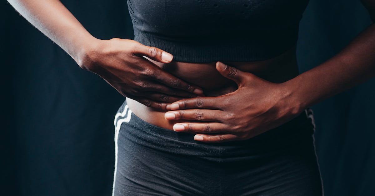
Relieving sharp pain beneath the right rib cage following a meal requires immediate dietary modification to strictly avoid lipid-rich foods, thereby preventing the gallbladder from contracting vigorously. This specific postprandial pain, clinically termed biliary colic, frequently indicates that a gallstone is temporarily obstructing the cystic duct when the gallbladder attempts to release bile for digestion. The primary clinical objective is to cease the chemical signals commanding the gallbladder to squeeze, allowing the obstructed stone to fall back into the hollow reservoir and rapidly restoring normal biliary flow.
The right upper quadrant of the abdomen houses several vital organs, primarily the liver and the gallbladder. When a sharp, agonizing pain reliably occurs thirty to sixty minutes after consuming a meal, the physiological mechanics of digestion are clearly implicated. The gallbladder serves as a storage sac for bile, a digestive fluid essential for breaking down dietary fats. When a mechanical blockage interrupts this fluid delivery system, the resulting pressure buildup causes immense localized distress.
Clinical assessment of right upper quadrant pain focuses on differentiating a temporary, benign gallbladder spasm from a severe, acute infection or adjacent gastrointestinal pathology. By understanding the precise hormonal triggers of digestion and utilizing specific resting modalities, patients can effectively manage a flare-up of biliary colic while pursuing definitive diagnostic imaging and medical guidance.
2. Anatomy of the Biliary Tract
To comprehend the mechanical nature of this pain, one must visualize the biliary tree. The liver continuously produces bile, a dark green fluid composed of bile salts, cholesterol, and bilirubin. This fluid travels down the hepatic ducts and flows into the gallbladder, a small, pear-shaped organ tucked neatly under the right lobe of the liver and protected by the lower right rib cage.
The gallbladder concentrates this bile, making it exceptionally potent, and stores it until it is needed for digestion. When a meal is consumed, the gallbladder contracts, squeezing the concentrated bile out through a narrow tube called the cystic duct.
The cystic duct merges with the common bile duct, which ultimately empties into the duodenum, the first section of the small intestine. This intricate plumbing system relies entirely on unobstructed tubes. Any solid obstruction within this pathway instantly disrupts the flow of fluid, leading to severe retrograde pressure.
3. Pathophysiology of Biliary Colic
Biliary colic is the classic presentation of symptomatic gallstone disease, known as cholelithiasis. Gallstones form when the chemical components of bile, typically cholesterol or bilirubin, become supersaturated and crystallize into solid, pebble-like structures within the gallbladder.
When a patient eats, the gallbladder contracts to expel bile. If a gallstone is swept up in this fluid wave, it can become physically wedged in the narrow opening of the cystic duct. As the gallbladder continues to squeeze against this fixed, solid blockage, the internal pressure within the organ skyrockets.
The wall of the gallbladder becomes distended and intensely stretched. The visceral nerve fibers lining the organ are exceptionally sensitive to this stretching. They fire rapid pain signals to the central nervous system, which the patient perceives as a deep, sharp, or gnawing pain localized firmly under the right rib cage, frequently radiating straight through to the right shoulder blade.
4. The Role of Cholecystokinin
The physical contraction of the gallbladder is entirely governed by a specific digestive hormone called cholecystokinin. When partially digested food leaves the stomach and enters the small intestine, specialized cells in the duodenal lining detect the exact macronutrient composition of the meal.
If the sensors detect a high concentration of dietary lipids (fats) or dense proteins, they release a massive surge of cholecystokinin into the bloodstream. This hormone acts directly on the smooth muscle receptors of the gallbladder, commanding a vigorous, sustained contraction to deliver enough bile to emulsify the heavy fats.
Therefore, the severity of a biliary colic attack is directly proportional to the fat content of the preceding meal. A meal consisting of fried foods, heavy cream, or dense cheese provokes a far more forceful contraction than a meal of simple carbohydrates, making the former infinitely more likely to wedge a gallstone tightly into the duct.
5. Fasting and Acute Bowel Rest
When acute biliary colic strikes, the most effective immediate intervention is absolute bowel rest. The patient must completely cease all oral intake of food and caloric liquids. Ingesting any further calories will only stimulate the continued release of cholecystokinin, forcing the gallbladder to continue squeezing against the blockage.
By initiating a strict fast, the hormonal signaling rapidly diminishes. The smooth muscle of the gallbladder gradually relaxes. Once the squeezing force is eliminated, the impacted gallstone will frequently dislodge from the narrow cystic duct and fall harmlessly back into the wide body of the gallbladder.
As soon as the stone falls back, the anatomical blockage is cleared. The trapped bile drains normally, the internal pressure normalizes, and the sharp, agonizing pain typically resolves completely within one to four hours, leaving behind only a dull, residual ache.
6. Thermal Therapy for Smooth Muscle Spasm
While waiting for the hormonal signals to subside and the stone to shift, thermal therapy can provide significant palliative relief. The severe distension of the gallbladder frequently causes the surrounding abdominal musculature to seize up in a protective reflex, adding secondary muscular cramping to the primary visceral pain.
Applying a hot water bottle or a medical heating pad directly over the right upper quadrant and the right flank induces deep, localized vasodilation. The thermal energy physically softens the tense abdominal wall and increases blood flow to the distressed region.
This warmth provides a soothing sensory input that helps override the visceral pain signals traveling to the spinal cord. It also encourages systemic relaxation, which actively lowers the sympathetic nervous system’s fight-or-flight response that inherently exacerbates the perception of severe pain.
7. Hydration and Biliary Sludge
Beyond solid stones, patients can experience biliary colic from biliary sludge. Sludge is a thick, microscopic suspension of cholesterol crystals and mucin that acts like wet sand within the gallbladder. When the organ contracts, this dense sludge struggles to push through the cystic duct, causing friction, sluggish flow, and resultant pressure pain.
Chronic systemic dehydration heavily contributes to the formation of biliary sludge. When the body lacks water, the liver produces bile that is inherently thicker and more prone to crystallization.
Maintaining aggressive daily hydration with clear water is crucial for preventing future attacks. Ample fluid intake ensures that the liver produces a thinner, more watery bile, allowing it to flow seamlessly out of the gallbladder and preventing the stagnation necessary for solid stones and sludge to precipitate.
8. Differentiating Peptic Ulcer Disease
A clinician must always differentiate biliary colic from peptic ulcer disease, as both pathologies cause severe postprandial upper abdominal pain. A peptic ulcer is a raw, open sore in the mucosal lining of the stomach or the duodenum.
Pain from a gastric ulcer typically presents as a burning, gnawing sensation slightly to the left of the midline or dead center in the epigastrium. Crucially, ulcer pain often begins almost immediately upon eating, as the physical presence of food and the surge of stomach acid irritate the raw, exposed tissue.
Conversely, biliary colic is localized sharply to the right side and is typically delayed by thirty minutes to an hour after eating, corresponding to the time it takes for the fat to enter the small intestine and trigger the release of cholecystokinin. Furthermore, biliary colic is characterized by a crescendo-decrescendo pattern of intense pressure, rather than the acidic burning typical of an ulcer.
9. Gastroesophageal Reflux Considerations
Severe gastroesophageal reflux can occasionally mimic right-sided rib pain, especially if the esophageal spasms radiate atypically. Reflux pain is generally described as a fiery, burning sensation that travels upward behind the breastbone toward the throat.
Reflux is frequently worsened by lying down completely flat immediately after a meal, whereas the physical posture of the patient does not typically alter the severe, mechanical pressure pain of a wedged gallstone.
If the pain is accompanied by a sour taste in the mouth or frequent belching, and is relieved by drinking alkaline water or taking antacids, the etiology is far more likely to be localized to the esophagus and stomach rather than the biliary tree.
10. Long-Term Dietary Management
Patients who experience a confirmed episode of biliary colic must implement strict, long-term dietary modifications to prevent recurrent attacks while awaiting definitive medical evaluation. The cornerstone of this management is a profoundly low-fat diet.
All fried foods, fatty cuts of meat, full-fat dairy products, and heavy oils must be strictly eliminated. By restricting dietary lipids, the patient essentially starves the body of the exact macronutrient required to trigger a massive cholecystokinin release.
Meals should consist of complex carbohydrates, lean proteins such as baked chicken or fish, and ample vegetable fiber. Consuming smaller, more frequent meals throughout the day is vastly superior to consuming large, heavy dinners, as small meals provoke only minor, gentle contractions of the gallbladder that are less likely to displace a resting stone.
11. Diagnostic Ultrasound and Blood Panels
Any patient experiencing recurrent right upper quadrant pain after eating requires a formal clinical evaluation. The gold standard diagnostic tool is a transabdominal ultrasound of the right upper quadrant.
Ultrasound is a fast, painless, and non-invasive modality that utilizes sound waves to create a precise image of the liver and biliary tree. It can definitively identify the presence of gallstones, measure the thickness of the gallbladder wall, and determine if the common bile duct is abnormally dilated.
Simultaneously, a physician will order a comprehensive metabolic blood panel to evaluate liver enzymes (AST, ALT, Alkaline Phosphatase) and total bilirubin. Elevated liver enzymes strongly suggest that a gallstone has escaped the cystic duct and is currently blocking the main drainage pathway of the liver itself, a condition requiring urgent intervention.
12. Red Flags for Acute Cholecystitis
While biliary colic is a temporary, mechanical blockage, it can rapidly evolve into a severe, life-threatening infection known as acute cholecystitis. This occurs when the gallstone becomes permanently wedged in the cystic duct and refuses to fall back into the gallbladder.
The trapped bile inside the blocked gallbladder becomes stagnant and rapidly infected by intestinal bacteria. The organ swells dangerously and risks rupturing. Acute cholecystitis represents a strict medical emergency.
The red flag symptoms indicating this transition include unrelenting pain that lasts longer than six hours, a high fever, severe chills, and a phenomenon known as Murphy’s sign, where the patient physically cannot take a deep breath when the physician palpates the right rib cage due to excruciating tenderness.
13. The Risk of Choledocholithiasis and Jaundice
Another severe complication of gallstones is choledocholithiasis. This occurs when a stone successfully passes through the cystic duct but becomes firmly lodged in the common bile duct, the final pathway that empties into the intestine.
A blockage here prevents all bile from leaving the liver. Because the bile cannot drain, bilirubin backs up into the systemic bloodstream. This systemic accumulation of bilirubin clinically presents as jaundice, a distinct yellowing of the skin and the sclera (whites) of the eyes.
Furthermore, a common bile duct stone can block the pancreatic duct, triggering acute pancreatitis. The onset of jaundice, accompanied by dark, tea-colored urine and pale, clay-colored stools, mandates immediate emergency endoscopic intervention to retrieve the obstructing stone.
14. Surgical Interventions and Cholecystectomy
For patients who suffer from recurrent, debilitating attacks of biliary colic, or who develop acute cholecystitis, the definitive medical cure is the surgical removal of the gallbladder, a procedure known as a cholecystectomy.
Because the gallbladder is simply a storage reservoir and not a vital organ for survival, its removal permanently eliminates the anatomical space where stones form and become trapped. The surgery is predominantly performed laparoscopically, utilizing small incisions and a camera, resulting in a rapid recovery.
Following a cholecystectomy, the liver simply drips bile continuously into the small intestine. Patients lead entirely normal lives, though some may need to moderate their intake of exceptionally heavy, fatty meals to avoid mild digestive urgency, as they no longer possess a storage sac to deliver large boluses of bile on demand.
| Clinical Condition | Pain Characteristics | Accompanying Symptoms |
|---|---|---|
| Biliary Colic | Sharp, right-sided, 30-60 mins after fatty meal. Resolves in 1-4 hours. | Nausea, restlessness, pain radiating to right shoulder. |
| Peptic Ulcer | Burning, central or left-sided, begins quickly after eating. | Relieved by antacids, worsened by acidic foods or stress. |
| Acute Cholecystitis | Unrelenting right-sided pain lasting >6 hours. | High fever, chills, exquisite tenderness to touch. |
15. Frequently Asked Questions
1. Does apple cider vinegar dissolve gallstones?
No. There is absolutely no clinical or biochemical evidence that consuming apple cider vinegar, or any specific fruit juice, can dissolve calcified or cholesterol gallstones inside the gallbladder. This is a persistent and dangerous myth that delays proper medical evaluation.
2. Why does the pain shoot all the way into my right shoulder?
The gallbladder and the right shoulder share sensory nerve pathways that enter the spinal cord at the exact same level. When the gallbladder becomes intensely distended, the brain misinterprets the origin of the distress signals, creating a phenomenon known as referred pain in the shoulder blade.
3. Can I live a normal life without a gallbladder?
Yes. The liver will continue to produce bile to digest your food. Instead of storing the bile, it will simply flow directly into your intestine in a slow, steady drip. Most patients digest normally, though some experience mild diarrhea if they consume very large, high-fat meals.
4. Will drinking coffee trigger a gallbladder attack?
While caffeine stimulates bowel motility, coffee itself is not high in fat and does not trigger the massive release of cholecystokinin required to cause a severe gallbladder contraction. However, adding heavy cream or whole milk to the coffee certainly provides the lipids necessary to trigger an attack.
5. How fast do gallstones form?
Gallstones form incredibly slowly, typically taking years to grow to a size that causes mechanical obstruction. They form when the chemical composition of the bile is chronically imbalanced, allowing microscopic crystals to fuse together over thousands of digestive cycles.
16. Bibliography
Disclaimer: The content is for informational purposes only and does not replace medical advice. Always consult your doctor for personalized treatment.