1. Introduction
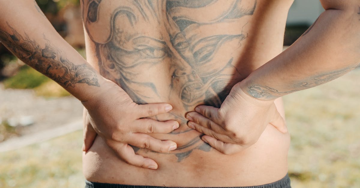
A sharp, electrical pain shooting down the lower spine and into the leg is the hallmark symptom of lumbar radiculopathy, commonly known as sciatica. This neuropathic pain occurs when a spinal nerve root in the lower back is mechanically compressed or chemically irritated, most often by a herniated intervertebral disc or a narrowing of the spinal canal. Addressing the structural impingement through targeted physical therapy and anti-inflammatory management is essential to relieve the nerve compression and restore pain-free mobility.
The sensation of electrical pain is distinct from the dull ache of a simple muscle strain. Peripheral nerves operate via precise electrochemical gradients. When a major nerve bundle is crushed or inflamed, it misfires chaotically, broadcasting a searing, shocking sensation along its entire anatomical pathway. Understanding the trajectory of the sciatic nerve reveals exactly why a problem originating in the lower back is felt so intensely in the calf or foot.
2. Anatomy Of The Sciatic Nerve
The sciatic nerve is the longest and thickest nerve in the human body, roughly the diameter of an adult thumb at its origin. It does not begin as a single structure; rather, it is formed by the convergence of five distinct nerve roots exiting the lower spine, specifically from the lumbar segments L4 and L5, and the sacral segments S1, S2, and S3.
These nerve roots bundle together deep within the pelvis to form the main sciatic nerve. The nerve then exits the pelvis, passes beneath the gluteal muscles, and travels directly down the posterior aspect of the thigh. Above the knee, it branches into smaller nerves that supply sensation and motor function to the calf, ankle, and toes.
3. The Pathophysiology Of Lumbar Radiculopathy
Radiculopathy refers to the clinical syndrome caused by the pinching or irritation of a nerve root as it exits the spinal column. The spine is constructed of stacked vertebrae separated by intervertebral discs. The nerve roots exit through small, bony windows called neural foramina.
If the space within a neural foramen decreases, the exiting nerve root is crushed against the surrounding bone. This mechanical compression obstructs the microscopic blood vessels supplying the nerve, causing immediate ischemia. Deprived of oxygen, the nerve becomes highly irritable and spontaneously discharges action potentials, which the brain interprets as a sharp, shooting electrical shock.
4. Intervertebral Disc Herniation
The most frequent cause of acute sciatica in young to middle-aged adults is a herniated lumbar disc. Intervertebral discs feature a tough, fibrous outer ring called the annulus fibrosus, enclosing a soft, gel-like center known as the nucleus pulposus.
Heavy lifting, sudden twisting, or gradual wear can cause the annulus to tear. The internal gel extrudes backward into the spinal canal or the neural foramen. This herniated material not only physically compresses the adjacent nerve root but also releases highly inflammatory proteins. This potent combination of mechanical crushing and chemical burning induces severe, unrelenting radicular pain.
5. Lumbar Spinal Stenosis
In older adults, shooting leg pain is frequently driven by lumbar spinal stenosis. As the spine ages, the intervertebral discs dehydrate and lose height, and the facet joints may develop osteoarthritis. The body responds to this joint instability by forming bone spurs, or osteophytes, to restrict excess movement.
These bone spurs, combined with a thickening of the spinal ligaments, slowly encroach upon the space within the spinal canal and the neural foramina. This structural narrowing acts like a tightening vice around the nerve roots. The pain associated with stenosis often worsens during walking or standing, as these upright postures further narrow the already compromised spinal spaces.
6. Piriformis Syndrome And Peripheral Entrapment
Not all shooting leg pain originates in the spinal column. The sciatic nerve must navigate a complex muscular pathway through the buttocks. In most individuals, the nerve passes directly beneath the piriformis muscle, a deep external rotator of the hip.
If the piriformis muscle becomes chronically tight, spasms, or hypertrophies due to overuse or poor biomechanics, it can compress the sciatic nerve against the underlying pelvic bone. This condition, known as piriformis syndrome, produces symptoms entirely identical to a herniated disc, though the site of the nerve entrapment is peripheral rather than central.
7. Sensory Distribution And Dermatomes
Neurologists utilize the specific location of the electrical pain to identify precisely which nerve root is being compressed. The skin is mapped into distinct sensory zones called dermatomes, with each zone supplied by a specific spinal nerve.
If the L4 nerve root is compressed, the shooting pain typically wraps around to the front of the thigh and the inner knee. Compression of the L5 root shoots pain down the outside of the calf and into the big toe. S1 root compression drives the electrical shock down the back of the hamstring, into the heel, and along the sole of the foot.
8. Motor Deficits And Muscle Weakness
In addition to transmitting sensory data, the sciatic nerve provides critical motor commands to the muscles of the leg. Severe or prolonged compression of a nerve root disrupts these motor signals, leading to profound, measurable weakness in specific muscle groups.
A compressed L5 nerve root frequently results in foot drop, where the patient is physically unable to lift the front of their foot, causing them to trip while walking. S1 compression impairs the calf muscles, making it impossible for the patient to stand on their tiptoes. Identifying these motor deficits is crucial, as they indicate a severe degree of nerve compromise.
9. Differentiating Neuropathic Pain Origins
Distinguishing the specific trigger of the shooting pain ensures an accurate anatomical diagnosis. The table below outlines key clinical differences.
| Pain Quality and Trigger | Associated Symptoms | Most Likely Origin |
|---|---|---|
| Sharp shock worse when sitting or bending forward | Pain radiates past the knee, coughing increases pain | Herniated lumbar disc |
| Ache and shock worse when walking, relieved by leaning forward | Heavy feeling in both legs, older adult | Lumbar spinal stenosis |
| Deep ache in the buttock, sharp shock down back of thigh | Pain worse when climbing stairs or sitting on hard surfaces | Piriformis syndrome |
| Dull, throbbing ache localized to the lower back | No pain radiating below the knee | Simple lumbar muscle strain |
10. The Straight Leg Raise Test
A classic diagnostic maneuver utilized by physicians is the straight leg raise test. While the patient lies flat on their back, the clinician slowly elevates the symptomatic leg while keeping the knee perfectly straight.
This movement applies direct tensile stretch to the sciatic nerve. If a nerve root is tethered by a herniated disc, lifting the leg between thirty and seventy degrees will reproduce the sharp, shooting electrical pain down the limb. A positive test strongly correlates with the presence of lower lumbar radiculopathy.
11. Spondylolisthesis And Vertebral Shifting
Spondylolisthesis occurs when one lumbar vertebra slips forward over the vertebra immediately beneath it. This slippage can be caused by a congenital defect in the bone, a stress fracture, or severe degenerative joint disease.
When the vertebra shifts out of proper alignment, it severely distorts the structural dimensions of the spinal canal. The shifting bone physically guillotines the exiting nerve roots. Patients with this condition often experience sharp electrical shocks that spike with sudden movements or when arching the back backward, which exacerbates the bony slippage.
12. Conservative Management And Physical Therapy
The vast majority of sciatica cases resolve with conservative management. The primary goal is to centralize the pain, drawing the electrical sensations out of the leg and back up toward the spine, indicating that the nerve compression is lessening.
Physical therapy employs specific movement protocols, such as the McKenzie method, which utilizes targeted spinal extension exercises to mechanically encourage the herniated disc material to retreat away from the affected nerve root. Core stabilization exercises are also critical to provide a muscular corset that prevents micro-movements from irritating the nerve further.
13. Pharmacological Modulators
Because sciatic pain is neuropathic, traditional painkillers are frequently ineffective. Pharmacological management relies heavily on nonsteroidal anti-inflammatory drugs to reduce the chemical inflammation burning the nerve root.
For severe, unrelenting electrical pain, physicians prescribe neuropathic modulators such as gabapentin or pregabalin. These medications act directly on the central nervous system to quiet the erratic, chaotic electrical firing of the damaged nerve, blunting the sharp shocks while the underlying structural issue heals.
14. Epidural Steroid Injections
If oral medications and physical therapy fail to provide relief, an epidural steroid injection is a potent intervention. Under fluoroscopic guidance, a physician injects a concentrated corticosteroid directly into the epidural space of the spine, bathing the exact nerve root that is being compressed.
This localized application of steroids provides massive, direct anti-inflammatory power, rapidly shrinking the swollen tissues surrounding the nerve. While it does not physically remove a herniated disc, it frequently breaks the cycle of acute pain, allowing the patient to actively participate in rehabilitation.
15. When To Seek Emergency Medical Care
While severe shooting pain is deeply uncomfortable, it is rarely a surgical emergency. However, a surgical evaluation is absolutely necessary if the shooting pain is accompanied by a progressive, profound loss of strength in the leg, indicating that the nerve is actively dying.
Immediate emergency medical care is mandatory if the severe back and leg pain is accompanied by a sudden loss of bowel or bladder control, or profound numbness in the groin and saddle area. These are the hallmark signs of cauda equina syndrome, a catastrophic compression of the entire terminal spinal cord that requires immediate emergency decompression surgery to prevent permanent paralysis.
16. Frequently Asked Questions (FAQ)
1. Can resting in bed for a week cure my sciatica?
No, prolonged bed rest is counterproductive. Immobility causes the supporting back muscles to weaken and the spinal joints to stiffen. You should maintain gentle, daily movement and walking, resting only when the pain becomes intolerable.
2. Does applying heat or ice help with nerve pain?
Applying ice to the lower back can numb the area and reduce superficial inflammation, while heat relaxes tight, spasming back muscles. However, neither will physically un-pinch the nerve deep inside the spine that is causing the electrical shock in your leg.
3. Can wearing a tight belt or heavy wallet cause sciatica?
Sitting on a thick wallet in your back pocket can directly compress the sciatic nerve against your pelvis, causing a peripheral entrapment similar to piriformis syndrome. A tight belt typically does not affect the sciatic nerve.
4. Will a herniated disc eventually heal on its own?
Yes, in the vast majority of cases, the body’s immune system recognizes the extruded disc gel as foreign material. Over several months, macrophages will slowly dissolve and reabsorb the herniated material, naturally relieving the pressure on the nerve root.
5. Why does coughing or sneezing make the shooting pain so much worse?
Coughing and sneezing cause a massive, instantaneous spike in the pressure inside your abdominal and spinal cavities. This sudden pressure wave forcefully jams the herniated disc material harder against the sensitive nerve root, producing a violent electrical shock.
17. Bibliography
Disclaimer: The content is for informational purposes only and does not replace medical advice. Always consult your doctor for personalized treatment.