1. Introduction to Solitary Fibrous Tumour
A solitary fibrous tumour is a rare, mesenchymal soft tissue neoplasm that originates from fibroblasts, the cells responsible for producing connective tissue. When classified as Grade 1, the tumor is considered benign and exhibits indolent, slow-growing behavior. Historically, these growths were primarily identified in the pleural cavity, the protective membrane surrounding the lungs, but they are now known to occur at almost any anatomical site in the body.
The discovery of a solitary fibrous tumour requires comprehensive clinical evaluation. Despite its benign Grade 1 designation, the tumor can grow to a significant size, compressing adjacent anatomical structures and causing localized symptoms. The cornerstone of management relies on accurate pathological identification and prompt surgical intervention.
Patients often experience high levels of anxiety when diagnosed with any tumor. It is a primary responsibility of the healthcare team to provide clear, factual reassurance regarding the non-aggressive nature of a Grade 1 lesion. With precise surgical resection, the long-term prognosis is excellent, and patients can anticipate a return to normal health.
2. The Historical Context of Haemangiopericytoma
The term “haemangiopericytoma” holds significant historical relevance in surgical pathology. Decades ago, it was used to describe a distinct category of vascular soft tissue tumors characterized by a dense network of branching blood vessels. However, advancements in molecular biology and genetics have fundamentally reshaped medical classification.
Today, modern pathology recognizes that the vast majority of tumors previously diagnosed as haemangiopericytoma are biologically and genetically identical to solitary fibrous tumours. Consequently, the World Health Organization has largely retired the term haemangiopericytoma, classifying these lesions uniformly under the solitary fibrous tumour spectrum.
Despite its obsolescence in official diagnostic manuals, patients may still encounter the term in historical medical literature or older clinical reports. Understanding that haemangiopericytoma and solitary fibrous tumour represent the same underlying biological entity helps eliminate confusion and streamlines the modern approach to treatment and patient education.
3. Pathophysiology and Molecular Genetics
The most significant breakthrough in understanding solitary fibrous tumours is the identification of their driving genetic mutation. The pathophysiology is unequivocally linked to a specific chromosomal inversion on chromosome 12. This genetic error fuses two genes together: the NAB2 gene and the STAT6 gene.
The resulting NAB2-STAT6 fusion gene acts as the central engine for tumor development. It produces an abnormal fusion protein that enters the nucleus of the cell and disrupts normal gene transcription, leading to the uncontrolled, yet slow, proliferation of spindle cells. This genetic anomaly is highly specific to solitary fibrous tumours and is present in almost all cases.
Because this mutation drives the disease, the tumor is not caused by lifestyle factors, diet, or environmental exposures. It is a spontaneous genetic event occurring within a localized cluster of connective tissue cells.
4. Histological Features of Grade 1 Tumours
To confirm a Grade 1 classification, a pathologist must analyze the tumor tissue under a microscope. A Grade 1 solitary fibrous tumour displays specific, reassuring features that confirm its non-aggressive nature. The tissue is composed of bland, uniform spindle cells lacking significant nuclear abnormalities.
These cells are arranged in a disorganized, “patternless pattern” interspersed within a matrix of thick, rope-like collagen bundles. Another classic histological hallmark is the presence of numerous, prominent blood vessels that branch out in a shape reminiscent of deer antlers, commonly referred to as “staghorn” vessels.
Crucially, to maintain a Grade 1 designation, the tumor must exhibit a very low mitotic rate, meaning the cells are dividing extremely slowly. Furthermore, there must be an absolute absence of tumor necrosis, which is cell death commonly seen in rapidly growing, malignant tumors.
5. Key Immunohistochemical Markers
The definitive diagnosis of a solitary fibrous tumour relies heavily on immunohistochemistry. This laboratory technique applies specific antibodies to the tissue sample to detect unique proteins, allowing the pathologist to differentiate this tumor from other spindle cell neoplasms.
The most critical diagnostic marker is the nuclear expression of the STAT6 protein. Because the NAB2-STAT6 genetic fusion drives the tumor, the STAT6 protein accumulates heavily in the nucleus of the tumor cells. Strong nuclear positivity for STAT6 is considered the gold standard for diagnosis.
Additionally, solitary fibrous tumours typically show diffuse positive staining for CD34, a marker of vascular and connective tissue cells. Conversely, they consistently test negative for proteins such as cytokeratins, desmin, and S100, which effectively rules out carcinomas, smooth muscle tumors, and nerve sheath tumors, respectively.
6. Common Anatomical Sites
While initially described as a lesion of the pleura, solitary fibrous tumours are ubiquitous and can arise anywhere in the body where connective tissue exists. Anatomical presentation is broadly divided into pleural and extrapleural sites.
Pleural tumors grow within the thoracic cavity, frequently attaching to the visceral pleura lining the lung via a small stalk or pedicle. Extrapleural tumors can emerge in a wide variety of locations. The most common extrapleural sites include the deep soft tissues of the lower extremities, the abdominal cavity, the retroperitoneum, and the head and neck region.
Meningeal solitary fibrous tumours, which grow from the protective membranes covering the brain and spinal cord, also occur and were historically a major category of lesions labeled as haemangiopericytomas. Regardless of the location, the underlying biology and cellular appearance of the Grade 1 tumor remain identical.
7. Clinical Symptoms and Presentation
Because Grade 1 solitary fibrous tumours grow very slowly, they are frequently asymptomatic during their initial stages. Many are discovered entirely by accident when a patient undergoes imaging, such as a chest X-ray or abdominal ultrasound, for an unrelated medical complaint.
When symptoms do present, they are almost entirely the result of the mechanical mass effect—the tumor pressing against adjacent organs, nerves, or blood vessels. A pleural tumor might cause a persistent cough, shortness of breath, or a feeling of heaviness in the chest. A retroperitoneal tumor might manifest as a palpable abdominal mass, vague pelvic discomfort, or urinary frequency due to bladder compression.
In exceptionally rare instances, solitary fibrous tumours can cause paraneoplastic syndromes. The most notable is Doege-Potter syndrome, where the tumor produces an insulin-like growth factor that causes the patient to experience episodes of severe hypoglycemia, or low blood sugar. These systemic symptoms resolve immediately upon surgical removal of the mass.
8. Diagnostic Imaging Approaches
Comprehensive diagnostic imaging is mandatory for the initial assessment and surgical planning of a solitary fibrous tumour. For intrathoracic lesions, a computed tomography scan of the chest with intravenous contrast is the standard modality. The tumor typically appears as a well-defined, smooth, lobulated mass that enhances intensely due to its rich vascular network.
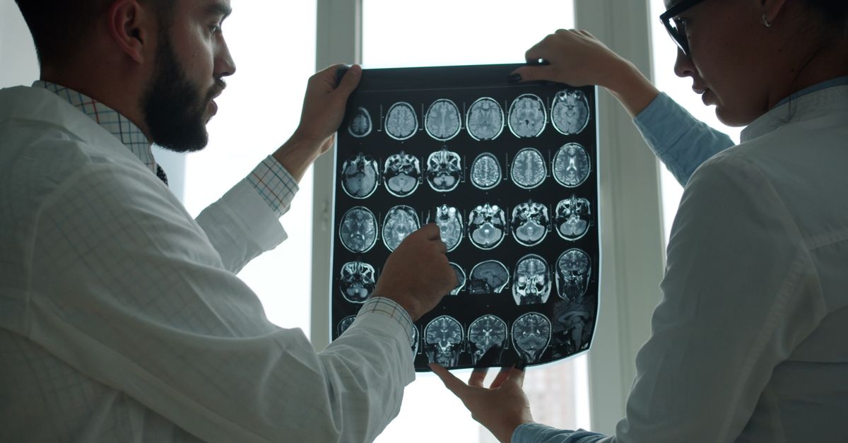
For extrapleural tumors in the extremities, pelvis, or head and neck, magnetic resonance imaging provides superior contrast resolution. On magnetic resonance imaging, the dense collagen content of the tumor often results in low signal intensity on T2-weighted images, while the prominent “staghorn” vessels may appear as distinctive flow voids within the mass.
Imaging not only defines the size and boundaries of the tumor but also evaluates its proximity to vital anatomical structures, which is critical information for the surgeon tasked with safely removing the lesion.
9. Differential Diagnosis
Accurately distinguishing a Grade 1 solitary fibrous tumour from other soft tissue masses requires expert clinical and pathological correlation.
| Tumor Type | Key Differentiating Features |
|---|---|
| Solitary Fibrous Tumour (Grade 1) | STAT6 nuclear positivity, CD34 positivity, prominent staghorn vasculature, low mitotic rate. |
| Synovial Sarcoma | Malignant, features t(X;18) genetic translocation, often positive for epithelial markers like cytokeratin. |
| Gastrointestinal Stromal Tumour | Occurs in the gastrointestinal tract, positive for KIT (CD117) and DOG1, negative for STAT6. |
| Schwannoma | Benign nerve sheath tumor, strongly and diffusely positive for S100 protein, arises directly from a nerve. |
Pathologists meticulously evaluate these markers to ensure the correct diagnosis and prevent the application of overly aggressive or inappropriate therapies. For related inquiries on persistent tiredness linked to abdominal masses, patients sometimes explore chronic fatigue symptoms.
10. Core Needle Biopsy and Pathology
While imaging provides a strong suspicion, a tissue sample is required for a definitive diagnosis. A core needle biopsy, performed under image guidance, is the preferred method for obtaining tissue without risking tumor spillage into surrounding anatomical compartments.
During this procedure, a specialized needle is used to extract small cylinders of tissue from the mass. The pathologist then subjects these samples to the histological and immunohistochemical analyses described earlier, specifically looking for the STAT6 and CD34 markers.
A fine-needle aspiration, which extracts only single cells rather than tissue architecture, is generally inadequate for diagnosing a solitary fibrous tumour due to the importance of observing the patternless pattern of cells and the structural collagen matrix.
11. Surgical Resection as the Standard of Care
The primary and most effective treatment for a Grade 1 solitary fibrous tumour is complete surgical resection. Because the tumor is benign and non-infiltrative, achieving negative surgical margins—meaning the entire tumor is removed with a surrounding layer of healthy tissue—is highly curative.
The surgical approach depends entirely on the tumor’s location. Pleural tumors may be removed via a thoracotomy or minimally invasive video-assisted thoracoscopic surgery, frequently requiring only the division of the pedicle attaching the tumor to the lung. Tumors in the abdomen or extremities require careful dissection to separate the fibrous mass from adjacent nerves and blood vessels.
Due to the prominent vascularity of these tumors, surgical planning must account for the risk of intraoperative bleeding. In cases where the tumor is exceedingly large and highly vascular, preoperative embolization—a procedure to deliberately block the tumor’s blood supply—may be performed a day prior to surgery to minimize blood loss.
12. Radiotherapy and Adjuvant Treatments
For Grade 1 solitary fibrous tumours that are completely resected with clear margins, adjuvant treatments such as radiation therapy or chemotherapy are typically unnecessary. The biological behavior of a Grade 1 lesion is benign, making the toxicity of systemic or localized radiation unwarranted.
However, clinical scenarios occasionally arise where the tumor is located in a highly complex anatomical area, such as the base of the skull or deep pelvis, making complete surgical removal impossible without causing unacceptable functional damage.
In these rare instances of incomplete resection, or if the tumor displays unexpected aggressive features on final pathology, localized adjuvant radiation therapy may be considered. Radiation aims to destroy any microscopic tumor cells left behind, thereby reducing the risk of local recurrence. Chemotherapy has proven largely ineffective against benign solitary fibrous tumours.
13. Long-Term Prognosis and Recurrence
The prognosis for a patient diagnosed with a Grade 1 solitary fibrous tumour following complete surgical excision is excellent. The ten-year survival rate is exceptionally high, and the majority of patients are considered permanently cured.
Despite the benign classification, long-term clinical surveillance is essential. There is a documented risk of local recurrence, particularly if the initial surgical margins were narrow or microscopically positive. Recurrences can manifest many years, sometimes over a decade, after the initial surgery.
Therefore, patients are typically enrolled in a long-term follow-up program. This involves routine physical examinations and periodic imaging studies, such as computed tomography scans or magnetic resonance imaging, tailored to the original anatomical site, to ensure early detection of any recurrent growth.
14. When to Consult an Oncologist
Patients should schedule an evaluation with a medical professional if they notice an unexplained, palpable mass anywhere on their body, or if they develop persistent symptoms such as an unyielding cough, unexplained shortness of breath, or a sensation of abdominal fullness.
If an imaging study incidentally reveals a mass in the chest, abdomen, or soft tissues, prompt referral to a surgical oncologist or a soft tissue specialist is imperative. Specialized centers possess the multidisciplinary expertise required to accurately diagnose and manage rare soft tissue tumors.
Early consultation facilitates a coordinated diagnostic approach, ensuring that biopsies and definitive surgeries are performed with precision, minimizing risks, and optimizing the patient’s long-term health outcome.
15. Frequently Asked Questions (FAQ)
1. Is a Grade 1 solitary fibrous tumour considered cancer?
No, a Grade 1 classification indicates that the tumor is benign. It grows slowly, does not destroy surrounding tissues aggressively, and has an exceptionally low risk of spreading to other parts of the body.
2. Why was this tumor previously called a haemangiopericytoma?
Historically, “haemangiopericytoma” was a descriptive term based on the prominent blood vessels seen under a microscope. Modern genetic testing has proven that most of these are actually solitary fibrous tumours, so the old term has been largely retired.
3. What causes the NAB2-STAT6 gene mutation?
The genetic fusion is considered a spontaneous, random error in cell division. It is not inherited from parents, nor is it caused by lifestyle factors, diet, or environmental exposures.
4. Will I need chemotherapy for a Grade 1 tumor?
Chemotherapy is virtually never used for Grade 1 solitary fibrous tumours. Complete surgical removal is the standard and most effective treatment.
5. Can the tumor come back after surgery?
While complete surgery is usually curative, there is a small risk of local recurrence, sometimes many years later. This is why long-term follow-up with periodic imaging is highly recommended.
16. Bibliography
Disclaimer: The content is for informational purposes only and does not replace medical advice. Always consult your doctor for personalized treatment.