1. Introduction
Spinal pain represents a complex, multidimensional clinical entity that spans the entire axial skeleton, from the highly mobile cervical segments to the robust, load-bearing lumbar and sacral regions. The human spine is a masterpiece of biomechanical engineering, providing the structural foundation for upright posture while housing and protecting the delicate spinal cord and exiting nerve roots. Because of the spine’s continuous, dynamic mechanical loading, its interlocking array of intervertebral discs, facet joints, robust ligaments, and deep stabilizing muscles is profoundly susceptible to acute trauma, repetitive strain, and progressive degenerative decline.
Addressing spinal pain requires a meticulous, region-specific diagnostic approach. A patient experiencing a sharp, neuropathic shock radiating down a limb requires entirely different clinical management than a patient suffering from a dull, localized, mechanical ache originating from an arthritic facet joint. By carefully mapping the precise anatomical pain generator and understanding the underlying pathophysiological mechanisms, clinicians can deploy targeted pharmacological, physical, and interventional therapies to resolve the acute distress, halt progressive tissue degradation, and restore functional spinal mobility.
2. Functional Anatomy of the Vertebral Column
The vertebral column is composed of 33 vertebrae, categorized into cervical, thoracic, lumbar, sacral, and coccygeal regions. Each distinct region exhibits unique structural adaptations. The cervical spine is optimized for a vast range of motion to support the head, rendering it vulnerable to acceleration-deceleration injuries. The thoracic spine is securely tethered to the rib cage, providing rigidity to protect the cardiopulmonary organs, and is subsequently the least common site for mechanical pain.
The massive lumbar vertebrae are engineered to bear the entire compressive weight of the upper body. The vertebrae are separated by fibrocartilaginous intervertebral discs, which feature a tough outer annulus fibrosus and a highly hydrated, gel-like nucleus pulposus. The posterior elements of the spine articulate via paired synovial facet joints. The intricate harmony between the shock-absorbing anterior discs and the motion-guiding posterior facet joints is the foundation of a pain-free spine.
3. Biomechanics of Spinal Load Distribution
The onset of mechanical spinal pain is intimately linked to altered biomechanics. In a healthy state, the intervertebral disc bears approximately 80% of the axial compressive load, while the posterior facet joints absorb the remaining 20%.
As an individual ages, the discs naturally lose hydration and elasticity, a process known as desiccation. As the disc flattens, its shock-absorbing capacity diminishes. This forces a biomechanical shift, transferring an abnormal, excessive load onto the posterior facet joints. This pathological redistribution of mechanical stress initiates a cascade of structural breakdown, triggering the formation of osteophytes (bone spurs), capsular hypertrophy, and chronic, mechanically driven pain.
4. Discogenic Pain and Annular Tears
Discogenic pain originates directly from the intervertebral disc itself, independent of nerve root compression. While the inner nucleus pulposus has no nerve supply, the outer third of the annulus fibrosus is heavily innervated by nociceptive nerve fibers.
When the annulus fibrosus sustains micro-tears or fissures due to excessive torsional strain or prolonged axial loading, these sensitive nerve endings are chemically irritated by the inflammatory proteins leaking from the nucleus pulposus. Patients with primary discogenic pain typically report a deep, centralized, aching pain that is reliably provoked by activities that increase intradiscal pressure, most notably prolonged sitting, bending forward, or lifting heavy objects.
5. Facet Joint Arthropathy
Facet joint arthropathy is the clinical manifestation of osteoarthritis occurring in the posterior synovial joints of the spine. Driven by the abnormal load redistribution from degraded discs, the articular cartilage of the facet joints erodes, and the joint capsule becomes severely inflamed.
Facet-mediated pain presents distinctly differently from discogenic pain. The pain is usually unilateral and is described as a deep, dull ache. Because the facet joints guide and restrict spinal extension, the pain is specifically exacerbated when the patient leans backward (extension) or twists the spine. It frequently causes referred pain down into the shoulders (from cervical facets) or into the upper buttocks and thighs (from lumbar facets), but it does not radiate past the knees.
6. Spinal Stenosis and Neurogenic Claudication
Spinal stenosis is a progressive narrowing of the central spinal canal or the lateral neural foramina. This condition is the cumulative result of advanced degenerative changes: the intervertebral discs bulge outward, the facet joints enlarge due to osteoarthritis, and the ligamentum flavum thickens.
The clinical hallmark of lumbar spinal stenosis is neurogenic claudication. As the patient walks or stands upright, the spinal canal anatomically narrows further, physically compressing and creating transient ischemia in the exiting nerve roots. The patient experiences a heavy, cramping, burning pain or weakness in the legs. Crucially, the pain is rapidly relieved when the patient sits down or leans forward over a shopping cart, as spinal flexion physically opens the spinal canal and restores local blood flow.
7. Muscular Strains and Myofascial Pain
The most prevalent cause of acute spinal pain is muscular strain. The deep erector spinae muscles and the complex paraspinal stabilizers are constantly engaged to maintain posture. Sudden, uncoordinated lifting, or cumulative fatigue from poor ergonomic positions, can cause the muscle fibers to stretch beyond their physiological limit.
The resulting micro-tears provoke a rapid inflammatory response. The body initiates a protective reflex, causing the surrounding musculature to enter a state of severe, involuntary spasm to immobilize the injured segment. The pain is intense, highly localized, and causes severe mechanical stiffness, but lacks the radiating, electrical characteristics of neurological impingement.
8. Inflammatory Spondyloarthropathies
When evaluating chronic spinal pain, particularly in younger adult patients, clinicians must consider systemic autoimmune etiologies. Ankylosing spondylitis is the prototype of inflammatory spondyloarthropathy. The immune system attacks the entheses—the sites where ligaments insert into the bone—primarily targeting the sacroiliac joints and the axial spine.
The clinical presentation is characterized by insidious, severe inflammatory back pain that is distinctly worse in the morning or after prolonged rest, and significantly improves with exercise and movement. Without aggressive systemic immunomodulatory treatment, the chronic inflammation causes the vertebral bodies to fuse completely, creating a brittle, immobile “bamboo spine” that is highly susceptible to traumatic fractures.
9. Osteoporotic Compression Fractures
In the elderly population, particularly postmenopausal women, the structural density of the vertebral bodies declines due to osteoporosis. The weakened, porous bone becomes incapable of supporting normal physiological loads.
An osteoporotic compression fracture occurs when the anterior portion of the vertebral body collapses under normal pressure, such as stepping off a curb, coughing, or bending to tie a shoe. The patient experiences a sudden, acute, severe band-like pain wrapping around the torso. Multiple compression fractures over time lead to a pronounced forward curvature of the thoracic spine (kyphosis) and a noticeable loss of overall body height.
10. Categorizing Spinal Pain Syndromes
Accurate diagnosis requires correlating the pain quality with the biomechanical triggers.
| Pain Generator | Quality of Pain | Aggravating / Relieving Factors |
|---|---|---|
| Discogenic Tear | Deep, centralized ache. | Worsens with sitting and bending forward. |
| Facet Arthropathy | Unilateral ache, no deep radiation. | Worsens with standing, leaning backward, and twisting. |
| Nerve Root (Radiculopathy) | Sharp, electrical, shooting down a limb. | Worsens with coughing, sneezing, or specific nerve stretching. |
| Inflammatory (Ankylosing) | Severe morning stiffness, deep ache. | Worsens with rest; significantly improves with physical activity. |
11. Comprehensive Clinical Evaluation
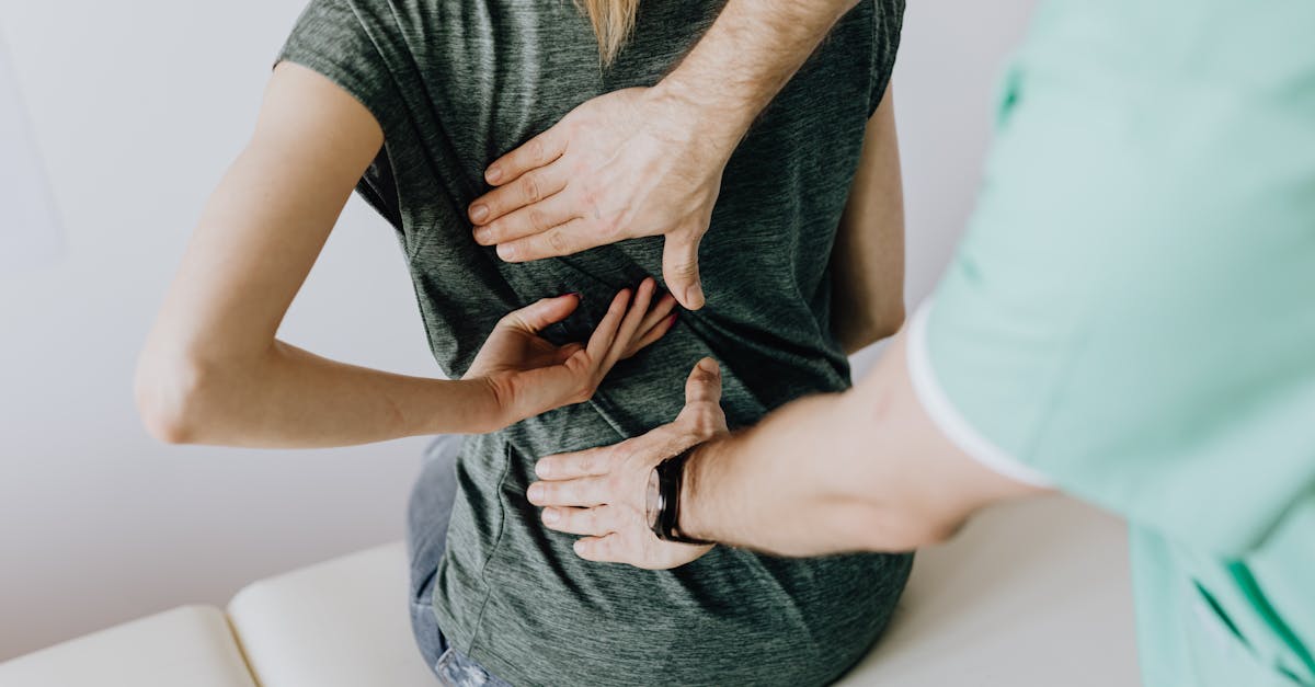
The physical examination is the cornerstone of spinal diagnosis. The clinician observes the patient’s gait, global posture, and active range of motion, noting any structural deformities or antalgic (pain-avoidant) lean. Palpation isolates focal muscular spasms or step-off deformities indicative of spondylolisthesis.
The clinician employs targeted provocative maneuvers. The Spurling test in the cervical spine and the Straight Leg Raise test in the lumbar spine apply mechanical tension to the nerve roots; a positive test reproduces the patient’s shooting, neuropathic limb pain, confirming radicular involvement.
12. Neurological Assessment
A detailed neurological examination is mandatory for any patient presenting with spinal pain to assess the functional integrity of the spinal cord and exiting nerve roots. The clinician tests dermatomal sensory perception to identify localized areas of numbness.
Motor strength is rigorously tested across all major muscle groups of the upper and lower extremities. A profound loss of motor strength (such as foot drop) indicates severe nerve compression requiring urgent intervention. Finally, testing deep tendon reflexes and screening for upper motor neuron signs (like a positive Babinski reflex) help differentiate peripheral nerve root compression from dangerous central spinal cord compression (myelopathy).
13. Diagnostic Imaging Modalities
Diagnostic imaging provides the structural roadmap. Plain radiographs are the first-line modality, efficiently demonstrating overall spinal alignment, severe degenerative disc height loss, osteophyte formation, and acute fractures.
Magnetic Resonance Imaging (MRI) is the definitive gold standard for evaluating the soft tissues of the spine. An MRI provides high-resolution, multi-planar visualization of the intervertebral discs, the neural foramina, the spinal cord, and the paraspinal musculature. It accurately delineates the extent of disc herniations and the precise degree of nerve root compression driving the patient’s radicular symptoms.
14. Pharmacological Pain Management
Pharmacological therapy focuses on reducing acute inflammation and modulating aberrant nerve signals. Nonsteroidal anti-inflammatory drugs (NSAIDs) represent the foundation of conservative care, effectively blunting the local prostaglandin response associated with muscular strains and facet joint inflammation. Short-course muscle relaxants are deployed to break the painful cycle of acute, severe paraspinal spasms.
For patients suffering from chronic radiculopathy or spinal stenosis, standard analgesics are often ineffective. Neuromodulators, specifically gabapentinoids (gabapentin or pregabalin), are prescribed to stabilize the hyperexcitable nerve membranes and significantly reduce the sharp, burning sensations associated with nerve compression.
15. Rehabilitation and Physical Therapy
Active physical rehabilitation is the most critical component for achieving long-term resolution of mechanical spinal pain. Passive modalities (like heat or ultrasound) provide only temporary relief. A specialized physical therapist guides the patient through a progressive, targeted exercise regimen.
Therapy focuses on restoring segmental spinal mobility, correcting postural imbalances, and aggressively strengthening the core and deep paraspinal stabilizing muscles. A robust, muscular core acts as a dynamic internal brace, absorbing mechanical shocks and effectively offloading the damaged intervertebral discs and arthritic facet joints during daily activities.
16. Interventional and Surgical Options
When conservative and pharmacological therapies are exhausted, interventional pain procedures offer targeted relief. Epidural steroid injections deliver potent anti-inflammatory medication directly to the inflamed nerve root space, often resolving acute radiculopathy. For facet-mediated pain, medial branch blocks followed by radiofrequency ablation can temporarily deactivate the tiny sensory nerves supplying the arthritic joints, providing profound, long-lasting relief.
Surgical intervention is strictly reserved for patients with intractable pain that fails extensive conservative care, or for those presenting with progressive neurological deficits. Procedures ranging from microdiscectomy (removing the herniated portion of a disc) to spinal fusion (permanently connecting unstable vertebrae) are highly successful in carefully selected patients, aiming to decompress vital neural structures and restore mechanical spinal stability.
17. Frequently Asked Questions (FAQ)
1. How do I know if my spinal pain is a muscle spasm or a disc problem?
A muscle spasm feels like a severe, tight, localized cramp that restricts movement but stays in the back. A disc problem, particularly a herniation, often pinches a nerve, causing a sharp, electrical or burning pain that shoots away from the spine and travels down your arm or leg.
2. Why does my back hurt much more when I sit at my desk?
Sitting, especially with a slouched posture, significantly increases the mechanical pressure inside the intervertebral discs of your lower back. If you have a small tear or degeneration in a disc, this increased pressure causes a deep, centralized, aching pain.
3. What is spinal stenosis, and why does leaning forward help the pain?
Spinal stenosis is a narrowing of the bone canal where your spinal cord and nerves travel. When you stand straight, the canal naturally tightens, pinching the nerves and causing your legs to feel heavy and painful. Leaning forward (like over a shopping cart) physically opens the canal up, relieving the pressure instantly.
4. Can an X-ray show why my back is hurting?
An X-ray only shows bone. It is excellent for finding arthritis, bone spurs, or fractures. However, it cannot show soft tissues like a herniated disc, a pinched nerve, or a torn muscle. To see those specific soft-tissue injuries, a Magnetic Resonance Imaging (MRI) scan is required.
5. When is spinal pain considered a medical emergency?
You must seek immediate emergency evaluation if your spinal pain is accompanied by a sudden, progressive weakness in your arms or legs, a loss of sensation in your groin (saddle area), or a sudden loss of bowel or bladder control. These are severe warning signs of dangerous spinal cord or major nerve root compression.
18. Bibliography
Disclaimer: The content is for informational purposes only and does not replace medical advice. Always consult your doctor for personalized treatment.