1. Introduction
A white coating on the back of the tongue typically indicates an accumulation of dead keratinized cells, trapped food debris, and bacterial biofilm, or an opportunistic fungal infection known as oral candidiasis. The rough, highly textured anatomy of the tongue’s posterior surface makes it uniquely susceptible to harboring foreign material, particularly when normal salivary flow is reduced or systemic health is compromised.
While the sudden appearance of a thick white layer can be visually alarming, the underlying mechanisms are often benign, relating to mild dehydration or poor mechanical oral hygiene. However, persistent, plaque-like white lesions that cannot be easily scraped away may signify cellular changes, such as leukoplakia or lichen planus, which require definitive clinical diagnosis.
Addressing this clinical symptom necessitates evaluating the patient’s immune status, hydration levels, and daily habits. Understanding whether the coating is a simple mechanical buildup or a true pathological lesion directs the implementation of appropriate targeted therapies, ranging from specialized mechanical debridement to systemic antifungal medications.
2. Anatomy of the Tongue Papillae
The surface of the human tongue is completely covered with thousands of microscopic, hair-like projections called papillae. The filiform papillae are the most numerous; they do not contain taste buds but serve a mechanical function, providing friction to help grip and maneuver food during chewing.
These papillae are composed of epithelial cells that are constantly growing and shedding. Under optimal physiological conditions, the friction of chewing solid foods and the continuous washing action of saliva keep the papillae short and free of debris.
The back of the tongue, however, experiences significantly less mechanical friction against the hard palate than the tip. Because of this anatomical positioning, the posterior papillae naturally tend to grow slightly longer and are inherently more prone to trapping biological material, leading to the visual presentation of a white coating.
3. Cellular Hypertrophy and Keratinization
When the normal shedding process of the filiform papillae is disrupted, they undergo cellular hypertrophy, growing abnormally long. As they elongate, the cells produce excess keratin, the same tough protein found in hair and nails. This process is clinically termed hyperkeratosis.
The elongated, keratinized papillae interlock, creating a dense, forest-like mat on the back of the tongue. This thick structural network traps massive amounts of dead epithelial cells, mucin from post-nasal drip, and microscopic food particles.
Because the trapped cellular debris is largely white or pale yellow, the entire posterior surface of the tongue takes on a distinctly coated appearance. This condition is often referred to as a “furred tongue” and is the most common, non-infectious cause of a white posterior coating.
4. Oral Candidiasis (Thrush)
If the white coating resembles thick, cottage cheese-like plaques that spread beyond the back of the tongue onto the cheeks or palate, the primary clinical suspicion is oral candidiasis, widely known as thrush. This is an opportunistic fungal infection caused by an overgrowth of Candida albicans.
Candida is a normal resident of the oral microbiome, kept in check by a healthy immune system and competing bacterial flora. When this balance is shattered—frequently following a course of broad-spectrum antibiotics, the use of inhaled corticosteroid inhalers for asthma, or systemic immunosuppression—the fungus multiplies rapidly.
Unlike simple keratin buildup, the fungal plaques of oral thrush adhere aggressively to the mucosal surface. If a clinician attempts to scrape the white patches away, the underlying tissue is often revealed to be deeply erythematous, raw, and prone to pinpoint bleeding, confirming the infectious nature of the coating.
5. Oral Lichen Planus
Oral lichen planus is a chronic inflammatory condition driven by an autoimmune response, where the body’s T cells mistakenly attack the mucosal lining of the mouth. This condition frequently manifests as white, lacy, web-like patches on the inside of the cheeks, but can also heavily involve the sides and back of the tongue.
The white lesions of lichen planus, known as Wickham striae, are structural changes within the tissue itself. They are not composed of superficial debris and therefore cannot be scraped or wiped off the tongue surface.
While the exact trigger for this autoimmune response is often idiopathic, it represents a permanent structural alteration of the mucosa. The white patches are typically painless, but if the condition progresses to an erosive form, the patient will experience significant burning pain when consuming acidic or spicy foods.
6. Leukoplakia and Cellular Dysplasia
Leukoplakia is a strict clinical diagnosis defined as a white patch or plaque on the oral mucosa that cannot be rubbed off and cannot be clinically characterized as any other specific disease. When leukoplakia presents on the lateral borders or the posterior aspect of the tongue, it requires immediate clinical attention.
The white appearance is due to a profound hyperkeratinization and thickening of the epithelium in response to chronic irritation. The most common irritants include heavy tobacco use, chronic alcohol consumption, or repeated mechanical trauma from a jagged tooth or ill-fitting dental appliance.
The clinical significance of leukoplakia lies in its potential for malignant transformation. A distinct percentage of leukoplakia lesions demonstrate cellular dysplasia under microscopic examination, meaning they are precancerous and carry a high risk of developing into squamous cell carcinoma if the source of irritation is not removed.
7. The Impact of Dehydration and Xerostomia
Adequate salivary flow is the absolute foundation of a healthy, clean oral cavity. Saliva continuously washes the tongue, physically dislodging trapped cells and maintaining a balanced pH that prevents bacterial and fungal overgrowth.
Severe systemic dehydration directly reduces the volume of saliva produced by the major salivary glands. When the mouth becomes dry, a condition known as xerostomia, the self-cleaning mechanism completely fails. The filiform papillae become dry, brittle, and highly retentive.
Consequently, dead cells and sticky mucin from the nasal passages rapidly accumulate on the posterior tongue, creating a thick white or yellow sludge. Restoring the moisture balance is the fastest and most effective intervention for resolving dehydration-induced white tongue coatings.
8. Dietary and Lifestyle Triggers
Dietary choices profoundly influence the appearance of the tongue mucosa. A diet composed entirely of soft, highly processed foods provides zero mechanical friction against the posterior tongue. Without the abrasive action of chewing raw, fibrous vegetables, the papillae fail to shed, accelerating the buildup of the white coating.
Heavy consumption of coffee and alcohol acts as a severe diuretic, promoting systemic dehydration and localized xerostomia. Furthermore, smoking tobacco introduces highly irritating chemical compounds to the mouth while simultaneously constricting the local blood vessels, impairing mucosal immune function.
Smoking forces the papillae to keratinize rapidly as a defense mechanism against the heat and chemicals, leading to a condition known as smoker’s tongue, characterized by a tough, leathery, white or grayish coating heavily concentrated on the back of the tongue.
9. Differentiating White Tongue Lesions
A clinician assesses the physical characteristics, adherence, and accompanying symptoms to differentiate the underlying cause of the white coating.
| Clinical Condition | Characteristics of the Coating | Can It Be Scraped Off? |
|---|---|---|
| Hyperkeratosis (Furred Tongue) | Diffuse, even white/yellow coating, concentrated at the back. | Yes, partially, reveals normal pink tissue. |
| Oral Candidiasis (Thrush) | Cottage-cheese like plaques, often patchy. | Yes, but reveals raw, bleeding red tissue underneath. |
| Oral Lichen Planus | Lacy, web-like white lines, often bilateral. | No, the lesion is structurally embedded in the tissue. |
| Leukoplakia | Thick, distinct, sharply defined white plaque. | No, cannot be scraped away. |
10. Clinical Diagnostic Swabs and Biopsies
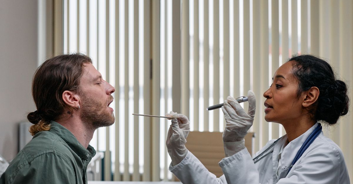
When the clinical diagnosis is ambiguous, targeted diagnostic testing is required. If a fungal infection is suspected, the clinician will perform a quick mucosal swab. The sample is treated with potassium hydroxide and examined microscopically for the presence of fungal hyphae, confirming candidiasis.
If the clinician suspects a systemic autoimmune issue or identifies a thick, non-removable white plaque indicative of leukoplakia, a tissue biopsy is strictly mandatory.
During a biopsy, a small piece of the white lesion is surgically excised under local anesthesia and sent to an oral pathologist. Microscopic evaluation is the only definitive method to assess the cells for malignant dysplasia and rule out early-stage oral cancer.
11. Antifungal Pharmacological Interventions
Confirmed cases of oral candidiasis require targeted antifungal therapy. For mild to moderate localized infections, topical antifungal agents are the first line of defense. Nystatin oral suspension is frequently prescribed; the patient swishes the liquid in their mouth for several minutes to coat the fungal plaques before swallowing.
Clotrimazole troches (lozenges) are another highly effective topical option. They dissolve slowly in the mouth, providing a sustained release of the antifungal medication directly to the posterior tongue and throat tissues over twenty minutes.
In severe cases, or in patients with significant systemic immunosuppression, oral systemic medications such as fluconazole are prescribed to eradicate the fungal pathogen from the bloodstream and deep mucosal tissues simultaneously.
12. Mechanical Debridement and Hygiene
For the vast majority of patients whose white coating is caused by cellular hypertrophy and trapped debris, aggressive mechanical debridement is the definitive cure. Standard toothbrush bristles are too soft and pliable to effectively dislodge the sticky keratin matrix.
Patients must utilize a dedicated tongue scraper. The scraper should be placed as far back on the posterior tongue as physically possible without triggering a severe gag reflex. By applying firm, downward pressure and dragging the scraper forward, the thick white sludge is physically sheared away from the papillae.
Performing this mechanical cleaning twice a day immediately disrupts the structural integrity of the biofilm, preventing the anaerobic bacteria from colonizing and rapidly restoring the normal pink, healthy appearance of the mucosal surface.
13. Restoring Salivary Gland Function
Resolving a white tongue requires correcting the underlying dry mouth. Patients must significantly increase their plain water intake to ensure the body has adequate fluid to produce saliva. Caffeinated and alcoholic beverages must be restricted, as they chemically inhibit salivary flow.
To actively stimulate the salivary glands, clinicians recommend chewing sugar-free gum sweetened exclusively with xylitol. The mechanical act of chewing forces the major salivary glands to pump fresh, oxygenated saliva across the posterior tongue, actively washing away dead cells.
If xerostomia is a permanent side effect of necessary systemic medications, patients must employ artificial saliva sprays or gels frequently throughout the day, and particularly at night, to maintain a synthetic protective barrier over the oral mucosa.
14. Nutritional Deficiencies Affecting Oral Mucosa
The rapid turnover of the epithelial cells on the tongue requires a robust supply of specific micronutrients. Deficiencies in Vitamin B12, iron, and folic acid profoundly disrupt the normal cellular regeneration cycle within the mouth.
While these deficiencies typically present as a completely smooth, red, and swollen tongue (atrophic glossitis), they can initially cause an irregular shedding pattern, leading to patchy white areas of retained keratin surrounded by angry red tissue, a condition clinically termed geographic tongue.
If a patient presents with a chronic, changing white and red map-like pattern on the tongue, alongside systemic fatigue, a comprehensive blood panel is ordered to identify and correct any underlying severe nutritional anemias.
15. When to Seek an Otolaryngologist Evaluation
A white coating on the back of the tongue is frequently managed successfully with improved mechanical hygiene and hydration. However, any distinct white patch or plaque that does not resolve within two weeks of rigorous tongue scraping demands professional evaluation by a dentist or Otolaryngologist.
Immediate specialist intervention is required if the white patch is unilaterally located on the side or back of the tongue, feels hard or indurated to the touch, or is accompanied by unexplained weight loss or a persistent sore throat.
Early detection and biopsy of leukoplakia are paramount, as removing the dysplastic tissue before it undergoes malignant transformation prevents the development of invasive, life-threatening oral carcinomas.
16. Frequently Asked Questions (FAQ)
1. Is it normal to have a white tongue when waking up in the morning?
Yes, it is highly common. Salivary flow decreases dramatically during sleep, causing the mouth to dry out. This extreme overnight xerostomia allows dead cells and bacteria to accumulate rapidly on the posterior papillae, creating a morning white coating that usually clears after hydration and breakfast.
2. Can taking antibiotics cause my tongue to turn white?
Yes, broad-spectrum antibiotics indiscriminately kill both bad and good bacteria in the mouth. This eliminates the natural competition for space and nutrients, allowing the opportunistic Candida fungus to multiply wildly, resulting in the thick white plaques of oral thrush.
3. Will mouthwash get rid of a white coated tongue?
Mouthwash alone is ineffective. The white coating is a thick, structurally sound biofilm of keratin and mucin. Liquid rinses cannot penetrate or dissolve this physical barrier. It must be mechanically scraped off before antimicrobial rinses can reach the underlying bacteria.
4. Why does scraping my tongue make me gag?
The posterior third of the tongue is heavily innervated by the glossopharyngeal nerve, which triggers the protective gag reflex to prevent choking. Relaxing the throat, breathing through the nose, and gently guiding the scraper further back over time can help desensitize the reflex.
5. Can acid reflux cause a white tongue?
Yes, severe gastroesophageal reflux brings stomach acid up into the throat and back of the mouth. This highly acidic fluid chemically irritates the tongue papillae, causing them to inflame, hypertrophy, and trap debris at a much faster rate, resulting in a persistent white coating.
17. Bibliography
Disclaimer: The content is for informational purposes only and does not replace medical advice. Always consult your doctor for personalized treatment.