
1. Introduction
A ganglion cyst of the wrist is a benign, fluid-filled mass arising from the complex ligaments and joint capsules that stabilize the carpal bones. Representing the most frequent soft tissue mass encountered in the hand and wrist, these cysts present as smooth, distinct nodules beneath the skin. The wrist is a biomechanical junction of immense complexity, enduring massive, repetitive mechanical loads while coordinating intricate multi-directional movements. When the dense connective tissue encapsulating these joints experiences micro-trauma or degenerative weakening, highly viscous synovial fluid leaks outward, forming a pressurized, localized sac.
While the sudden appearance of a lump on the wrist frequently generates patient anxiety, it is crucial to establish that ganglion cysts lack any malignant potential. The clinical priority is confirming the benign nature of the mass and assessing its impact on surrounding anatomy. Depending on their size and specific location, wrist ganglia can compress major arteries, irritate sensory nerves, and significantly limit mechanical joint function. Effective management spans a spectrum from patient reassurance and temporary immobilization to image-guided aspiration and meticulous surgical excision of the underlying capsular stalk.
2. Complex Carpal Anatomy
The human wrist is not a single joint, but an intricate arrangement of eight small carpal bones tightly bound by a labyrinth of stout ligaments. These bones articulate with the radius and ulna of the forearm, forming the radiocarpal and midcarpal joints. Every joint space within this architecture is enclosed by a fibrous capsule lined with a synovial membrane, which continuously secretes lubricating synovial fluid.
The stability of the wrist relies heavily on these intrinsic and extrinsic ligaments. The scapholunate ligament, located centrally between the scaphoid and lunate bones, is one of the most critical stabilizers. The immense mechanical stress placed on this specific ligament during daily activities makes it the most frequent site of structural weakening and subsequent fluid herniation in the entire upper extremity.
3. Pathophysiology of Wrist Ganglia
The formation of a wrist ganglion cyst is driven by a localized breakdown in the joint capsule or supporting ligaments. Histological studies suggest a process called myxoid degeneration, where repetitive mechanical stress causes the dense collagen fibers to fray and degenerate, forming small pools of mucin.
These microscopic pools coalesce, creating a defect or a one-way valve in the capsular wall. As the patient extends and flexes the wrist, the normal biomechanical pressure generated inside the joint forces synovial fluid through this valve and into the surrounding subcutaneous tissue. The body encapsulates this fluid in a fibrous sac. Over time, the fluid inside the cyst concentrates, becoming a remarkably dense, jelly-like substance rich in hyaluronic acid, giving the cyst its characteristic hard, rubbery texture.
4. Dorsal Wrist Ganglia
Dorsal wrist ganglia are the most prevalent type, accounting for approximately seventy percent of all wrist cysts. They present as a visible, smooth lump on the back of the wrist.
These cysts predominantly originate from the scapholunate ligament. Because of their location on the extensor surface, they become distinctly more prominent and tense when the patient aggressively flexes the wrist downward. Patients typically complain of a dull, continuous ache at the base of the cyst, which often sharpens into a localized pain when bearing weight through an extended wrist, such as when performing a push-up or pushing up from a chair.
5. Volar Wrist Ganglia
Volar wrist ganglia occur on the palmar surface of the wrist, most frequently developing on the radial side, near the base of the thumb. These cysts typically arise from the radiocarpal joint or the scaphotrapezial joint.
The clinical significance of a volar ganglion lies in its immediate anatomical proximity to the radial artery. The cyst often grows entirely around or directly adjacent to this major blood vessel. Consequently, patients frequently report feeling a distinct pulse transmitted through the mass. The cyst can also compress the median nerve or branches of the radial nerve, generating symptoms of tingling, numbness, or radiating pain into the palm and thumb.
6. Occult Ganglion Cysts
Not all wrist ganglia present as a visible, palpable lump on the skin surface. An occult ganglion cyst is a small fluid collection that remains hidden deep within the complex architecture of the carpal bones.
Because these cysts are confined within the tight spaces of the joint, even a minute accumulation of pressurized fluid stretches the highly innervated joint capsule, causing severe, disproportionate pain. Patients with occult cysts present with unexplained, chronic wrist pain, profound weakness during gripping, and localized tenderness, often leading to a frustratingly prolonged diagnostic process until advanced imaging is obtained.
7. Clinical Presentation and Fluctuation
The clinical behavior of a wrist ganglion is notoriously dynamic. Patients frequently report that the size of the cyst fluctuates dramatically based on their level of physical activity.
Following a period of heavy lifting, typing, or rigorous sports like gymnastics, the increased mechanical pumping action of the joint forces more fluid into the sac, causing the cyst to enlarge, harden, and become increasingly painful. Conversely, after several days of complete rest or wearing a supportive brace, the fluid production decreases, and the cyst may visibly shrink or soften, though it rarely disappears entirely without intervention.
8. Diagnostic Physical Examination
The diagnosis is primarily established through a targeted physical examination. The clinician assesses the precise anatomical location, mobility, and consistency of the mass. A classic ganglion cyst is smooth, firmly tethered to the deep joint structures, and generally unattached to the overlying skin.
The physician will perform a transillumination test. By pressing a focused, bright penlight against the side of the mass in a darkened room, the clinician looks for a uniform glow. Because the cyst is filled with clear mucin, light transmits easily through it. Solid tumors, such as giant cell tumors or lipomas, will block the light entirely.
9. Vascular Assessment for Volar Cysts
When evaluating a volar wrist cyst, assessing the integrity of the radial artery is a mandatory clinical step before any intervention is planned. The clinician performs the Allen test.
The patient is instructed to make a tight fist to drain blood from the hand. The physician then uses their thumbs to firmly compress both the radial and ulnar arteries at the wrist. The patient opens their blanched hand, and the physician releases only the ulnar artery. A rapid return of color to the entire hand confirms that the ulnar artery is capable of supplying the hand independently. This ensures that if the radial artery, which is intimately entangled with the cyst, is accidentally compromised during aspiration or surgery, the hand will still receive adequate blood flow.
10. Diagnostic Imaging Modalities
While the physical examination is robust, imaging is frequently utilized to confirm the diagnosis, rule out occult bone pathology, and provide a roadmap for surgical intervention.
| Imaging Modality | Clinical Utility in Wrist Ganglia |
|---|---|
| Radiography (X-ray) | Does not visualize the fluid cyst, but rules out underlying osteoarthritis, occult carpal fractures, or bone tumors. |
| Diagnostic Ultrasound | Excellent for confirming the fluid nature of the mass, visualizing the vascular flow of the radial artery near volar cysts, and guiding needle aspiration. |
| Magnetic Resonance Imaging | The definitive test for diagnosing deep, occult cysts causing unexplained wrist pain and evaluating the integrity of the scapholunate ligament. |
11. Conservative Management and Immobilization
Because wrist ganglia are entirely benign, the primary recommendation for asymptomatic or mildly annoying cysts is watchful waiting and reassurance. Many cysts will spontaneously resolve over several months or years.
When a cyst becomes painful due to activity-related expansion, temporary immobilization is highly effective. The physician prescribes a rigid, custom-fitted wrist splint. The splint prevents the continuous flexion and extension of the wrist, effectively shutting down the mechanical fluid pump. Wearing the splint for several weeks typically reduces the fluid volume, shrinking the cyst and alleviating the compressive pain.
12. Needle Aspiration and Corticosteroids
For cysts causing significant pain or aesthetic distress, needle aspiration offers immediate, localized relief in an outpatient setting. After injecting local anesthesia, the physician utilizes a large-bore needle to puncture the thick fibrous wall and forcefully draw out the dense, clear mucin.
The physician may inject a small volume of a corticosteroid into the collapsed cavity before removing the needle to suppress localized inflammation. While aspiration is rapid and instantly gratifying for the patient, it does not remove the underlying one-way valve in the joint capsule. Consequently, the recurrence rate for aspirated wrist ganglia is high, often exceeding fifty percent, as normal joint mechanics inevitably pump new fluid back into the defect.
13. Surgical Excision Principles
Surgical excision is the definitive treatment for symptomatic cysts that fail conservative management or rapidly recur after aspiration. The procedure is typically performed by a specialized hand surgeon under regional or general anesthesia.
The critical objective of the surgery is not simply removing the visible bump on the wrist. The surgeon must perform a meticulous, deep dissection between the extensor or flexor tendons to trace the cyst entirely down to its root. The surgeon excises the cyst wall, the stalk, and a small, contiguous portion of the joint capsule or ligament where the herniation originated. Completely removing this one-way valve significantly drops the surgical recurrence rate to below ten percent.
14. Arthroscopic Excision Techniques
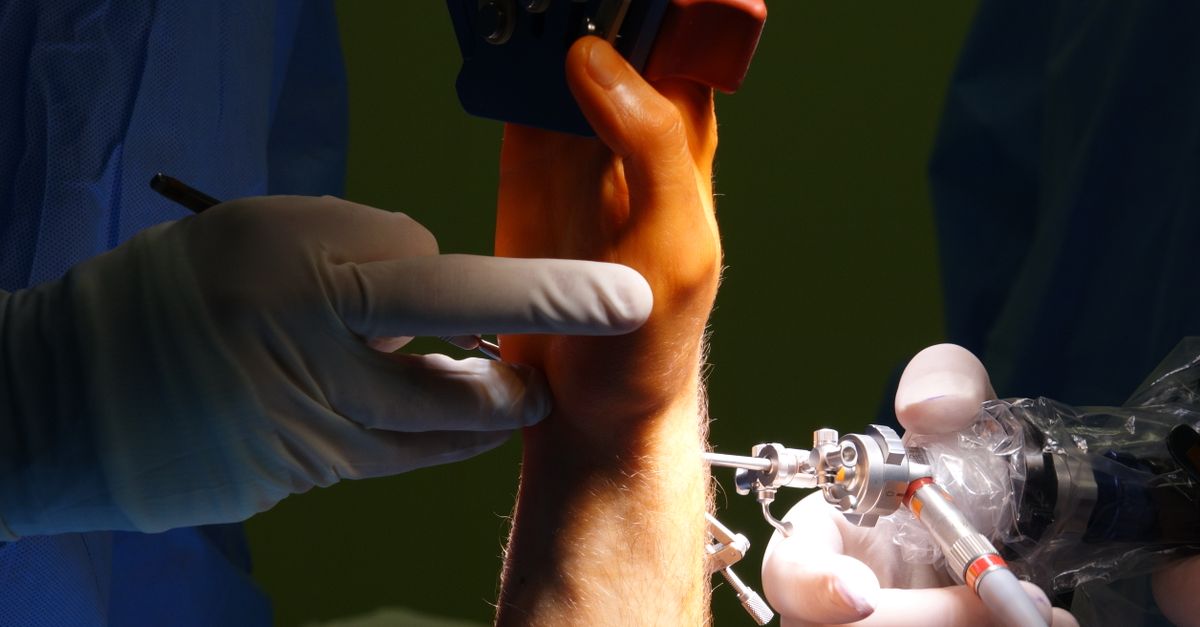
For dorsal wrist ganglia, an arthroscopic approach is an increasingly preferred alternative to open surgery. The surgeon inserts a tiny camera and micro-instruments through keyhole incisions in the wrist joint.
Working entirely from the inside of the joint, the surgeon locates the base of the cyst stalk on the scapholunate ligament. The surgeon then uses a specialized shaving instrument to core out the base of the cyst from the inside, allowing the fluid to drain back into the joint and removing the one-way valve mechanism. Arthroscopic excision minimizes damage to the overlying skin and tendons, often resulting in less postoperative stiffness and a faster return to normal activities.
15. Postoperative Rehabilitation and Prognosis
Following surgical excision, the wrist is placed in a bulky dressing and a protective splint for a short period to allow the capsular incision to heal. However, prolonged immobilization is strictly avoided.
Because the surgery involves manipulating the highly complex joint capsule and surrounding tendons, wrist stiffness is the most common postoperative complication. Patients are referred to a certified hand therapist to begin early, aggressive range-of-motion exercises and scar mobilization techniques. With dedicated physical therapy, patients reliably regain full, pain-free wrist mobility and structural stability, restoring their complete occupational and functional capacity.
16. When to Seek Immediate Medical Attention
A wrist ganglion cyst is a slow-growing, benign mass. However, you must seek immediate medical evaluation if a lump on your wrist appears suddenly following a severe fall or high-impact trauma, as this may represent a dislocated carpal bone or a severe ligament rupture rather than a simple cyst. Furthermore, if the cyst causes sudden, sharp shooting pain, intense burning, or profound numbness extending into your palm and fingers, prompt clinical assessment is required to address acute nerve compression.
17. Frequently Asked Questions FAQ
1. Can I pop the wrist cyst myself by hitting it with a heavy object?
No. The dangerous practice of striking a “Bible bump” with a heavy book causes severe, blunt force trauma to the delicate bones, arteries, and tendons of the wrist. It can cause permanent damage and frequently fails to cure the cyst.
2. Why does the doctor check my pulse before draining a cyst on the front of my wrist?
Cysts on the palm side of the wrist (volar cysts) grow intimately close to the radial artery, which supplies blood to your hand. The doctor must ensure your other arteries are functioning perfectly before inserting a needle near this major blood vessel.
3. Will the cyst on my wrist turn into cancer?
No. Ganglion cysts are completely benign fluid collections. They possess absolutely no malignant cells and have a zero percent chance of turning into cancer or spreading to other parts of your body.
4. Why is my wrist so stiff after the surgery to remove the cyst?
The surgery requires cutting deep down into the thick joint capsule and moving tendons aside. As the body heals, it creates dense scar tissue. You must perform daily physical therapy stretches to break up this scar tissue and restore your normal flexibility.
5. If the needle draining doesn’t cure it permanently, why do it?
Needle aspiration provides immediate, profound relief from pain and pressure. For many patients, the cyst may take months or years to grow back to a bothersome size, allowing them to avoid surgery for a long period of time.
Bibliography
Disclaimer: The content is for informational purposes only and does not replace medical advice. Always consult your doctor for personalized treatment.
