
1. Introduction
A fetal monitoring scalp injury is a specialized form of birth trauma directly resulting from the application of an internal fetal scalp electrode during labor. While continuous electronic fetal monitoring is essential for identifying physiological distress in the unborn child, traditional external monitors strapped to the maternal abdomen frequently suffer from signal loss. When maternal movement, obesity, or rapid fetal shifts compromise the external signal, the obstetrical team may deploy a fetal scalp electrode. This device guarantees an uninterrupted, highly accurate electrocardiogram of the fetal heart rate, providing the critical data required to manage a high-risk labor safely.
To acquire this pristine signal, the tiny spiral wire of the electrode must physically breach the epidermal layer of the fetal scalp, anchoring itself directly into the presenting part of the fetus. Consequently, a mild, localized injury is an inherent and unavoidable aspect of its correct anatomical application. The vast majority of these injuries are superficial, presenting as minor abrasions or small puncture marks that heal rapidly without clinical intervention.
However, the invasive nature of the device introduces a distinct spectrum of potential complications. Ranging from localized bacterial infections to severe mechanical traumas and the potential transmission of maternal blood-borne viruses, the risks associated with internal monitoring must be meticulously weighed against the necessity of obtaining accurate fetal heart rate data. Recognizing, managing, and preventing these specific neonatal injuries is a fundamental component of immediate postpartum pediatric care.
2. Indications for Internal Fetal Monitoring
The decision to transition from non-invasive external monitoring to invasive internal monitoring is driven by the absolute clinical necessity for continuous, precise fetal heart rate data. The external Doppler ultrasound transducers utilized on the maternal abdomen are highly sensitive to movement. If the mother frequently changes positions, or if the fetus is highly active, the external monitor “drops” the signal, leaving the medical team blind to the physiological status of the fetus.
This loss of signal becomes critically dangerous during a high-risk labor. If the fetus is already demonstrating signs of distress, such as deep variable decelerations or a rising baseline heart rate, the physician cannot afford momentary gaps in data. Furthermore, maternal obesity adds a thick layer of adipose tissue between the external monitor and the fetal heart, profoundly dampening the acoustic signal and rendering external monitoring highly unreliable.
In these precarious scenarios, the application of a fetal scalp electrode provides a direct, R-wave to R-wave electrocardiogram. This data is impervious to maternal movement or tissue thickness, allowing the obstetrician to accurately interpret the subtlest changes in fetal cardiovascular stability and make informed, immediate decisions regarding the necessity of an emergency surgical delivery.
3. The Anatomy of the Scalp Electrode
The fetal scalp electrode is a highly specialized, sterile medical device designed to establish a direct electrical connection with the fetus while minimizing physical trauma. The device consists of a long, flexible guide tube that protects the maternal vaginal and cervical tissues during insertion. Housed within this tube is the electrode wire, which terminates in a tiny, sharp, corkscrew-like spiral tip, typically measuring one and a half millimeters in depth.
During application, the physician or midwife performs a sterile vaginal examination, guiding the plastic tube through the dilated cervix until it rests firmly against the presenting part of the fetus. The practitioner then rotates the inner wire mechanism, driving the tiny spiral tip directly into the epidermal and dermal layers of the fetal scalp.
Because the tip is exceptionally shallow, it is designed to lodge firmly in the superficial layers of the skin, deliberately avoiding the deeper aponeurosis, the periosteum covering the skull bone, and the skull bone itself. The wire then trails out of the maternal vagina and connects to a leg plate on the mother’s thigh, which transmits the electrical signal directly to the monitoring console.
4. Normal Physiological Application Site
The most frequent and preferred site for the application of the electrode is the fetal scalp over the parietal or occipital bones, as the vast majority of fetuses present in a head-down, cephalic position. The thick skin over these large cranial bones provides a safe, solid anchoring point for the spiral wire.
However, the physician must exercise extreme anatomical precision during application. The device must absolutely avoid the fontanelles—the large, soft spots on the top of the infant skull where the cranial bones have not yet fused. Applying the electrode over a fontanelle risks driving the sharp wire directly through the thin protective membranes and into the fragile underlying brain tissue or crucial venous sinuses.
Furthermore, if the fetus presents in a breech position (buttocks down), the electrode must be placed carefully on the fleshy portion of the fetal buttocks, strictly avoiding the fetal genitalia, anus, and spine. While the device is referred to as a “scalp” monitor, its application to the buttocks carries the identical risks of localized trauma and infection.
5. Common and Minor Scalp Injuries
Because the device functions by intentionally breaching the skin, a minor physical wound is an expected, guaranteed outcome of its use. Following delivery and the careful unscrewing and removal of the electrode, the neonate will invariably present with a small, localized mark on the presenting part.
The most common presentation is a tiny, distinct puncture wound or a small, circular abrasion corresponding to the diameter of the spiral wire. This area may exhibit mild, localized erythema (redness) and a tiny spot of dried blood. Occasionally, if the fetus shifted significantly during labor or if the electrode was accidentally snagged during delivery, the spiral wire may cause a shallow, linear scratch or laceration across the scalp.
These minor injuries are generally benign. They cause minimal discomfort to the newborn, require only routine, gentle cleansing with soap and water, and typically heal completely within a few days to a week without leaving a permanent scar or requiring any specific medical intervention.
6. Moderate Complications: Localized Infection
The most frequent clinically significant complication arising from a fetal scalp electrode is a localized bacterial infection. By piercing the protective epidermal barrier of the fetal scalp, the electrode creates a direct portal of entry. During labor, the amniotic fluid and the vaginal canal are heavily colonized by diverse maternal bacterial flora.
Bacteria, such as Staphylococcus aureus, Escherichia coli, or Group B Streptococcus, can easily migrate down the wire of the electrode and inoculate the puncture wound. This inoculation leads to localized cellulitis, characterized by spreading redness, warmth, and firm swelling around the electrode site. If the infection localizes further, it forms a scalp abscess—a painful, raised, pus-filled nodule.
An infected scalp wound requires prompt pediatric evaluation. Mild cellulitis may be managed with topical or oral antibiotics. However, a distinct scalp abscess frequently requires the pediatrician to perform a minor bedside procedure to lance and drain the pus. The wound fluid is cultured to identify the specific bacteria, ensuring the targeted antibiotic therapy is appropriately destroying the pathogen.
7. Severe Complications: Cephalohematoma
While the spiral tip of the electrode is designed to be very shallow, improper application technique or abnormal pressure during delivery can cause the device to inflict deeper mechanical trauma. If the electrode is driven too deeply, or if the traction applied to the wire pulls the scalp violently away from the underlying skull bone, it can tear the fragile blood vessels coursing beneath the periosteum.
The tearing of these blood vessels causes a cephalohematoma—a collection of blood trapped tightly between the skull bone and the tough periosteal membrane. This presents clinically as a firm, distinct, localized lump on the infant head that strictly does not cross the bony suture lines of the skull.
While a cephalohematoma is typically a consequence of the crushing forces of the pelvic bones or the use of a vacuum extractor, the localized trauma of an incorrectly placed scalp monitor can directly initiate this internal bleeding. The trapped blood eventually resorbs over several weeks, but as the large volume of red blood cells breaks down, the infant faces a substantially elevated risk of developing severe neonatal jaundice requiring phototherapy.
8. Subgaleal Hemorrhage
The most profound and life-threatening mechanical complication potentially associated with scalp monitor trauma is a subgaleal hemorrhage. This catastrophic event occurs when the emissary veins—which connect the scalp tissues to the venous sinuses inside the skull—are sheared or torn.
The blood hemorrhages into the subgaleal space, a vast, loose layer of tissue that spans the entire surface of the head from the eyebrows to the nape of the neck. Because this space is immense and unconfined by suture lines, a newborn can rapidly bleed out a massive percentage of their total blood volume into this compartment.
While overwhelmingly caused by the aggressive use of a vacuum extractor during delivery, a fetal scalp electrode applied with excessive force or accidentally ripped from the scalp during an emergency extraction can precipitate this profound vascular tear. An infant with a subgaleal hemorrhage presents with a massive, fluid-like, boggy swelling across the entire head and rapidly descends into severe, life-threatening hypovolemic shock.
9. Transmission of Maternal Viral Pathogens
Beyond physical trauma and bacterial infection, the most insidious risk associated with an internal scalp electrode is the facilitation of vertical transmission of severe maternal viral pathogens. The electrode physically breaches the fetal skin, directly exposing the fetal bloodstream to the maternal blood and cervical fluids present in the birth canal.
For a mother infected with the Human Immunodeficiency Virus (HIV) or the Hepatitis B or C viruses, the use of a fetal scalp electrode is generally strictly contraindicated. Breaching the fetal skin barrier exponentially increases the risk of the infant acquiring these lifelong, severe viral infections during the labor process.
Similarly, if a mother has a history of Genital Herpes Simplex Virus (HSV) and exhibits active lesions or premonitory symptoms, the use of a scalp monitor is absolutely forbidden. Inoculating the highly destructive herpes virus directly into the fetal scalp leads to a catastrophic, systemic neonatal herpes infection that frequently attacks the central nervous system, causing severe viral encephalitis and high neonatal mortality.
10. Structured Data: Complications of Fetal Scalp Electrodes
Categorizing the severity of the complications guides immediate pediatric monitoring and intervention.
| Severity Level | Specific Injury / Complication | Clinical Management |
|---|---|---|
| Minor (Expected) | Small puncture mark, superficial scratch | Routine cleansing with soap and water, observation |
| Moderate | Scalp cellulitis or localized abscess | Incision and drainage, oral or topical antibiotics |
| Severe (Mechanical) | Cephalohematoma | Monitor for severe neonatal jaundice and hyperbilirubinemia |
| Critical (Mechanical) | Subgaleal Hemorrhage | Immediate massive blood transfusion to prevent lethal shock |
| Critical (Infectious) | Transmission of HSV or HIV | Requires aggressive antiviral protocols and specialized infectious disease care |
11. Systemic Neonatal Sepsis
A localized scalp abscess caused by the electrode is a concerning but generally manageable issue. However, if the localized bacterial infection is not recognized and treated promptly, it can escalate into a profound, life-threatening systemic crisis. The rich vascular network of the infant scalp provides bacteria with direct, rapid access to the systemic circulation.
If aggressive pathogens, particularly Group B Streptococcus or Methicillin-Resistant Staphylococcus aureus (MRSA), enter the bloodstream, the newborn rapidly develops early-onset neonatal sepsis. The fragile infant immune system is quickly overwhelmed by the circulating bacteria.
An infant developing sepsis will exhibit significant clinical instability. They frequently present with profound temperature instability—often hypothermia rather than a high fever—severe lethargy, a refusal to feed, and subtle changes in skin color, appearing pale or mottled. This rapid deterioration requires immediate admission to the neonatal intensive care unit for comprehensive blood cultures and the rapid administration of broad-spectrum intravenous antibiotics to prevent septic shock and multi-organ failure.
12. Contraindications for Application
Because the risks of invasive monitoring are tangible, obstetrical protocols dictate strict contraindications for the use of a fetal scalp electrode. The most absolute contraindications involve maternal infectious diseases. As previously noted, mothers with known HIV, active Hepatitis B or C, or a suspected active Genital Herpes outbreak must not undergo internal monitoring to protect the fetus from direct viral inoculation.
Fetal bleeding disorders also present an absolute contraindication. If there is a known family history or a confirmed prenatal diagnosis of fetal hemophilia or alloimmune thrombocytopenia (a condition where the fetus has dangerously low platelets), puncturing the scalp can lead to severe, uncontrollable, and potentially fatal bleeding from the tiny wound site.
Anatomical considerations are equally critical. The physician must definitively identify the presenting part. If the presentation is a face or a brow, applying an electrode is strictly forbidden, as driving the wire into the delicate facial bones, the eyes, or the fragile cartilage of the nose will cause severe, permanent structural and cosmetic damage.
13. Clinical Evaluation of the Newborn
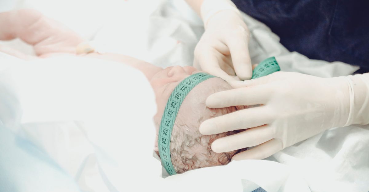
The management of a newborn who was monitored with a fetal scalp electrode requires a proactive, structured clinical assessment immediately following delivery. The pediatrician or neonatal nurse meticulously examines the exact site where the electrode was attached. They document the size, depth, and appearance of the puncture wound or abrasion in the medical record.
The scalp is gently palpated to ensure the surrounding tissue is soft and normal, actively ruling out the presence of a developing cephalohematoma or the alarming, fluid-like wave of a subgaleal hemorrhage. The neurological status of the infant is evaluated, checking muscle tone, pupil reactivity, and normal reflexes to ensure no deep cranial trauma occurred.
This thorough initial assessment establishes a baseline. Because the signs of a localized infection or a slow internal bleed may not manifest until twenty-four to forty-eight hours after birth, the initial documentation is critical for identifying subtle, progressive changes during the newborn hospital stay.
14. Neonatal Wound Care Protocols
For the vast majority of infants, the post-removal wound care is simple and non-invasive. The primary goal is to keep the tiny puncture site clean and dry to facilitate rapid epithelialization and prevent secondary bacterial colonization. During the initial newborn bath, the nurse gently cleanses the scalp with a mild, neutral soap and warm water, avoiding rigorous scrubbing over the electrode site.
The application of strong, caustic antiseptic solutions, such as harsh iodine preparations or concentrated rubbing alcohol, is strictly avoided, as these chemicals can damage the delicate, newly forming skin cells and delay the natural healing process. Parents are instructed to keep the area exposed to the air as much as possible and to avoid placing tight hats or bows directly over the healing wound.
If the area shows mild redness, the pediatrician may recommend the application of a simple, over-the-counter topical antibiotic ointment, such as bacitracin, applied in a very thin layer twice daily to provide a mild antimicrobial barrier while the puncture closes completely.
15. Parental Education and Discharge Planning
Before discharging an infant who had a scalp electrode applied, the medical team must provide comprehensive education to the parents regarding the signs of delayed complications. Parents are often alarmed to discover a scab or a small bald spot on their newborn head. The physician must reassure them that this is a standard, expected result of necessary medical monitoring that will heal perfectly over time.
Parents are instructed to monitor the site daily. They must be explicitly taught the warning signs of a developing scalp abscess. If they notice the redness expanding outward from the scab, if the area becomes unusually warm to the touch, or if they observe a distinct, firm bump filled with yellow or white pus forming under the skin, they must contact their pediatrician immediately.
Furthermore, parents are educated on the systemic signs of neonatal infection. If the infant develops a rectal temperature of 38.0 degrees Celsius (100.4 degrees Fahrenheit) or higher, becomes profoundly lethargic, or entirely refuses to feed, they must proceed immediately to a pediatric emergency department, as these are critical signs of potential systemic sepsis requiring urgent blood cultures and intravenous antibiotics.
16. Obstetrical Best Practices for Prevention
Minimizing the incidence and severity of scalp monitoring injuries relies on meticulous obstetrical technique. The use of a fetal scalp electrode should never be routine; it must be reserved strictly for situations where continuous monitoring is clinically mandated, and the external Doppler signal is definitively inadequate.
When application is necessary, the physician or midwife must confirm the fetal position with absolute certainty, utilizing a bedside ultrasound if the physical examination is ambiguous, to ensure the electrode is placed securely on the robust parietal bones and safely away from the fontanelles or the delicate facial structures.
During delivery, the removal of the electrode requires precision. The practitioner must carefully rotate the inner wire counter-clockwise to smoothly unthread the spiral from the fetal skin. Abruptly pulling or yanking the wires to detach the monitor is the primary cause of deep lacerations and severe vascular tears, and this aggressive action must be strictly avoided under all circumstances.
17. When to Seek Urgent Pediatric Care
Once the infant is home, parents must remain vigilant regarding the healing of the scalp wound. The scab should dry out and eventually flake off naturally. If the wound begins to actively ooze thick, foul-smelling yellow or green fluid, or if the skin around the scab becomes distinctly bright red and swollen, a prompt visit to the pediatrician is required to evaluate for cellulitis or an abscess.
Immediate emergency medical attention is absolutely required if the infant begins to display any systemic signs of illness. A newborn under twenty-eight days old who develops a fever, or conversely, feels unusually cold and clammy, must be evaluated in an emergency department.
Additionally, if parents notice a large, soft, fluid-like swelling developing across the top of the baby head several days after birth, or if the infant exhibits any unusual, rhythmic twitching of the limbs or a blank, unblinking stare, they must call emergency services immediately. These are severe warning signs of delayed internal bleeding or neonatal seizures related to cranial trauma.
18. Frequently Asked Questions (FAQ)
1. Why did the doctor put a wire in my baby’s head instead of using the belt monitor?
The external belt monitors frequently lose the signal if the baby is moving rapidly or if you change positions. If the doctor was concerned about the baby heart rate, they used the internal wire to get a perfect, uninterrupted signal to ensure the baby was safe.
2. Will the small scab on my baby’s head leave a permanent bald spot or scar?
No. The tiny puncture is very superficial. Once the scab falls off, the hair follicles beneath the skin will continue to grow normally, and it is extremely rare for the tiny mark to leave any noticeable permanent scar.
3. Can the wire poke into my baby’s brain?
No. The spiral tip is designed to be extremely short, only about one and a half millimeters deep. It only pierces the very top layer of the skin and does not come anywhere near the skull bone or the brain tissue when applied correctly.
4. How should I wash my baby’s head while the scab is still there?
During bath time, use a soft washcloth with warm water and a mild baby shampoo. Gently pat the area clean; do not rub or scrub the scab. Let it air dry completely before putting on a hat.
5. What should I do if the spot looks red and feels hot?
Expanding redness, warmth, or a bump filled with pus are clear signs of a local bacterial infection. You must call your pediatrician right away so they can prescribe an antibiotic ointment or an oral antibiotic to clear the infection before it spreads.
19. Bibliography
Disclaimer: The content is for informational purposes only and does not replace medical advice. Always consult your doctor for personalized treatment.

