1. Introduction
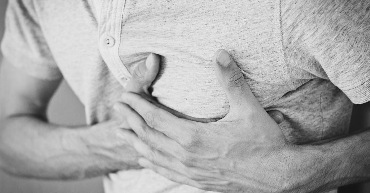
A rib injury involves trauma to the skeletal cage that encloses and protects the vital organs of the thoracic cavity, primarily the heart and the lungs. Injuries to this bony framework range from simple, localized contusions to multiple, severe fractures that destabilize the entire chest wall. The primary clinical concern in managing rib trauma is not the bone injury itself, but the secondary physiological impact on the patient ability to breathe deeply and effectively.
The ribs are designed to be somewhat flexible to accommodate the expansion of the lungs, but they possess a definitive breaking point under sufficient mechanical stress. When a rib fractures, the intense pain triggers a protective biological response where the patient instinctively avoids taking deep breaths or coughing. This shallow breathing pattern sets the stage for severe pulmonary complications.
Medical management focuses heavily on aggressive pain control, rigorous respiratory therapy, and, in specific severe cases, surgical stabilization. By eliminating the pain, physicians ensure the patient can perform the deep mechanical ventilation necessary to clear the airways and prevent life-threatening pneumonias from developing during the long healing process.
2. Anatomy of the Thoracic Cage
The human thoracic cage is composed of twelve pairs of ribs, the sternum centrally, and the twelve thoracic vertebrae posteriorly. The upper seven pairs, known as the true ribs, attach directly to the sternum via resilient costal cartilage. Ribs eight through ten attach to the cartilage of the rib above them, while the eleventh and twelfth are floating ribs, possessing no anterior attachment.
Between each adjacent rib lies an intercostal space containing three layers of intercostal muscles. These muscles contract to elevate the rib cage during active inhalation. Critically, running along the inferior groove of every single rib is an intercostal neurovascular bundle, comprising an artery, a vein, and a highly sensitive nerve.
This intimate anatomical relationship explains why rib fractures are exceptionally painful; the jagged edges of a broken rib constantly scrape against the adjacent intercostal nerve with every single breath. Furthermore, the sharp, fractured ends are positioned dangerously close to the delicate pleural membrane that wraps the lungs, introducing the risk of internal lacerations.
3. Mechanisms of Chest Wall Trauma
Rib injuries are almost exclusively the result of mechanical force applied to the thorax. Clinicians categorize these injuries based on the direction and magnitude of the trauma, which helps predict the severity of the fractures and the likelihood of associated internal injuries.
| Mechanism of Injury | Common Etiology | Typical Fracture Pattern |
|---|---|---|
| Blunt Frontal Impact | Steering wheel collision, CPR compressions | Fractures of the sternum and anterior costal cartilage |
| Lateral Compression | Side-impact vehicle crashes, heavy falls | Mid-shaft fractures of multiple consecutive ribs |
| Direct Blunt Force | Assault, sports collisions, falls on objects | Localized fractures at the specific point of impact |
| Repetitive Stress | Chronic severe coughing, repetitive sports motions | Stress fractures, typically insidious in onset |
The location of the fracture is clinically significant. Fractures of the first and second ribs indicate an impact of immense, life-threatening force, as these ribs are heavily protected by the clavicle and shoulder musculature. Fractures of the lower ribs raise suspicion for concurrent damage to the spleen or liver situated just below the diaphragm.
4. Simple Fractures and Contusions
A simple rib fracture involves a crack or break in the bone without significant displacement of the fractured ends. The structural integrity of the chest wall as a whole remains intact. A rib contusion is a severe bruise to the bone and the surrounding intercostal muscles, occurring when the trauma is insufficient to crack the bone but strong enough to rupture superficial blood vessels.
Clinically, a severe contusion is virtually indistinguishable from a simple fracture. Both conditions produce intense, sharp pain directly over the injury site that worsens drastically with movement, deep inspiration, or coughing.
For the purposes of immediate treatment, the distinction between a simple fracture and a contusion is largely irrelevant. The primary medical threat from both injuries stems entirely from the resulting pain and the subsequent impairment of normal respiratory mechanics.
5. The Pathophysiology of Flail Chest
A flail chest is a critical, life-threatening orthopedic emergency resulting from massive blunt trauma. It occurs when three or more consecutive ribs are fractured in two or more places. This pattern creates a freely floating segment of the chest wall that is completely disconnected from the rest of the skeletal cage.
This disconnected segment exhibits paradoxical motion during the respiratory cycle. During inhalation, as the chest expands and internal pressure drops, the flail segment is sucked inward. During exhalation, it is pushed outward. This paradoxical movement severely impairs the mechanical efficiency of breathing, preventing the lungs from expanding fully.
Furthermore, a flail chest is always accompanied by a massive underlying pulmonary contusion. The combination of structural instability and crushed, bleeding lung tissue rapidly leads to profound hypoxemia and respiratory failure, demanding immediate critical care intervention.
6. Pulmonary Complications: The Real Danger
The ribs themselves will eventually heal, but the secondary pulmonary complications are what cause significant morbidity and mortality, particularly in elderly patients. The severe pain of a rib injury causes a patient to “splint” their chest, taking rapid, shallow breaths and suppressing the natural urge to cough.
When a patient stops taking deep breaths, the small airways at the base of the lungs, known as alveoli, begin to collapse, a condition called atelectasis. This collapse creates an environment of warm, stagnant air and trapped mucus deep within the lungs.
This environment is ideal for bacterial proliferation. Without the mechanical force of a strong cough to clear the mucus, patients rapidly develop post-traumatic pneumonia. This is the most common cause of delayed death in patients suffering from multiple rib fractures, highlighting the absolute necessity of aggressive pain management.
7. Associated Internal Injuries
Physicians must maintain a high index of suspicion for internal injuries when evaluating a fractured thoracic cage. The sharp, jagged edge of a displaced rib acts like an internal blade. If forced inward, it can easily lacerate the delicate pleural lining, allowing air into the chest cavity and causing a pneumothorax, or collapsed lung.
If the laceration extends into the vascular tissue of the lung, a hemothorax occurs, filling the chest with blood. In cases involving lower rib fractures on the left side, the spleen is at high risk of puncture or rupture, leading to massive abdominal hemorrhage. Similarly, lower right rib fractures can lacerate the liver.
Consequently, any patient with significant chest trauma must undergo a comprehensive evaluation of their respiratory status and a thorough examination of the abdomen to rule out concealed, life-threatening hemorrhage.
8. Clinical Presentation and Evaluation
The classic presentation of a rib injury is localized, sharp back pain or chest pain that is exquisitely sensitive to movement. The physician will note that the patient avoids twisting their torso and exhibits an involuntary wincing response when asked to breathe deeply.
During palpation, the physician gently presses along the course of the ribs to locate the point of maximal tenderness. They may feel a distinct “step-off” where the bone fragments are malaligned, or feel crepitus, a grating sensation caused by the broken bone ends rubbing against each other.
The clinician will also auscultate the lungs carefully. Decreased breath sounds on the side of the injury suggest that the patient is splinting severely, or worse, that a pneumothorax has developed. Any signs of respiratory distress, such as rapid breathing, cyanosis, or low oxygen saturation, indicate significant internal complications requiring urgent imaging.
9. Diagnostic Imaging Strategies
Standard anterior-posterior and lateral chest X-rays are the initial diagnostic tool. While they are useful for identifying large pneumothoraces, massive hemothoraces, or wide structural displacements, standard X-rays are notoriously poor at identifying simple, non-displaced rib fractures or fractures involving the costal cartilage.
Computed Tomography is the definitive imaging modality for severe chest trauma. A CT scan provides a highly detailed, three-dimensional reconstruction of the thoracic cage. It accurately maps the exact number and location of all fractures, identifies flail segments, and vividly displays underlying pulmonary contusions or small pockets of trapped air.
However, if the clinical examination points strongly to a simple, isolated rib fracture without signs of respiratory distress, advanced imaging may not be strictly necessary. Confirming a single cracked rib on an X-ray does not fundamentally change the treatment plan, which remains focused entirely on pain control.
10. The Cornerstones of Pain Management
The overarching goal of rib injury management is not skeletal alignment, but pain control to facilitate deep breathing. The pharmacological approach is often multimodal, utilizing different classes of medications to interrupt pain signals at various points in the nervous system.
Initial therapy involves scheduled, rather than as-needed, administration of non-steroidal anti-inflammatory drugs like ibuprofen or ketorolac, combined with acetaminophen. This baseline therapy reduces the inflammatory response directly at the fracture site.
For more severe injuries, physicians prescribe short-acting opioid analgesics to handle breakthrough pain. However, opioids must be used cautiously, as high doses depress the central respiratory drive, exactly counteracting the clinical goal of promoting deep, forceful mechanical ventilation.
11. Regional Anesthesia Techniques
When oral or intravenous medications are insufficient to control the pain of multiple fractures, clinicians employ advanced regional anesthesia techniques. By targeting the nervous system locally, they can provide profound pain relief without the systemic side effects of heavy narcotics.
Intercostal nerve blocks involve injecting a long-acting local anesthetic directly beneath the specific rib, numbing the individual nerve that is being irritated by the fracture. This provides excellent, though temporary, relief.
For extensive, multi-level fractures, an epidural catheter may be placed in the thoracic spine. This allows for the continuous infusion of numbing medication, effectively blocking all pain signals from the entire affected side of the chest. Epidural analgesia is widely considered the gold standard for severe chest wall trauma, significantly reducing the incidence of post-traumatic pneumonia.
12. Respiratory Therapy and Pulmonary Hygiene
Alongside aggressive pain control, proactive respiratory therapy is non-negotiable. Patients are provided with an incentive spirometer, a small mechanical device that encourages slow, deep, sustained inhalations. They are instructed to use this device every hour while awake.
The visual feedback of the spirometer ensures that the patient is expanding their lungs fully, forcing the collapsed alveoli back open. Furthermore, patients are taught specific coughing techniques. By holding a pillow firmly against the injured area while coughing, they provide external mechanical support, reducing the movement of the broken bones and making the cough less painful and more effective.
Early mobilization is also critical. Sitting upright and walking around the hospital ward increases the metabolic demand for oxygen, naturally forcing the patient to breathe deeper and preventing the pooling of respiratory secretions in the lower lung lobes.
13. Surgical Plating and Stabilization
Historically, all rib fractures were allowed to heal conservatively. However, modern orthopedic trauma surgery has introduced surgical stabilization of rib fractures for specific, severe cases, such as a large flail chest or multiple severely displaced fractures that fail to respond to pain management.
The procedure, known as open reduction and internal fixation, involves exposing the fractured ribs and realigning the segments. The surgeon then secures the bones together using specialized, low-profile titanium plates and screws designed to match the natural curvature of the ribs.
Surgical stabilization immediately eliminates the mechanical grinding of the bone ends, drastically reducing pain and instantly restoring the structural integrity of the chest wall. Studies indicate that for severe trauma, surgical fixation significantly decreases the duration of mechanical ventilation and the length of the intensive care stay.
14. Frequently Asked Questions (FAQ)
1. Should I wrap my chest tightly for a broken rib?
Absolutely not. Wrapping or binding the chest restricts the expansion of the lungs. This prevents deep breathing, severely increases the risk of pneumonia, and is no longer recommended in modern medical practice.
2. How long does a broken rib take to heal?
The bone itself typically takes six to eight weeks to fuse and heal completely. However, the acute, sharp pain usually begins to subside significantly after the third or fourth week.
3. Why does my doctor want me to cough if it hurts?
Coughing is the only mechanical way to clear mucus out of the lower lungs. If you avoid coughing because of pain, the mucus becomes trapped and rapidly leads to a severe bacterial lung infection.
4. Is there a cast for a broken rib?
No, it is impossible to cast the chest because the ribs must move continuously to allow for breathing. Treatment relies entirely on pain medication and deep breathing exercises while the bone heals naturally.
5. How do I sleep with broken ribs?
Most patients find it most comfortable to sleep in a recliner or propped up with several pillows in a semi-upright position. Lying completely flat increases the pressure on the chest and makes breathing more difficult.
15. Bibliography
Disclaimer: The content is for informational purposes only and does not replace medical advice. Always consult your doctor for personalized treatment.