1. Introduction
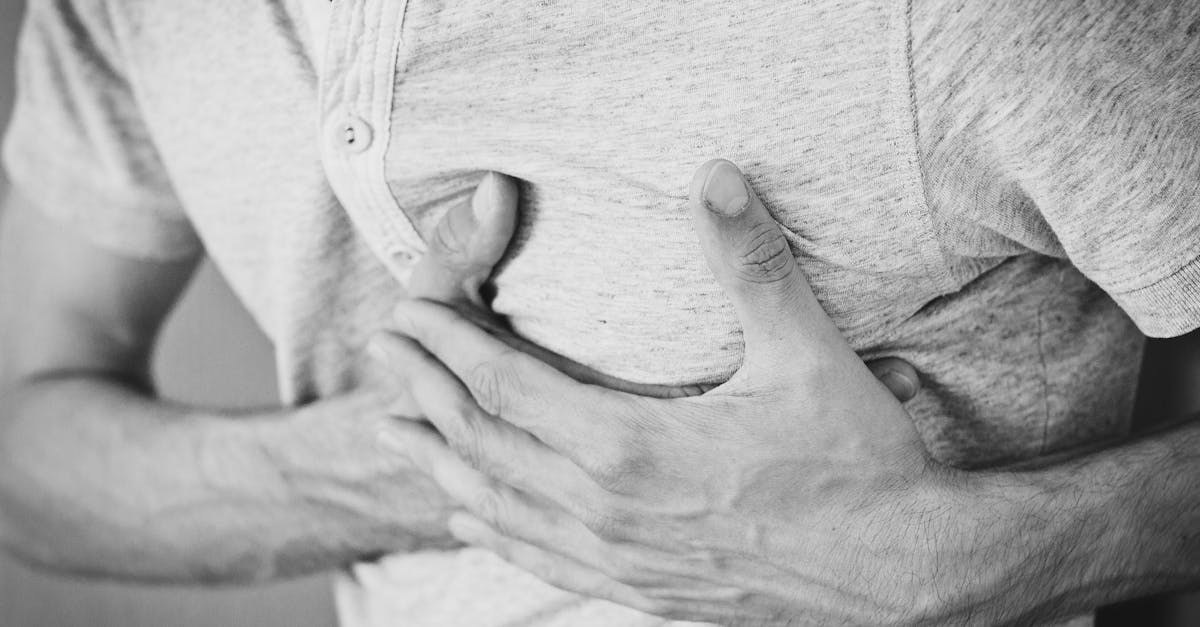
A chest wall injury constitutes a significant structural disruption of the thoracic cage, encompassing damage to the ribs, sternum, intercostal musculature, and the underlying pleural membranes. The thoracic cavity is a dynamic, pressure-regulated enclosure responsible for safeguarding the cardiopulmonary system while facilitating the complex mechanics of respiration. When an external force compromises this structural integrity, it disrupts both the physical protection of vital organs and the physiological process of ventilation.
Chest trauma is a leading consequence of motor vehicle collisions, substantial falls, and direct physical impacts. The clinical spectrum of these injuries ranges from isolated, uncomplicated rib fractures to profound structural failures such as flail chest, which precipitate severe respiratory distress. The localized pain generated by a compromised chest wall significantly inhibits normal breathing patterns, creating a cascading risk for secondary pulmonary complications.
Effective clinical management requires a thorough understanding of thoracic biomechanics and respiratory physiology. Rapid identification of hidden internal injuries, aggressive pain control, and targeted respiratory support are essential to stabilize the patient, restore adequate oxygenation, and prevent the rapid deterioration associated with severe chest wall trauma.
2. Anatomy of the Thoracic Cage
The thoracic cage is a resilient, bony framework composed of twelve pairs of ribs, the sternum anteriorly, and the twelve thoracic vertebrae posteriorly. This structure exhibits a unique balance of rigidity and flexibility. The ribs are attached to the sternum via pliable costal cartilages, allowing the chest wall to expand and contract symmetrically during the respiratory cycle.
Between each rib lies the intercostal space, housing the external and internal intercostal muscles, which elevate and depress the ribs during breathing. Crucially, the neurovascular bundle, containing the intercostal vein, artery, and nerve, courses along the inferior groove of each rib. This anatomical positioning renders the intercostal vessels highly susceptible to laceration by jagged bone fragments following a fracture.
The interior surface of the chest wall is lined by the parietal pleura, a delicate membrane that interfaces seamlessly with the visceral pleura covering the lungs. The preservation of the potential space between these two membranes is an absolute requirement for generating the negative pressure necessary for lung expansion.
3. Biomechanics of Blunt Thoracic Trauma
Blunt thoracic trauma accounts for the vast majority of chest wall injuries. The damage sustained is determined by the kinetic energy transferred during the impact and the elasticity of the patient’s skeletal structure. When a substantial force strikes the thorax, the ribs compress inward. If the compressive force exceeds the tensile strength of the bone, a fracture occurs.
The location of the fracture provides valuable diagnostic information. The upper ribs (one through three) are thick and protected by the shoulder girdle; a fracture here implies a transfer of profound kinetic energy, often correlating with severe injury to the underlying great vessels or the brachial plexus. The middle ribs (four through nine) are the most frequently fractured due to their exposed anatomical position.
Fractures of the lower ribs (ten through twelve) are highly concerning due to their proximity to solid abdominal organs. A displaced fracture in this region frequently masks concurrent lacerations to the liver on the right side or the spleen on the left side, necessitating a comprehensive evaluation of the abdominal cavity.
4. Penetrating Thoracic Injuries
Penetrating trauma involves an object breaching the integrity of the chest wall, directly exposing the intrathoracic contents to the external environment. These injuries are typically caused by sharp implements, ballistic projectiles, or severe industrial accidents.
The severity of a penetrating wound is dictated by the anatomical trajectory of the object. A simple puncture can lacerate an intercostal artery, causing significant localized bleeding into the chest cavity. More deeply penetrating injuries directly threaten the pulmonary parenchyma, the myocardium, and the major thoracic blood vessels.
High-velocity projectiles, such as bullets, generate a rapid shockwave that creates extensive cavitation within the tissue. This blast effect destroys pulmonary tissue far beyond the physical track of the projectile. Managing penetrating trauma requires immediate surgical evaluation to halt internal hemorrhage and restore the airtight seal of the thoracic cavity.
5. Rib Fractures and Flail Chest
Simple rib fractures are the most common manifestation of blunt chest trauma. While typically non-life-threatening on their own, the intense pain associated with the fracture severely limits chest wall expansion. Patients frequently adopt shallow, rapid breathing patterns to avoid discomfort, which prevents adequate lung inflation and impairs the clearance of pulmonary secretions.
A flail chest represents a critical structural failure of the thoracic cage. It occurs when three or more consecutive ribs are fractured in two or more places, creating a localized segment of the chest wall that is completely detached from the bony continuity of the rib cage.
This detached segment exhibits paradoxical motion during respiration. When the patient inhales and the intact chest wall expands outward, the negative intrathoracic pressure pulls the flail segment inward. Upon exhalation, the segment pushes outward. This mechanical inefficiency severely compromises the volume of air moving in and out of the lungs, leading to profound respiratory failure and requiring immediate ventilatory support.
6. Pulmonary Contusion Dynamics
A pulmonary contusion is a bruise directly to the lung tissue, almost invariably accompanying severe blunt chest wall trauma, particularly flail chest injuries. The rapid compression and decompression of the chest wall transmit shockwaves through the lung parenchyma, rupturing delicate alveolar capillaries.
This microvascular disruption causes blood and inflammatory fluid to flood into the alveolar spaces and the surrounding interstitial tissue. The accumulation of this fluid creates a physical barrier, severely impeding the diffusion of oxygen from the lungs into the bloodstream and preventing the exhalation of carbon dioxide.
Clinically, a pulmonary contusion is an evolving pathology. The localized edema and inflammation typically worsen over the first twenty-four to forty-eight hours following the trauma. Patients may initially appear stable but subsequently develop profound hypoxia, crackles upon lung auscultation, and the clinical signs of acute respiratory distress syndrome.
7. Pleural Space Complications
The integrity of the pleural space is frequently compromised in chest wall trauma, leading to specific, potentially life-threatening conditions that impair lung expansion.
| Pleural Complication | Pathophysiology | Clinical Presentation |
|---|---|---|
| Simple Pneumothorax | Air enters the pleural space through a lung laceration, causing partial lung collapse. | Shortness of breath, decreased breath sounds on the affected side. |
| Tension Pneumothorax | Air enters the pleural space but cannot escape, building immense pressure and shifting the heart. | Severe distress, absent breath sounds, distended neck veins, dangerously low blood pressure. |
| Hemothorax | Blood accumulates in the pleural space, typically from a lacerated intercostal vessel. | Dullness to percussion, signs of hemorrhagic shock, diminished breath sounds. |
A tension pneumothorax is an absolute clinical emergency. The rising pressure completely collapses the lung and physically compresses the superior vena cava, halting blood return to the heart. Immediate intervention using needle decompression is required to release the trapped air and restore cardiovascular function.
8. Clinical Assessment of Chest Trauma
The clinical evaluation of a patient with a chest wall injury prioritizes the immediate identification of life-threatening respiratory or cardiovascular compromise. The provider observes the patient’s breathing pattern, noting the respiratory rate, the symmetry of chest wall expansion, and the presence of paradoxical motion indicative of a flail segment.
Palpation of the thorax is conducted systematically. The clinician searches for point tenderness over the ribs, the presence of crepitus (a grinding sensation indicating broken bone ends rubbing together), or subcutaneous emphysema. Subcutaneous emphysema feels like bubble wrap beneath the skin and definitively indicates that air is leaking from the lung or airway into the surrounding soft tissues.
Auscultation of lung sounds is critical. The absence or significant reduction of breath sounds on one side of the chest strongly suggests a pneumothorax or a massive hemothorax, directing the clinician toward rapid diagnostic imaging and the preparation for chest tube insertion.
9. Diagnostic Imaging Modalities
Standard anterior-posterior chest radiography remains the initial diagnostic imaging tool for suspected chest trauma. A plain chest X-ray rapidly identifies significant pneumothoraces, large hemothoraces, and displaced rib fractures. It also helps detect gross abnormalities in the mediastinal contours that may suggest severe vascular injury.
However, a Computed Tomography scan of the chest provides vastly superior diagnostic detail. It is highly sensitive for detecting subtle rib fractures missed on plain films, identifying the precise extent of a pulmonary contusion, and locating small collections of air or blood within the pleural space.
Furthermore, a Computed Tomography scan with intravenous contrast provides a detailed evaluation of the thoracic aorta and the great vessels, which is absolutely essential in high-energy deceleration injuries to rule out a traumatic aortic tear, a condition that carries an exceptionally high mortality rate.
10. Acute Pain Management Strategies
Aggressive pain management is the cornerstone of treating non-surgical chest wall injuries. Uncontrolled pain limits ventilation, suppresses the cough reflex, and rapidly leads to atelectasis (collapse of the microscopic air sacs) and subsequent pneumonia.
Systemic pharmacological management typically utilizes a multimodal approach. This includes the scheduled administration of acetaminophen and non-steroidal anti-inflammatory drugs to reduce bone and soft tissue inflammation, combined with carefully titrated opioids for breakthrough pain relief.
For patients with multiple rib fractures or severe localized pain, regional anesthesia is highly effective. Thoracic epidural analgesia or continuous paravertebral nerve blocks provide profound, localized pain relief without causing the systemic sedation or respiratory depression associated with high-dose intravenous opioids, thereby facilitating aggressive respiratory therapy.
11. Respiratory Therapy and Pulmonary Hygiene
Maintaining clear airways and ensuring adequate lung expansion are vital components of recovery following a chest wall injury. Because the natural cough reflex is blunted by pain, patients require structured pulmonary hygiene protocols to clear secretions and prevent infection.
Incentive spirometry is a fundamental therapeutic tool. Patients are instructed to take slow, deep breaths, utilizing a visual gauge to monitor their inspiratory volume. This mechanical expansion opens collapsed alveoli, improving oxygen exchange and preventing the pooling of mucus in the lower lung lobes.
If the patient develops respiratory failure secondary to a severe pulmonary contusion or flail chest, non-invasive positive pressure ventilation may be utilized. In severe cases, endotracheal intubation and mechanical ventilation are required to provide the continuous positive airway pressure necessary to push fluid out of the alveoli and internally stabilize a flail chest segment.
12. Surgical Stabilization of Rib Fractures
Historically, the management of rib fractures was entirely conservative. However, surgical stabilization of rib fractures has become the standard of care for specific, severe injuries. The procedure involves realigning the displaced bone fragments and securing them with specialized titanium plates and screws.
Surgical fixation is strongly indicated for patients presenting with a flail chest that prevents liberation from mechanical ventilation. It is also considered for patients with severely displaced fractures that cause continuous, unmanageable pain, or bone fragments that immediately threaten to puncture the underlying lung tissue.
By restoring the structural rigidity of the chest wall, surgical stabilization significantly decreases the duration of mechanical ventilation required, reduces the incidence of intensive care unit complications, and drastically improves long-term chest wall mechanics and patient comfort.
13. Management of the Pleural Space
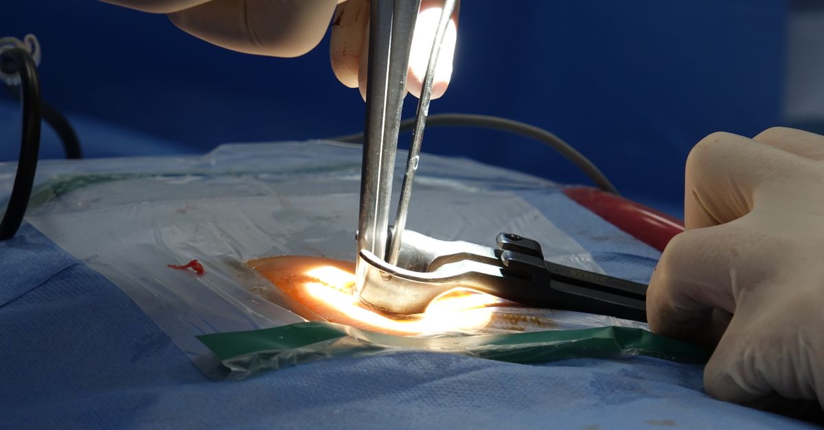
When a pneumothorax or hemothorax compromises respiratory function, the immediate placement of a thoracostomy tube, commonly known as a chest tube, is required. The clinician inserts a sterile, flexible tube through the intercostal space directly into the pleural cavity.
The chest tube is connected to a closed drainage system that applies continuous negative pressure. This mechanism actively evacuates the trapped air or blood, allowing the collapsed lung to immediately re-expand and adhere to the interior surface of the chest wall, restoring normal ventilatory mechanics.
The volume and nature of the chest tube output are meticulously monitored. If a hemothorax drains a massive initial volume of blood, or if continuous, heavy bleeding persists over several hours, it indicates a major internal vascular laceration requiring an emergency thoracotomy to surgically halt the intrathoracic hemorrhage.
14. Long-Term Prognosis and Recovery
The recovery trajectory following a significant chest wall injury is prolonged. Even with optimal clinical management, rib fractures require a minimum of six to eight weeks to achieve primary bony union. During this period, patients frequently experience a persistent, aching pain that intensifies during deep breathing, coughing, or sudden changes in posture.
Elderly patients possess an exceptionally high risk of morbidity following chest wall trauma. Their reduced physiological reserve and the high prevalence of preexisting pulmonary conditions make them highly susceptible to developing severe pneumonia secondary to minor rib fractures.
Comprehensive discharge planning involves educating the patient on the critical importance of continued pulmonary exercises, graduating their pain management regimen, and recognizing the early clinical signs of respiratory infection, ensuring a safe transition from the acute trauma setting back to daily functional activities.
15. Frequently Asked Questions (FAQ)
1. Why does it hurt so much to breathe after fracturing a rib?
Every time you take a breath, the muscles between your ribs contract and move the broken bone fragments. Because bones have a very rich nerve supply, this constant movement causes severe, sharp pain, which makes you want to take shallow breaths.
2. What is a flail chest?
A flail chest occurs when multiple ribs are broken in several places, creating a loose segment of the chest wall. When you breathe in, this broken segment sinks inward instead of expanding outward, severely restricting your ability to get enough oxygen.
3. Will my broken ribs need to be put in a cast?
No, you cannot put a cast on the chest because it would prevent you from breathing. Rib fractures are mostly treated with strong pain medications and breathing exercises. Only in very severe cases do surgeons use metal plates to fix the ribs.
4. What is a pulmonary contusion?
A pulmonary contusion is a deep bruise on the lung itself caused by the force of the impact. The bruised lung tissue swells and fills with fluid, making it very difficult for oxygen to pass from the lungs into the bloodstream.
5. Why is a chest tube inserted after some chest injuries?
If a broken rib punctures the lung or tears a blood vessel, air or blood gets trapped in the chest cavity and crushes the lung. A chest tube is a flexible plastic tube placed between the ribs to drain the air or blood, allowing the lung to re-expand and function normally.
16. Bibliography
Disclaimer: The content is for informational purposes only and does not replace medical advice. Always consult your doctor for personalized treatment.