1. Introduction to Traumatic Intracranial Hemorrhage
An intracranial hemorrhage following an injury, broadly categorized under traumatic brain injury (TBI), is a catastrophic medical event where blunt or penetrating physical force causes bleeding within the confines of the skull. This trauma disrupts the intricate vascular network of the brain or its protective coverings, introducing pressurized blood into an enclosed, non-expandable bony vault. The immediate consequence is a rapid escalation in intracranial pressure, which violently compresses adjacent, healthy neural tissue.
Unlike spontaneous strokes driven by chronic disease, traumatic hemorrhages are the direct result of kinetic energy transfer. Motor vehicle accidents, significant falls, severe sports impacts, and physical assaults are the primary mechanisms. The type of bleeding—whether it pools outside the brain, on its surface, or deep within the tissue—is dictated entirely by the biomechanics of the impact and exactly which blood vessels were sheared or torn by the force.
Medical management of a traumatic brain bleed operates on an extremely tight timeline. The primary clinical objective is to prevent the secondary cascade of brain injury caused by swelling and hypoxia. This requires immediate trauma resuscitation, rapid high-resolution neuroimaging, and often, emergency neurosurgical intervention to evacuate the expanding blood clot, restoring normal intracranial pressure and preserving the patient’s long-term neurological function.
2. The Mechanics of Traumatic Brain Injury
Understanding how a physical impact translates into internal bleeding requires a review of cranial biomechanics. The human brain is a soft, gelatinous organ floating in a shallow pool of cerebrospinal fluid, encased within the rigid, unyielding bone of the skull.
When a sudden, violent force is applied to the head, two distinct mechanical events occur. Contact loading is the direct physical impact of an object against the skull, causing localized fractures and bruising of the brain directly beneath the bone. Inertial loading is the rapid acceleration and sudden deceleration of the head.
During significant inertial loading, the skull abruptly stops moving, but the momentum causes the soft brain to violently slosh forward, slamming into the rough, bony ridges of the inner skull, and then rebounding backward. This intense shifting physically stretches, tears, and shears the delicate bridging veins, cortical arteries, and microscopic neural axons, initiating widespread internal hemorrhage.
3. Primary vs. Secondary Brain Injury
Traumatic brain injury must be conceptualized in two distinct, sequential phases: primary injury and secondary injury. The primary injury occurs at the exact millisecond of impact. It encompasses the immediate mechanical tearing of blood vessels, the shearing of nerve fibers, and the direct crushing of brain tissue. This damage is instantaneous, irreversible, and entirely prevented only by avoiding the trauma itself.
The secondary injury represents a complex, delayed pathological cascade that unfolds over minutes, hours, and days following the impact. The accumulating blood from the torn vessels forms an expanding hematoma that physically crushes healthy brain tissue.
Furthermore, the initial trauma triggers a massive release of excitatory neurotransmitters and inflammatory cytokines. These chemicals induce profound cellular swelling (cytotoxic edema) and cause the blood-brain barrier to leak fluid into the tissues (vasogenic edema). The combined effect of the expanding blood clot and the massive tissue swelling drastically raises intracranial pressure, starving the remaining healthy brain of oxygen and causing a secondary wave of cellular death.
4. Acute Subdural Hematoma
An acute subdural hematoma is one of the most common and devastating types of traumatic intracranial hemorrhage. It involves venous bleeding into the potential space between the tough outer covering of the brain (dura mater) and the delicate middle layer (arachnoid mater).
The primary mechanism is an acceleration-deceleration injury that violently shifts the brain within the skull. This shifting places immense traction on the “bridging veins”—small, fragile veins that drain blood from the surface of the brain into the large dural sinuses. When these veins are stretched beyond their tensile limit, they snap and bleed freely into the subdural space.
Because the bleeding is venous, it spreads diffusely in a crescent shape over the entire hemisphere of the brain. While venous pressure is lower than arterial pressure, acute subdural hematomas are notorious for their high mortality rate. This is because the sheer force required to tear the bridging veins almost always inflicts severe, underlying bruising (contusions) and swelling directly to the brain tissue itself.
5. Epidural Hematoma
An epidural hematoma occurs between the inner table of the skull and the dura mater. Unlike a subdural bleed, an epidural hematoma is overwhelmingly arterial in origin and is almost inextricably linked to a direct, localized fracture of the skull vault.
When blunt trauma strikes the side of the head (the temporal or parietal region), the resulting bone fracture frequently lacerates the middle meningeal artery, which runs through a groove on the inner surface of the skull. The torn artery pumps blood under high systemic pressure, rapidly stripping the tightly adherent dura mater away from the bone.
This creates a dense, lens-shaped (biconvex) pocket of blood. Epidural hematomas are notorious for the “lucid interval.” The patient is knocked unconscious by the initial blow, awakens and appears neurologically intact, but then rapidly deteriorates into a deep coma as the high-pressure arterial bleed quickly reaches a critical volume, crushing the brainstem. It is a profound surgical emergency requiring immediate evacuation.
6. Traumatic Subarachnoid Hemorrhage (tSAH)
Traumatic subarachnoid hemorrhage involves bleeding into the subarachnoid space—the fluid-filled compartment where cerebrospinal fluid flows over the surface of the brain. While spontaneous SAH is typically caused by a ruptured aneurysm, traumatic SAH is caused by the tearing of small pial vessels directly on the brain’s surface due to severe rotational or shearing forces.
Blood in the subarachnoid space physically impedes the normal circulation and reabsorption of cerebrospinal fluid. This can rapidly lead to post-traumatic hydrocephalus, an abnormal accumulation of fluid that further escalates intracranial pressure.
Additionally, the breakdown products of the red blood cells act as severe chemical irritants to the cerebral blood vessels. This irritation can cause the arteries to undergo severe, delayed constriction (vasospasm) days after the injury, restricting vital blood flow and causing secondary ischemic strokes in already traumatized brain tissue.
7. Intraparenchymal Contusions and Hemorrhage
Intraparenchymal hemorrhages are essentially severe bruises (contusions) occurring directly within the functional tissue (parenchyma) of the brain. These occur when the brain forcefully impacts the rough, bony ridges of the skull base, particularly in the frontal and temporal lobes, during an acceleration-deceleration event.
These contusions consist of crushed neural tissue, edema, and multiple small, coalescing areas of hemorrhage. A unique and dangerous characteristic of cerebral contusions is their propensity to “blossom” or expand significantly in the first 24 to 72 hours after the initial injury.
As the bruised tissue undergoes necrosis and intense inflammation, microscopic bleeding continues, causing a small contusion on the initial CT scan to evolve into a massive, space-occupying intraparenchymal hemorrhage days later. This delayed expansion requires patients with seemingly small contusions to undergo rigorous neurocritical care monitoring and serial imaging.
8. Neurological Signs and Symptoms
The clinical presentation of a traumatic brain bleed is dictated by the severity of the impact and the specific location of the hematoma. A sudden, prolonged loss of consciousness immediately following the trauma is a universal red flag indicating severe force transmission to the brainstem.
As the hematoma expands, the patient exhibits signs of rising intracranial pressure. They may awaken severely disoriented, exhibiting profound confusion, agitation, and repetitive questioning. A severe, worsening headache accompanied by projectile vomiting is a classic sign of pressure build-up.
Focal neurological deficits emerge as specific areas of the brain are crushed. The patient may develop acute weakness or paralysis on one side of their body (hemiparesis). An extremely ominous, late-stage sign of brainstem compression is the development of a blown pupil—one pupil becomes massively dilated and completely unresponsive to light, indicating that the brain is actively herniating downward into the spinal canal.
9. Emergency Department Assessment
Upon arrival at the trauma center, the patient undergoes a rapid, structured evaluation guided by Advanced Trauma Life Support (ATLS) protocols. The immediate focus is securing the airway, ensuring adequate breathing, and maintaining cardiovascular circulation, as the brain cannot survive hypoxia or low blood pressure.
Simultaneously, the physician performs a rapid neurological assessment using the Glasgow Coma Scale (GCS). The GCS evaluates eye-opening, verbal responses, and motor commands, assigning a score from 3 to 15. A score of 8 or less defines a severe traumatic brain injury and mandates immediate endotracheal intubation to protect the airway and control ventilation.
The medical team quickly assesses the patient’s pupils for symmetry and reactivity, and checks for abnormal motor posturing (decorticate or decerebrate posturing), which indicates severe structural damage to the deep motor tracts of the brain.
10. Advanced Neuroimaging Protocols
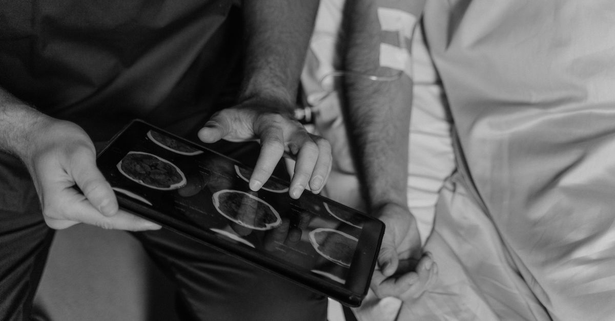
Definitive diagnostic imaging must be obtained without delay. The absolute gold standard for acute head trauma is a non-contrast Computed Tomography (CT) scan of the head. It is the fastest and most sensitive modality for detecting acute bleeding and skull fractures.
The radiologist analyzes the CT scan to categorize the type of hemorrhage (epidural, subdural, subarachnoid, or contusion). They measure the volume of the hematoma and, crucially, assess the degree of “mass effect.” This involves measuring the “midline shift”—how far the blood clot has physically pushed the brain off its central axis.
A midline shift greater than 5 millimeters is generally considered a critical finding that correlates strongly with an imminent need for neurosurgical decompression. Serial CT scans are routinely performed at 12 or 24-hour intervals to ensure that contusions are not blossoming and expanding.
11. Hemorrhage Types and Surgical Urgency
Differentiating the types of traumatic bleeds is essential for surgical planning. The table below outlines the key differences.
| Hemorrhage Type | Origin of Bleeding | CT Scan Appearance | Clinical Urgency |
|---|---|---|---|
| Epidural Hematoma | Torn meningeal artery | Biconvex (lens shape) | Extreme emergency; rapid expansion threatens brainstem. |
| Subdural Hematoma | Torn bridging veins | Crescent shape along skull | High urgency; often accompanied by severe underlying brain swelling. |
| Intraparenchymal | Crushed cortical vessels | Scattered dense blood in tissue | Requires serial imaging; may require surgery if it “blossoms.” |
| Traumatic SAH | Torn surface pial vessels | Blood in sulci/CSF spaces | Medical management for pressure; risk of hydrocephalus. |
12. Reversal of Anticoagulants and Coagulopathy
A massive complicating factor in traumatic brain injury is the presence of systemic coagulopathy. The severe trauma itself triggers a massive release of tissue factor from the crushed brain, which rapidly consumes the body’s clotting factors, leading to a state where the blood can no longer clot, worsening the intracranial bleeding.
Furthermore, a significant portion of trauma patients, particularly the elderly, are actively taking anticoagulant medications (blood thinners) for cardiac conditions. The presence of warfarin, apixaban, or clopidogrel turns a minor head bump into a catastrophic, unstoppable fatal hemorrhage.
Emergency medicine physicians must instantly identify these medications and administer powerful, targeted reversal agents. Intravenous Vitamin K, fresh frozen plasma, prothrombin complex concentrates (PCC), or specific monoclonal antibodies (like idarucizumab) are deployed rapidly to artificially restore the blood’s ability to form a clot and halt the expansion of the hematoma before surgery.
13. Neurosurgical Evacuation Techniques
When a hematoma is sufficiently large, causing significant midline shift or a rapid decline in the patient’s GCS score, emergency neurosurgery is the only definitive treatment to save the patient’s life and preserve neurological function.
The standard procedure is a craniotomy. The neurosurgeon removes a large flap of the skull bone over the site of the hemorrhage. The tough dura mater is opened, and the pressurized blood clot is carefully suctioned and irrigated away. The surgeon identifies the actively bleeding vessels and uses electrocautery to achieve meticulous hemostasis.
In cases where the underlying brain tissue is massively swollen, simply removing the blood is insufficient. The surgeon must perform a decompressive craniectomy. In this procedure, the bone flap is not replaced; it is stored in a freezer or surgically implanted in the patient’s abdomen. Leaving the skull open provides the severely swollen brain room to expand outward, preventing it from crushing inward and destroying the brainstem. The bone is replaced months later after the swelling has resolved.
14. Neurointensive Care and ICP Monitoring
Patients surviving a severe traumatic brain hemorrhage require prolonged, meticulous care in a specialized neuro-intensive care unit (Neuro-ICU). The absolute focus of the ICU team is preventing secondary brain injury by tightly controlling intracranial pressure (ICP) and ensuring adequate cerebral oxygenation.
An ICP monitor, typically a small catheter, is placed directly into the brain to provide real-time pressure data. If the pressure rises dangerously, a tiered medical protocol is initiated. The head of the bed is elevated to facilitate venous drainage.
Hyperosmolar therapy (intravenous mannitol or hypertonic saline) is administered to draw excess water out of the swollen brain tissue via osmosis. The patient is deeply sedated to reduce the brain’s metabolic demand for oxygen, and their core body temperature is strictly controlled to prevent fevers, which drastically worsen brain swelling and secondary injury.
15. Long-Term Prognosis and Rehabilitation
The trajectory of recovery from a severe traumatic intracranial hemorrhage is highly unpredictable. The initial GCS score, the patient’s age, and the swiftness of surgical intervention are the strongest predictors of long-term outcomes. Many patients face a prolonged period of unconsciousness or altered mental status.
Recovery requires a massive, multidisciplinary neurorehabilitation effort. Because the brain tissue physically destroyed by the trauma cannot regenerate, the brain must attempt to rewire itself through neuroplasticity.
Physical therapists work rigorously to overcome paralysis and severe spasticity. Occupational therapists focus on relearning basic activities of daily living. Cognitive and speech therapists address profound deficits in memory, executive functioning, emotional regulation, and communication. The psychological impact on the patient and their family is profound, as traumatic brain injuries frequently cause permanent, severe alterations in personality and functional independence.
16. When to Seek Emergency Care After Trauma
Any significant impact to the head requires immediate professional evaluation, as the symptoms of a lethal brain bleed can be delayed. It is a critical error to assume a patient is safe simply because they did not immediately lose consciousness.
Immediate emergency transport via ambulance is absolutely mandated if an individual experiences a worsening, severe headache following a trauma. Repeated vomiting, extreme lethargy (inability to stay awake), or acute confusion and agitation are major red flags indicating rapidly rising intracranial pressure.
Furthermore, the sudden onset of slurred speech, unequal pupil sizes, weakness in the arms or legs, or any seizure activity following a head injury demands immediate trauma center evaluation. These are clear neurological signs of an actively expanding internal hemorrhage crushing vital brain structures.
17. Frequently Asked Questions (FAQ)
1. How long after a head injury can a brain bleed happen?
While most severe bleeds present symptoms within the first few hours, it is possible for a slow venous bleed (subdural hematoma) to take days or even weeks to accumulate enough blood to cause noticeable symptoms. This is why strict monitoring for subtle cognitive changes is required after any head trauma.
2. Does a concussion mean I have a brain bleed?
No. A concussion is a mild traumatic brain injury caused by the brain shaking inside the skull, leading to temporary chemical and functional changes. While a concussion is serious, it typically does not involve macroscopic bleeding that shows up on a CT scan.
3. Will the doctor put the piece of skull back on immediately?
If a craniotomy is performed just to remove the clot, the bone is usually replaced immediately. However, if the brain is massively swollen (decompressive craniectomy), the bone is left off to allow the brain to swell outward safely. The bone is stored and replaced in a second surgery weeks or months later.
4. Can a person make a full recovery from a traumatic brain bleed?
It depends entirely on the severity of the initial brain tissue destruction. While young patients with rapid surgical intervention can make remarkable recoveries, severe traumatic brain injuries frequently leave patients with some degree of permanent physical, cognitive, or emotional impairment.
18. Bibliography
Disclaimer: The content is for informational purposes only and does not replace medical advice. Always consult your doctor for personalized treatment.