1. Introduction to Skull Vault Fractures with Intracranial Injury
A closed fracture of the cranial vault complicated by an intracranial injury constitutes a complex and severe form of head trauma. The cranial vault serves as a rigid, bony shield designed to protect the delicate neural tissues of the brain. When a biomechanical force is significant enough to fracture this robust bone without tearing the overlying scalp, the patient sustains a closed fracture. While the skin remains intact, masking the visual severity of the wound, the kinetic energy transferred through the bone inevitably damages the structures beneath it.
The primary clinical concern in this scenario is not the broken bone itself, but the associated internal injury. The impact forces physical deformation of the skull, which can contuse the brain surface, tear the protective meningeal linings, and sever critical blood vessels. This results in localized tissue destruction and the rapid accumulation of blood within the closed cranial cavity.
Because the skull cannot expand, any internal bleeding or cellular swelling exponentially increases the pressure against the healthy brain tissue. This rising pressure threatens to cut off vital cerebral blood flow and compress the brainstem. Prompt medical recognition, rapid high-resolution neuroimaging, and decisive neurocritical care or surgical intervention are essential to mitigate secondary brain damage and preserve neurological function.
2. Anatomy of the Cranial Vault
To appreciate the mechanics of this specific trauma, a thorough understanding of the cranial vault architecture is required. The vault, or calvaria, forms the upper and lateral portions of the skull. It is composed of several broad, curved bones, primarily the frontal, parietal, temporal, and occipital bones. These bones are firmly locked together by immovable fibrous joints known as sutures.
The bones of the vault possess a specialized, highly resilient structure. They consist of two dense, solid plates of cortical bone—the inner and outer tables—separated by a central layer of softer, spongy bone called the diploë. This specific design allows the skull to absorb and dissipate substantial amounts of kinetic energy, protecting the brain from routine impacts.
The inner table of the cranial vault is not entirely smooth. It features distinct grooves and channels that closely house the meningeal arteries, which supply blood to the coverings of the brain. The middle meningeal artery, running along the inner surface of the temporal and parietal bones, is particularly prominent. Due to its intimate physical attachment to the bone, this artery is highly vulnerable to laceration when the vault fractures.
3. Biomechanics of a Closed Fracture
A closed fracture of the cranial vault occurs when blunt force trauma overwhelms the structural elasticity of the bone. Motor vehicle collisions, falls from significant heights onto hard surfaces, and high-velocity sports impacts are the most frequent mechanisms. The designation of a “closed” fracture signifies that the overlying layers of the scalp remain intact, meaning the bone is not exposed to the external environment, significantly lowering the immediate risk of a direct bacterial infection.
These fractures are typically categorized into linear and depressed variants. A linear fracture presents as a single, distinct crack extending through the bone, without the displacement of bone fragments. While the structural alignment remains normal, the crack itself can sever blood vessels traversing its path.
A depressed fracture results from a more focused impact, driving a fragment of the fractured skull inward, below the normal anatomical curvature of the vault. In a depressed fracture, the sharp, jagged edges of the displaced bone fragment can directly pierce the tough dura mater and physically lacerate the underlying cerebral cortex, causing immediate localized bleeding and direct neural tissue destruction.
4. Associated Brain Trauma: Coup and Contrecoup
The kinetic energy sufficient to fracture the robust cranial vault invariably transmits destructive force directly into the brain tissue. This internal trauma frequently manifests in a distinct coup and contrecoup pattern.
The “coup” injury occurs exactly at the point of impact. The inward deformation of the skull, even momentarily, bruises the underlying brain tissue, resulting in a cerebral contusion. This localized area of crushed tissue, edema, and microscopic bleeding disrupts the normal function of that specific cortical region.
The “contrecoup” injury occurs on the exact opposite side of the brain. The impact accelerates the skull, but the gelatinous brain lags slightly behind due to inertia. As the head abruptly stops or changes direction, the brain sloshes violently forward, striking the hard, irregular internal ridges on the opposite side of the cranial vault. This secondary impact causes severe bruising and shearing injuries far removed from the original fracture site, compounding the overall neurological deficit.
5. Epidural Hemorrhage Mechanisms
The most classic and rapidly fatal intracranial injury specifically associated with a skull vault fracture is the epidural hematoma. This life-threatening condition occurs when a fracture, usually a linear fracture of the temporal or parietal bone, crosses the anatomical groove housing the middle meningeal artery.
The sharp edges of the fractured bone tear this major artery. Because the bleeding is arterial, blood escapes under high systemic pressure. This forceful bleeding acts as a wedge, aggressively peeling the tough dura mater away from the inner table of the skull.
The accumulating blood creates a rapidly expanding, lens-shaped pocket of pressure between the bone and the brain. An epidural hematoma is an absolute neurosurgical emergency. The rapid arterial expansion quickly drives up intracranial pressure, leading to a swift decline in consciousness and imminent brainstem herniation if the blood clot is not surgically evacuated.
6. Subdural Hemorrhage and Venous Tearing
A closed vault fracture can also be accompanied by a subdural hematoma. While an epidural hematoma is driven by arterial bleeding outside the dura, a subdural hematoma involves venous bleeding beneath the dura mater, specifically in the space between the dura and the delicate arachnoid membrane.
This injury is primarily caused by the severe acceleration and deceleration forces associated with the impact, rather than a direct laceration from the bone fracture itself. The violent shifting of the brain within the skull stretches and snaps the fragile “bridging veins” that drain blood from the brain’s surface into the large dural sinuses.
These torn veins bleed more slowly than an artery, causing blood to spread diffusely in a crescent shape over the cerebral hemisphere. However, acute subdural hematomas in the setting of severe trauma are notoriously lethal, as the sheer force required to tear these veins almost always inflicts substantial, widespread bruising and swelling to the underlying brain tissue itself.
7. Clinical Symptoms and the Lucid Interval
The clinical presentation of a patient suffering from a closed skull fracture with an internal injury depends entirely on the specific nature and volume of the underlying bleeding or tissue damage. A classic hallmark presentation of an expanding epidural hematoma involves a phenomenon termed the “lucid interval.”
Following the initial trauma, the patient often experiences a brief loss of consciousness due to the concussive force. They subsequently awaken, appearing relatively lucid, speaking clearly, and interacting normally. However, this period is highly deceptive. During this interval, the severed artery is actively pumping blood into the rigid cranial vault.
As the hematoma reaches a critical volume, the intracranial pressure spikes abruptly. The patient’s clinical status deteriorates with alarming speed. They develop a severe, unrelenting headache, projectile vomiting, and acute confusion, rapidly descending into a deep coma. A classic, late-stage physical sign is a dilated, unreactive pupil on one side, signaling that the expanding mass is actively compressing the brainstem.
8. Assessing Altered Consciousness
Medical professionals utilize the Glasgow Coma Scale to rapidly and objectively quantify the severity of a patient’s altered consciousness following head trauma. This standardized clinical tool evaluates the patient based on their eye-opening response, verbal response, and motor capabilities.
Scores range from 3, indicating a state of deep coma, to 15, representing a fully alert and oriented individual. A score of 8 or lower defines a severe traumatic brain injury and generally mandates immediate securing of the patient’s airway via endotracheal intubation.
In the context of a closed skull fracture, dynamic changes in the Glasgow Coma Scale are more important than a single reading. A patient whose score drops significantly over a short period—for instance, declining from a 14 to a 9 within an hour—provides the clearest clinical indication of an actively expanding intracranial hemorrhage requiring urgent intervention.
9. Diagnostic Neuroimaging
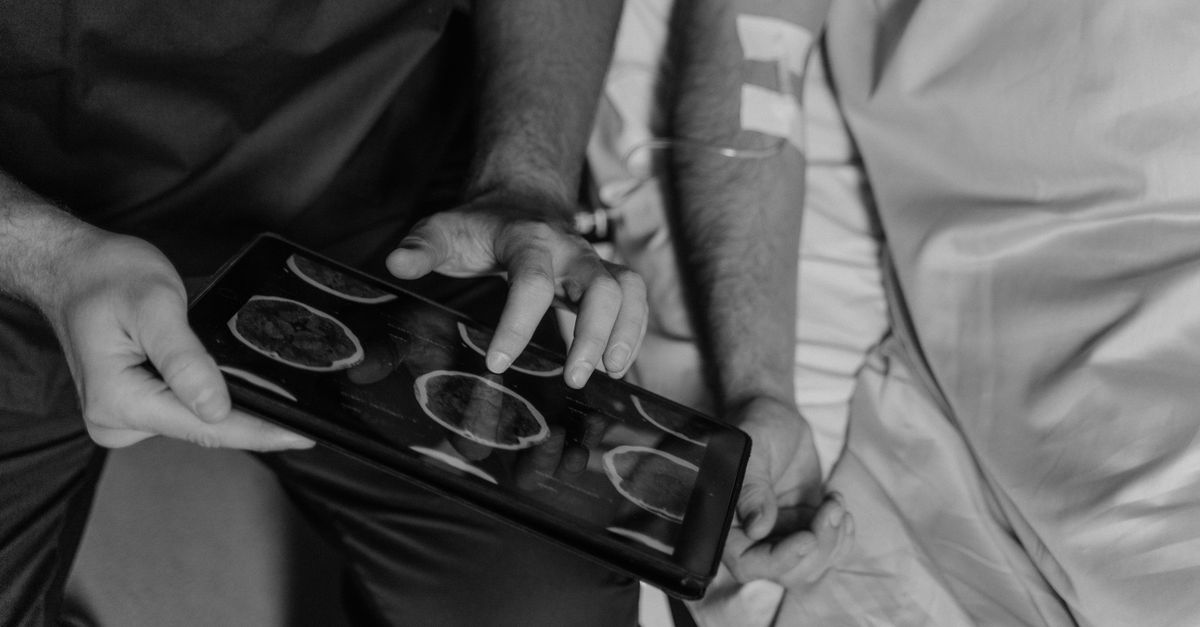
Upon a patient’s arrival at a trauma center, rapid and definitive diagnostic imaging is the absolute priority. The gold standard modality for evaluating acute head trauma is a non-contrast Computed Tomography scan of the head.
A Computed Tomography scan is exceptionally fast and highly sensitive to acute bleeding, which appears bright white against the darker tissue of the brain. The radiologist carefully analyzes the images using different settings. A “bone window” setting clearly delineates the cranial vault, confirming the presence, location, and degree of depression of any skull fractures.
Simultaneously, the scan reveals the extent of the intracranial injury. It visually differentiates between the lens shape of an epidural hematoma and the crescent shape of a subdural hematoma. Crucially, the scan allows the physician to measure the “midline shift,” determining exactly how far the expanding blood clot has physically pushed the normal brain structures off their central axis, indicating the severity of the internal pressure.
10. Comparing Associated Intracranial Injuries
Differentiating the internal injuries associated with vault fractures dictates the urgency and type of medical intervention. The table below outlines key distinctions.
| Intracranial Injury | Primary Mechanism | Radiological Appearance |
|---|---|---|
| Epidural Hematoma | Torn meningeal artery by skull fracture | Biconvex (lens-shaped) hyperdense collection |
| Subdural Hematoma | Torn bridging veins from acceleration/deceleration | Crescent-shaped hyperdense collection |
| Cerebral Contusion | Brain impacting the inner skull (Coup/Contrecoup) | Localized, mixed-density bruising in brain tissue |
| Depressed Fracture | Focal blunt force pushing bone inward | Bone fragments displaced below the cranial vault line |
11. Initial Emergency Resuscitation
The management of a severe cranial fracture with internal injury follows strict Advanced Trauma Life Support protocols. Because the injured brain is exquisitely sensitive to secondary insults, specifically a lack of oxygen (hypoxia) or low blood pressure (hypotension), systemic stabilization is paramount.
Airway control is secured immediately for patients with a depressed level of consciousness to ensure constant oxygen delivery and prevent the aspiration of gastric contents. Maintaining robust systemic blood pressure with intravenous fluids is critical to ensure that oxygenated blood can push against the rising intracranial pressure to reach the brain tissue.
If the patient is taking systemic anticoagulant medications, emergency reversal agents, such as prothrombin complex concentrates or fresh frozen plasma, must be administered rapidly. Restoring the blood’s normal clotting cascade is a critical, immediate step to halt the continuous expansion of the internal hematoma before surgery can be performed.
12. Management of Intracranial Pressure
For patients recovering post-operatively, or for those whose injuries do not immediately require surgery but are causing significant brain swelling, the neurocritical care unit focuses entirely on managing intracranial pressure.
A specialized monitor may be placed directly into the brain parenchyma to provide continuous pressure data. If the intracranial pressure rises to dangerous levels, hyperosmolar therapy is initiated. Intravenous mannitol or concentrated hypertonic saline is administered; these solutions draw excess water out of the swollen brain tissue and into the vascular system, physically shrinking the brain and relieving pressure.
The patient is kept with the head of their bed elevated to promote venous drainage from the brain. Deep pharmacological sedation is frequently utilized to reduce the brain’s metabolic demand for oxygen, and strict temperature control is maintained to prevent fevers, which can drastically worsen cerebral edema.
13. Neurosurgical Interventions
Emergency neurosurgery is indicated when a Computed Tomography scan reveals a significant hematoma causing a dangerous midline shift, or if a depressed skull fracture has driven bone fragments deep into the brain tissue.
The primary surgical procedure is a craniotomy. The neurosurgeon removes a section of the skull vault to access the underlying injury. For a depressed fracture, the surgeon carefully elevates the impacted bone fragments, removes any debris, and meticulously repairs the torn dura mater to create a watertight seal, preventing cerebrospinal fluid leaks.
For an epidural or subdural hematoma, the pressurized blood clot is suctioned out, immediately relieving the compression on the brain. The surgeon identifies the bleeding artery or vein and uses bipolar electrocautery to permanently stop the hemorrhage. The bone flap is then typically replaced and secured with low-profile titanium plates and screws.
14. Long-Term Neurological Complications
Even following successful surgical decompression, patients sustaining a skull fracture with significant intracranial injury face a high risk of prolonged neurological complications. The cerebral cortex directly bruised by the trauma frequently heals with a distinct glial scar. This scar tissue alters normal electrical pathways, making post-traumatic seizures a common complication, often requiring long-term anticonvulsant therapy.
Cognitive and psychiatric complications are profound. The sheer force required to fracture the skull often induces diffuse axonal injury—the microscopic stretching and tearing of neural connections throughout the brain.
This widespread microscopic damage manifests as persistent memory deficits, slowed processing speeds, emotional lability, and profound changes in executive functioning and personality. These invisible injuries often represent the most challenging aspects of recovery, significantly altering the patient’s daily life and interpersonal relationships.
15. Neurorehabilitation and Recovery
The brain tissue physically destroyed during the primary impact cannot regenerate. However, through the principle of neuroplasticity, the brain possesses a remarkable ability to reorganize its remaining healthy neural networks to compensate for the damaged areas over time.
This recovery relies entirely on intensive, multidisciplinary neurorehabilitation, beginning as soon as the patient attains medical stability. Physical therapists work rigorously to overcome motor deficits, restore balance, and improve ambulation. Occupational therapists assist the patient in relearning the necessary skills for independent daily living.
Speech-language pathologists focus on recovering complex language skills, reading comprehension, and swallowing function. Concurrently, neuropsychologists provide essential cognitive therapies and emotional support, helping the patient and their family navigate the complex psychological adjustments following a severe brain injury.
16. When to Seek Emergency Medical Care
Any substantial blunt force trauma to the head demands immediate, professional medical evaluation. A closed skull fracture presents no outward signs of bone damage, and the symptoms of a lethal internal hemorrhage can be deceptively delayed.
Immediate transport via ambulance to a trauma center is absolutely mandated if an individual experiences any loss of consciousness, regardless of how quickly they appear to recover. A progressively worsening, severe headache, repeated episodes of vomiting, or sudden, extreme lethargy are major clinical red flags indicating rapidly rising intracranial pressure.
Furthermore, if the patient develops acute neurological signs such as unequal pupil sizes, slurred speech, clear fluid draining from the ears or nose, or new weakness on one side of the body, these indicate severe, active internal damage that requires immediate life-saving intervention.
17. Frequently Asked Questions (FAQ)
1. Can a closed skull fracture heal without surgery?
Yes, a simple, linear closed skull fracture that is not depressed and is not associated with an internal blood clot will generally heal on its own over a period of months, much like other bone fractures. However, the patient must be monitored closely to ensure no delayed bleeding occurs.
2. Does having a skull fracture mean I have brain damage?
Not always, but the risk is very high. The skull fracture itself is just a broken bone. The critical issue is the force of the impact. It is entirely possible to fracture the skull and only sustain a mild concussion, just as it is possible to suffer a devastating brain injury without cracking the skull at all.
3. Why do doctors put someone with a head injury in a coma?
A medically induced coma is utilized to completely rest the injured brain. By administering deep sedation, doctors drastically reduce the brain’s metabolic need for oxygen and energy. This decreased demand helps to minimize blood flow requirements, which significantly reduces dangerous brain swelling during the acute healing phase.
4. Will someone fully recover from an epidural hematoma?
If an epidural hematoma is diagnosed rapidly and surgically evacuated before it crushes the brainstem, the patient has an excellent chance of a full neurological recovery, because the underlying brain tissue itself is often not severely bruised. Delaying surgery, however, frequently leads to permanent damage or death.
18. Bibliography
Disclaimer: The content is for informational purposes only and does not replace medical advice. Always consult your doctor for personalized treatment.