1. Introduction
Juvenile osteochondritis dissecans is a localized, highly specific joint condition where a segment of subchondral bone—the bone located directly beneath the protective cartilage of a joint—loses its blood supply and begins to undergo necrosis. As this small area of bone dies, the overlying hyaline cartilage loses its crucial structural support. This structural failure places the affected segment at a severe risk of separating entirely from the main bone, potentially creating a loose fragment that floats freely within the joint cavity.
This condition primarily affects children and young adolescents whose skeletal structures are still actively growing and developing. The knee is by far the most commonly afflicted joint, though the elbow and the ankle can also be involved. Early clinical detection is paramount. When diagnosed before the cartilage completely separates, conservative treatments can often coax the bone into healing itself. If separation occurs, aggressive surgical intervention is required to prevent early-onset degenerative joint disease.
2. Anatomy of Subchondral Bone and Cartilage
To understand the nature of this lesion, one must examine the microanatomy of a healthy joint surface. The end of a bone within a joint is capped by a thick, resilient layer of articular cartilage, which provides a perfectly smooth, frictionless surface for movement. Because this cartilage possesses no blood vessels of its own, it relies heavily on the underlying subchondral bone for physical support and chemical nutrient diffusion.
The subchondral bone is a dense, highly vascularized plate that absorbs the massive mechanical shocks of walking and jumping. A delicate biological balance exists between the bone and the cartilage. If the blood supply to a segment of the subchondral bone is abruptly compromised, that specific piece of bone softens and crumbles. Without a solid foundation, the overlying cartilage inevitably cracks and fails under normal physical stress.
3. Pathophysiology of the Lesion
The exact initiating event that chokes off the blood supply to the localized area of subchondral bone remains a topic of intense orthopedic debate, but the resulting pathological cascade is well understood. The localized cellular death, known as avascular necrosis, creates a weak, dead zone within the otherwise healthy bone structure.
The body attempts to repair this dead zone by forming fibrous scar tissue around the necrotic bone segment. This scar tissue acts as a physical barrier, preventing new blood vessels and fresh bone-building cells from reaching the dying fragment. As mechanical forces continue to pound against the joint, the dead fragment gradually loosens, threatening to break away entirely and plunge into the synovial fluid.
4. The Role of Repetitive Microtrauma
The most widely accepted primary cause of this condition in modern pediatric orthopedics is repetitive microtrauma. Young athletes involved in high-impact, repetitive sports such as gymnastics, competitive running, or baseball pitching subject their developing joints to immense, relentless mechanical stress.
These repeated physical impacts cause microscopic fractures in the subchondral bone before the skeleton is mature enough to withstand such forces. If the child does not receive adequate rest between intense training sessions, the bone cannot heal these microfractures. The cumulative damage eventually severs the fragile microscopic blood vessels supplying the bone, triggering the localized necrotic event.
5. Genetic and Vascular Factors
While biomechanical overload is the dominant factor, strong evidence suggests a concurrent genetic predisposition. Some children possess unique anatomical variations in the blood vessel architecture of their joints, leaving certain areas of the subchondral bone inherently vulnerable to vascular disruption.
Additionally, certain endocrine factors and rapid periods of skeletal growth during early puberty may alter the density and structural resilience of the subchondral plate, rendering it highly susceptible to injury. This multifactorial etiology explains why the condition sometimes appears simultaneously in both knees of a young athlete, despite only one side being subjected to the dominant mechanical stress.
6. Common Anatomical Locations
The disease has highly specific target locations within the skeletal system. The knee is overwhelmingly the most frequent site, with approximately seventy to eighty percent of all lesions occurring on the lateral aspect of the medial femoral condyle. This specific area bears the brunt of the rotational and compressive forces during activities involving running and sudden pivoting.
In the elbow, the capitellum is the classic site of involvement, almost exclusively seen in adolescent baseball pitchers or gymnasts who subject their arms to immense valgus forces. In the ankle, the domed surface of the talus is frequently affected, often linked to a history of severe or recurrent ankle sprains.
7. Recognizing the Symptoms
The clinical presentation in a child is often vague and develops insidiously over several months. Initially, the patient complains of a dull, poorly localized ache in the affected joint that is distinctly aggravated by physical activity and relieved by rest. There is rarely a specific history of a massive, sudden traumatic event.
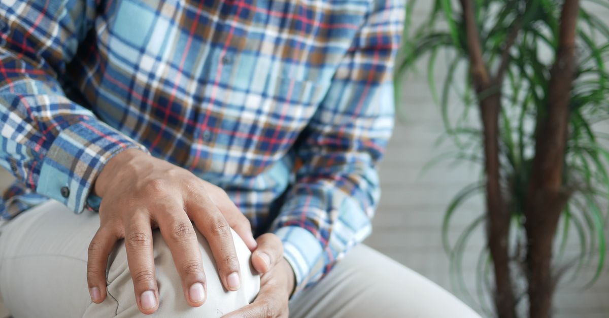
As the underlying bone softens and the lesion progresses, the joint may intermittently swell after practice or competition. The child may naturally alter their gait, walking with the foot slightly turned outward to shift weight away from the painful aspect of the femoral condyle. Any persistent knee pain in an active adolescent warrants a thorough clinical evaluation.
8. Mechanical Symptoms and Joint Locking
A critical shift in symptoms occurs if the necrotic bone fragment and its overlying cartilage physically detach from the main skeletal structure. This creates a loose body, colloquially known as a joint mouse, floating freely within the synovial fluid.
When this loose fragment becomes wedged between the moving surfaces of the joint, it causes sudden, severe, sharp pain. The joint may dramatically catch, click, or lock entirely in a bent position. The child will be physically unable to straighten their limb until the fragment randomly dislodges. The presence of these acute mechanical symptoms usually indicates a late-stage, unstable lesion.
9. Clinical Examination Techniques
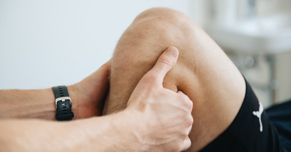
The orthopedic evaluation involves meticulous palpation to locate precise areas of tenderness. For a knee lesion, the physician may press firmly on the medial femoral condyle while bending the joint, which frequently elicits a sharp pain response identical to what the child experiences during sports.
The physician will also evaluate the joint for any effusion, which is an abnormal accumulation of inflammatory fluid. Specific stress tests, such as Wilson’s test for the knee, involve internally rotating the tibia while extending the leg to deliberately rub against the classic lesion site, seeking to reproduce the characteristic mechanical discomfort.
10. The Importance of Early Diagnosis
Diagnosing the condition while the lesion is still perfectly stable in its anatomical bed provides the absolute best chance for natural, non-surgical healing. Because the growth plates of a juvenile patient are still open, their skeletal system possesses a massive capacity for cellular regeneration and structural repair.
If a stable lesion is identified and completely protected from mechanical stress, new blood vessels can penetrate the fibrous barrier, and fresh osteoblasts can replace the dead bone. If the diagnosis is delayed until after the child reaches full skeletal maturity and the growth plates fuse, the biological healing capacity drops precipitously, drastically increasing the need for major surgery.
11. Radiographic Imaging and MRI
Standard plain x-rays are the initial diagnostic tool and clearly reveal the presence of a lesion. The x-ray typically shows a distinct, crescent-shaped radiolucent shadow outlining a small, dense fragment of subchondral bone.
However, x-rays cannot assess the health of the overlying cartilage or determine if the fragment is physically loose. Magnetic resonance imaging is the gold standard for comprehensively staging the disease. The magnetic resonance scan provides exceptional detail of the cartilage integrity and detects fluid tracking directly behind the bone fragment, which is a definitive radiological sign that the lesion has become unstable and is preparing to detach.
12. Stages of Lesion Progression
Orthopedic specialists classify the disease into four distinct progressive stages, which directly dictate the treatment protocol. Stage one involves a small area of compressed bone with completely intact, normal-appearing cartilage. Stage two shows a partially detached lesion, but the cartilage remains firmly in place.
Stage three is a highly unstable lesion. A clear, physical gap is visible around the entire bone fragment, and the overlying cartilage is severely cracked or frayed, though the fragment remains resting in its crater. Stage four represents a catastrophic detachment, where the bone and cartilage have fallen completely out of the crater, creating a large defect and a free-floating loose body.
13. Conservative Management Strategies
For stage one and stage two stable lesions in children with open growth plates, conservative therapy is the strict standard of care. The absolute priority is removing the mechanical stress that caused the initial vascular failure. The child must immediately halt all sports, running, and jumping activities.
Depending on the size and location of the lesion, the physician may require complete non-weight-bearing status using crutches, or utilize a specialized hinged brace to restrict the joint from bending into the specific angle that places pressure on the damaged bone. This strict rest period is lengthy, often requiring three to six months of absolute compliance before imaging shows signs of structural healing.
14. Physical Therapy and Activity Modification
Once follow-up magnetic resonance imaging confirms that the dead bone is successfully revascularizing and healing back into the main structure, a highly supervised physical therapy program begins. The goal is to safely restore muscle mass that atrophied during the prolonged rest period.
Physical therapists focus heavily on core stability, proprioceptive balance, and correcting any biomechanical flaws in the athlete’s running or throwing mechanics. A gradual, highly structured return to sports protocol is implemented. The child is closely monitored, and any recurrence of pain or swelling demands an immediate halt to physical activity.
15. Surgical Intervention Options
Surgery is mandatory for unstable stage three and stage four lesions, or for stable lesions that completely fail to heal after six months of rigorous conservative care. Minimally invasive arthroscopic surgery is utilized to address the defect.
If the fragment is loose but structurally sound, the surgeon will clean out the fibrous scar tissue beneath it and use specialized bio-absorbable screws to pin the fragment tightly back into its crater, forcing it to heal. If the fragment is hopelessly crushed or already floating free, it is removed, and the surgeon performs a microfracture procedure—drilling tiny holes into the underlying bone to stimulate the bone marrow to form a new, synthetic cartilage patch over the defect.
16. Summary of Disease Staging
The stage of the lesion directly determines the aggressiveness of the clinical intervention. The table below outlines these crucial stages.
| Disease Stage | Cartilage/Bone Status | Primary Clinical Treatment |
|---|---|---|
| Stage I | Bone compressed, cartilage intact | Strict rest, activity cessation, bracing |
| Stage II | Partial separation, cartilage intact | Strict rest, prolonged non-weight bearing |
| Stage III | Fluid tracking behind unstable fragment | Surgical fixation with bio-screws |
| Stage IV | Complete detachment, loose body in joint | Surgical removal and cartilage restoration |
17. Frequently Asked Questions
1. Will my child develop arthritis because of this condition?
If a stable lesion heals completely with conservative rest, the risk of future arthritis is minimal. However, if the fragment detaches and leaves a large, permanent crater in the articular cartilage, the risk of developing early-onset osteoarthritis in adulthood increases significantly.
2. Can my child just play through the dull pain?
Absolutely not. Playing through the pain is exactly what causes a minor, stable injury to progress into a severe, detached lesion requiring major surgery. Pain is the biological warning sign that the bone is actively dying.
3. Does physical therapy alone fix the dead bone?
No. Physical therapy cannot increase blood flow to the dead bone segment. Strict rest is the only mechanism that allows the bone to heal. Physical therapy is vital afterward to rebuild the surrounding muscle strength safely.
4. Are the surgical screws left in the joint forever?
Modern orthopedic surgeons primarily use bio-absorbable screws and pins. These specialized devices hold the bone tightly in place while it heals, and then slowly dissolve and are absorbed entirely by the body over several months, requiring no second surgery for removal.
5. How long will my child be out of sports?
Treating this condition requires immense patience. Even for mild, non-surgical cases, a child is typically prohibited from running and jumping for a strict minimum of three to six months to ensure the subchondral bone has fully regenerated.
18. Bibliography
Disclaimer: The content is for informational purposes only and does not replace medical advice. Always consult your doctor for personalized treatment.