1. Introduction
Pain in the lumbar spine, universally recognized as lower back pain, is one of the most pervasive and debilitating musculoskeletal conditions affecting the global adult population. The lumbar spine serves as the fundamental mechanical pillar of the human body, engineered to bear immense compressive axial loads while simultaneously providing the flexibility required for multidirectional truncal movement. This continuous, high-stress biomechanical environment renders the lumbar region highly susceptible to acute mechanical trauma, repetitive strain injuries, and progressive degenerative pathologies.
The clinical presentation of lumbar spine pain is remarkably heterogeneous. A patient may present with a benign, self-limiting muscular spasm or with severe, agonizing radiculopathy indicating critical nerve root compression. Effective clinical management requires a meticulous diagnostic process to differentiate between simple mechanical back pain, structural neurological impingement, and dangerous systemic diseases. By accurately isolating the specific anatomical pain generator, clinicians can deploy targeted pharmacological, physical, and interventional therapies to alleviate distress and restore functional spinal biomechanics.
2. Anatomy of the Lumbar Spine
The lumbar spine is composed of five massive, robust vertebrae designated L1 through L5. These osseous segments are stacked upon the sacrum and are separated by intervertebral discs, which function as critical fibrocartilaginous shock absorbers. Each disc consists of a tough, fibrous outer ring known as the annulus fibrosus, enclosing a gel-like center called the nucleus pulposus.
Posteriorly, the vertebrae articulate via paired synovial facet joints, which guide spinal motion and prevent excessive rotation. The spinal canal runs centrally through the vertebrae, housing the lower aspect of the spinal cord and the cauda equina nerve roots. The stability of this complex structure is maintained by a dense network of powerful paraspinal muscles and thick ligamentous bands. Pathology affecting any of these intricately connected structures will generate significant localized or referred pain.
3. Muscular Strains and Ligamentous Sprains
The vast majority of acute lumbar spine pain episodes are attributable to simple muscular strains or ligamentous sprains. The heavy muscles of the lower back, particularly the erector spinae group, are vulnerable to microscopic tearing when subjected to sudden, explosive lifting, twisting motions under load, or prolonged postural fatigue.
When these soft tissues are injured, the body responds with a localized inflammatory cascade. This inflammation triggers a powerful, protective reflex arc, resulting in severe, involuntary muscle spasms designed to completely immobilize the injured segment. The patient experiences intense, localized, dull or cramping pain that sharply exacerbates with any movement of the torso, though neurological symptoms such as leg numbness are entirely absent.
4. Degenerative Disc Disease
Degenerative disc disease is a natural, progressive consequence of aging and mechanical wear, though it can become intensely painful in some individuals. Over time, the intervertebral discs lose their hydration and elasticity, a process known as desiccation. As the discs flatten and lose height, they compromise the intrinsic shock-absorbing capacity of the spine.
This loss of disc height dramatically alters the biomechanics of the lumbar segment, shifting abnormal, increased loads onto the posterior facet joints. The annulus fibrosus also develops micro-tears, which are highly innervated and serve as a direct source of deep, axial back pain. The pain is typically described as a chronic, low-grade ache that worsens with prolonged sitting or forward flexion, as these positions increase intradiscal pressure.
5. Lumbar Disc Herniation and Radiculopathy
A lumbar disc herniation occurs when the gel-like nucleus pulposus pushes through a significant tear in the outer annulus fibrosus, protruding into the spinal canal or intervertebral foramen. This most frequently occurs at the L4-L5 or L5-S1 spinal levels.
When the extruded disc material physically compresses or chemically inflames the adjacent spinal nerve root, it generates severe neuropathic pain, clinically termed lumbar radiculopathy or sciatica. Patients report an agonizing, sharp, electrical, or burning sensation that originates in the lower back and shoots down the posterior or lateral aspect of the leg, often reaching the foot. This radiating pain is frequently accompanied by paresthesias and localized motor weakness in the specific dermatome supplied by the compressed nerve.
6. Lumbar Spinal Stenosis
Lumbar spinal stenosis is characterized by the pathological narrowing of the central spinal canal or the lateral nerve root canals, predominantly affecting the older adult population. This narrowing is the cumulative result of advanced degenerative changes, including hypertrophic facet joint osteoarthritis, thickening of the ligamentum flavum, and bulging of the intervertebral discs.
The hallmark clinical presentation of spinal stenosis is neurogenic claudication. Patients experience a heavy, cramping, or burning pain in the buttocks and thighs that reliably occurs after walking or prolonged standing. Crucially, the pain is rapidly relieved when the patient sits down or leans forward, as spinal flexion physically opens the spinal canal and temporarily relieves the mechanical pressure on the ischemic nerve roots.
7. Spondylolisthesis and Spinal Instability
Spondylolisthesis occurs when one lumbar vertebra slips forward relative to the vertebra immediately below it. This structural instability can be congenital, the result of a stress fracture in the pars interarticularis (isthmic spondylolisthesis), or due to severe degenerative facet arthritis.
The anterior translation of the vertebra stretches the stabilizing ligaments and alters the alignment of the spinal canal. Patients typically experience deep, mechanical back pain that worsens with standing or activities requiring spinal extension. If the slippage is severe enough to narrow the neural foramen, it can compress the exiting nerve roots, generating concurrent radicular leg pain.
8. Sacroiliac Joint Dysfunction
While technically located just below the lumbar spine, pathology in the sacroiliac joint is a prolific generator of pain that closely mimics lower back pain. The sacroiliac joint connects the sacrum to the ilium and is designed for stability and load transfer rather than mobility.
Dysfunction in this joint, whether due to hypermobility, mechanical inflammation, or degenerative arthritis, produces a deep, aching pain located unilaterally in the lower back and upper buttock. The pain frequently intensifies when transitioning from a seated to a standing position, or when bearing weight asymmetrically on one leg, differentiating it from purely lumbar discogenic pain.
9. Differentiating Lumbar Pain Syndromes
Accurate clinical diagnosis requires mapping the pain characteristics and mechanical triggers to the corresponding anatomical structure.
| Condition | Primary Pain Quality and Location | Key Exacerbating Factors |
|---|---|---|
| Muscular Strain | Dull, cramping, localized to lower back. | Truncal movement; no radiating leg pain. |
| Disc Herniation | Sharp, electrical, shooting down one leg. | Sitting, bending forward, coughing, sneezing. |
| Spinal Stenosis | Heavy, cramping in buttocks/legs (Neurogenic claudication). | Walking, standing straight; relieved by sitting/leaning forward. |
| Sacroiliac Dysfunction | Aching, localized to one side of very low back/upper buttock. | Standing up from a chair, asymmetrical weight-bearing. |
10. Systemic and Inflammatory Etiologies
The clinician must always consider systemic causes of lumbar pain, particularly in younger patients presenting with chronic symptoms. Ankylosing spondylitis is a chronic autoimmune inflammatory arthritis that primarily targets the axial skeleton. It causes profound inflammation at the entheses where ligaments attach to bone.
Patients with ankylosing spondylitis experience severe lower back pain and stiffness that is paradoxically worse in the morning or after prolonged rest, and significantly improves with physical activity. Without aggressive systemic immunomodulatory treatment, the chronic inflammation eventually leads to complete bony fusion of the spinal vertebrae, resulting in a rigid, fragile spine.
11. Clinical Evaluation and Neurological Testing
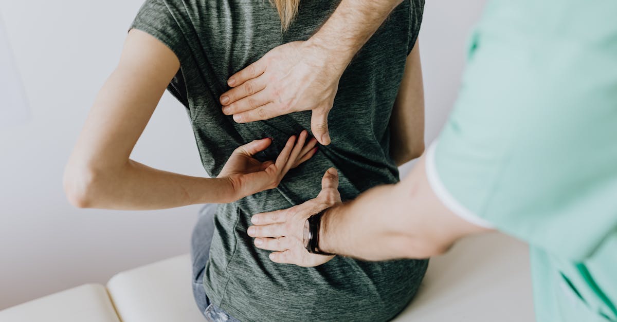
A thorough physical and neurological examination is paramount. The clinician observes the patient’s posture and gait, checking for antalgia (a pain-avoidance posture). Palpation identifies localized muscular spasms or extreme bony tenderness over the spinous processes.
Neurological testing is critical to assess the integrity of the spinal nerve roots. The clinician tests deep tendon reflexes (patellar and Achilles), sensory perception across the dermatomes, and muscle strength in the lower extremities. The Straight Leg Raise test is a highly sensitive provocative maneuver used to detect nerve root tension; lifting the extended leg of a supine patient will reliably reproduce the shooting leg pain characteristic of severe sciatica.
12. Diagnostic Imaging and Diagnostics
For acute, non-radiating lumbar pain without neurological deficits, diagnostic imaging is generally unwarranted in the first few weeks, as most cases are muscular and self-limiting. However, if red flag symptoms are present or radiculopathy persists, imaging is indicated.
Standard plain radiographs visualize bony alignment, identifying fractures, severe disc height loss, or spondylolisthesis. Magnetic Resonance Imaging is the definitive gold standard for evaluating the lumbar spine. It provides exquisite detail of the soft tissues, clearly visualizing the extent of disc herniations, the degree of central canal stenosis, and any chemical inflammation or physical compression affecting the spinal nerve roots.
13. Pharmacological Management
Pharmacological therapy targets pain modulation and inflammation reduction. Nonsteroidal anti-inflammatory drugs are the foundation of conservative care, effectively mitigating the localized inflammatory response associated with acute strains or discogenic flare-ups. Muscle relaxants provide short-term relief for severe, debilitating paraspinal muscle spasms.
For patients suffering from neuropathic radicular pain, standard analgesics lack efficacy. Neuromodulators, such as gabapentin or pregabalin, or certain tricyclic antidepressants are prescribed to dampen the aberrant, hyperexcitable electrical signals generated by the compressed nerve root, providing significant symptomatic relief.
14. Physical Therapy and Active Rehabilitation
Passive treatments offer only temporary relief; active physical rehabilitation is the cornerstone of long-term lumbar spine health. A targeted physical therapy protocol aims to restore normal spinal biomechanics. Therapy initially focuses on directional preference exercises, determining which movements centralize the radiating pain.
As acute pain subsides, the focus shifts to aggressive strengthening of the core musculature, particularly the transversus abdominis and multifidus muscles. A strong, responsive core acts as a dynamic internal corset, stabilizing the spinal segments and reducing the mechanical load transmitted through the vulnerable intervertebral discs and facet joints during daily activities.
15. Interventional Pain Procedures
When conservative measures fail, interventional pain procedures offer targeted relief. Lumbar epidural steroid injections are highly effective for managing severe radiculopathy and spinal stenosis. A potent corticosteroid is injected under fluoroscopic guidance directly into the epidural space, bathing the inflamed nerve root to rapidly suppress the localized immune response and reduce swelling.
For chronic facet joint pain, medial branch blocks are utilized diagnostically to isolate the pain generator. If successful, radiofrequency ablation can be performed to temporarily deactivate the small sensory nerves supplying the arthritic joint, providing months to years of substantial pain relief.
16. Surgical Indications and Red Flags
Surgery is considered a last resort for chronic pain, utilized when structural pathology causes intractable pain or progressive neurological decline. A microdiscectomy is performed to remove the herniated portion of a disc compressing a nerve root. For severe spinal stenosis, a laminectomy creates space by removing the bony roof of the spinal canal.
Immediate surgical evaluation is mandated if specific “red flag” symptoms arise. A patient requiring an evaluation for sudden, excruciating lower back pain accompanied by severe bilateral leg weakness, numbness in the saddle region, and bowel or bladder incontinence must be assessed for cauda equina syndrome. Alternatively, patients with severe back pain who also complain of buttock pain and progressive neurological deficits require urgent clinical investigation to prevent permanent nerve damage.
17. Frequently Asked Questions (FAQ)
1. How do I know if my back pain is just a pulled muscle or a slipped disc?
A pulled muscle generally causes a dull, cramping ache that stays entirely within your lower back and worsens when you twist or bend. A slipped (herniated) disc usually pinches a nerve, causing a sharp, electrical pain that shoots past your buttocks and down your leg, often accompanied by tingling or numbness.
2. Why does my lower back and legs hurt more when I stand and walk, but feel better when I sit?
This is a classic symptom of lumbar spinal stenosis. When you stand up straight, the spinal canal narrows slightly, pinching the nerves. When you sit or lean forward over a shopping cart, the canal physically opens up, relieving the pressure and stopping the pain.
3. Is bed rest the best thing for severe lower back pain?
No. While a day or two of rest is acceptable for extreme pain, prolonged bed rest actually makes back pain worse. It causes your supporting muscles to weaken and stiffen rapidly. Gentle, continuous movement like walking is essential for healing.
4. When should I insist on getting an MRI for my back pain?
An MRI is usually not needed for a standard backache, as most resolve in a few weeks. You should request an MRI if the pain is accompanied by severe weakness in your leg, numbness, loss of bladder control, a history of cancer, or if severe pain persists without improvement for over six weeks.
5. Will poor posture permanently damage my lower back?
Chronic poor posture, such as prolonged slouching, places extreme, continuous mechanical pressure on your lumbar discs and spinal ligaments. Over years, this accelerates degenerative disc disease and weakens your stabilizing muscles, significantly increasing your risk of developing chronic, painful spinal conditions.
18. Bibliography
Disclaimer: The content is for informational purposes only and does not replace medical advice. Always consult your doctor for personalized treatment.