1. Introduction
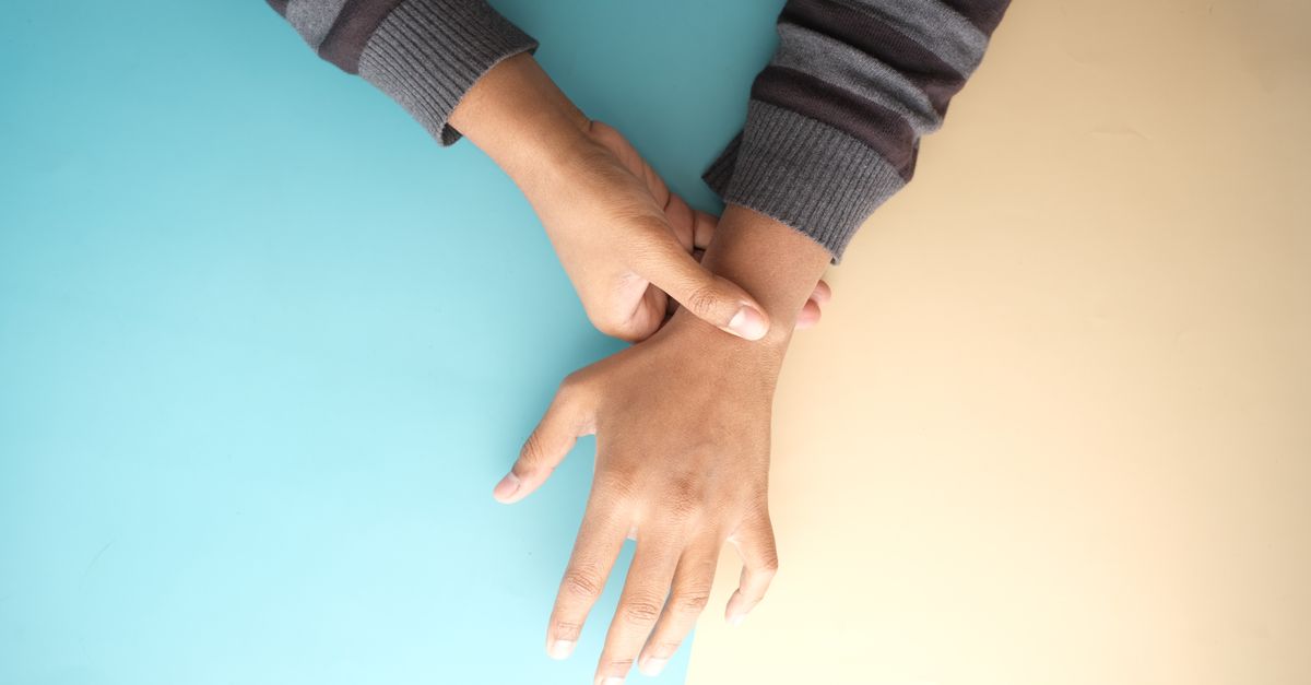
Wrist region pain arises from structural damage or inflammation within the complex network of bones, ligaments, tendons, and nerves connecting the hand to the forearm. The most frequent causes include tendinopathy, median nerve compression, and acute ligamentous sprains. Initial management focuses on absolute rest, rigid splinting, and the application of anti-inflammatory modalities to reduce local tissue edema.
The wrist is not a single joint, but a highly intricate mechanical transition zone. It facilitates complex multi-directional movements necessary for fine motor tasks and forceful grasping. Because of its constant use in daily activities, it is uniquely susceptible to both acute traumatic injuries and chronic repetitive strain.
Accurate diagnosis requires isolating the specific anatomical structure causing the discomfort. Differentiating between neurological compression, joint degeneration, and soft tissue inflammation is vital. Implementing the correct therapeutic strategy early prevents chronic stiffness and long-term functional impairment.
2. Anatomy of the Wrist Complex
The structural foundation of the wrist involves the distal radius and ulna of the forearm articulating with eight small carpal bones. These carpal bones are arranged in two distinct rows, forming a flexible arch. Intricate intrinsic and extrinsic ligaments hold these bones tightly together, allowing synchronized movement.
Multiple tendons cross the wrist joint to control the fingers and thumb. These tendons are encased in synovial sheaths that secrete lubricating fluid, allowing them to glide smoothly under restrictive ligamentous bands known as retinacula. Any inflammation in this crowded space restricts tendon excursion.
Major neurological structures, including the median, ulnar, and radial nerves, traverse the wrist to supply sensory and motor function to the hand. The median nerve, in particular, passes through a tight, rigid anatomical tunnel created by the carpal bones and the transverse carpal ligament, making it highly vulnerable to compression.
3. Pathophysiology of Wrist Discomfort
Pain in the wrist predominantly stems from mechanical overload or acute structural failure. In repetitive strain injuries, constant microtrauma outpaces the body’s natural tissue repair mechanisms. This leads to the breakdown of collagen fibers within tendons and ligaments, sparking a localized inflammatory response.
When synovial sheaths become inflamed, a condition known as tenosynovitis develops. The inflamed tissue swells, increasing friction during tendon gliding. This vicious cycle of friction and swelling produces the characteristic burning pain and crepitus associated with overuse syndromes.
Neuropathic pain develops through ischemia. When swelling increases pressure within confined anatomical spaces, such as the carpal tunnel, the microvascular blood supply to the entrapped nerve is compromised. Ischemic nerves misfire, transmitting signals perceived as profound pain, numbness, and tingling.
4. De Quervain Tenosynovitis
De Quervain tenosynovitis is a painful inflammation affecting the tendons on the thumb side of the wrist. Specifically, it involves the extensor pollicis brevis and the abductor pollicis longus tendons as they pass beneath the extensor retinaculum. It is classically associated with repetitive thumb abduction and grasping.
Patients present with sharp, localized pain over the radial styloid process, which often radiates up the forearm. The pain is severely aggravated by forming a fist, grasping objects, or twisting the wrist. Swelling is frequently palpable, and a distinct catching sensation may be felt during thumb movement.
This condition is highly prevalent in new mothers, often triggered by the repetitive, awkward lifting of an infant. Ergonomic modifications, rigid thumb-spica splinting, and targeted corticosteroid injections are highly effective at resolving the acute inflammatory phase.
5. Carpal Tunnel Syndrome
Carpal tunnel syndrome is the most common compressive neuropathy of the upper extremity. It occurs when the median nerve is compressed within the rigid carpal tunnel at the base of the palm. The compression is typically secondary to hypertrophy of the adjacent flexor tendon synovium.
The clinical presentation involves a distinct pattern of sensory changes. Patients experience numbness, tingling, and deep aching pain in the thumb, index, and middle fingers. Symptoms are classically worse at night, frequently awakening the patient from sleep. Shaking the hands temporarily relieves the ischemic discomfort.
As the condition progresses, motor deficits become apparent. Patients report dropping objects due to decreased grip strength. In advanced, neglected cases, visible atrophy of the thenar eminence muscles at the base of the thumb occurs, signifying severe, irreversible nerve damage.
6. Triangular Fibrocartilage Complex Tears
The triangular fibrocartilage complex is a critical stabilizing structure located on the ulnar side of the wrist, functioning similarly to the meniscus of the knee. It cushions the impact between the distal ulna and the carpal bones and provides primary stability for forearm rotation.
Tears of this complex result from acute twisting injuries under heavy load, or from progressive degenerative wear. Patients report deep, achy ulnar-sided wrist pain that dramatically worsens during forceful forearm rotation, such as turning a doorknob or using a screwdriver.
Physical examination often reveals localized tenderness just distal to the ulnar styloid. A painful click or snap may be felt during end-range rotation. Because the central portion of the complex lacks a blood supply, tears in this region heal poorly and frequently require arthroscopic surgical debridement.
| Condition | Primary Location of Pain |
|---|---|
| De Quervain Tenosynovitis | Radial side of wrist, near the base of the thumb. |
| Carpal Tunnel Syndrome | Volar wrist, with radiation to the thumb, index, and middle fingers. |
| TFCC Tear | Ulnar side of wrist, worsened by twisting motions. |
| Wrist Osteoarthritis | Deep, diffuse joint pain, often involving the base of the thumb. |
| Ganglion Cyst | Localized, prominent swelling, often on the dorsal aspect of the wrist. |
7. Osteoarthritis and Rheumatoid Arthritis
Osteoarthritis of the wrist often develops secondary to previous trauma, such as an old scaphoid fracture or chronic ligamentous instability. The progressive loss of articular cartilage leads to painful bone-on-bone articulation. The most common site for wrist osteoarthritis is the basal joint of the thumb.
Rheumatoid arthritis is a systemic autoimmune disease that aggressively targets the synovial lining of joints. The wrist is frequently the first area affected, with symmetric, bilateral involvement. The profound inflammation destroys cartilage and weakens supporting ligaments, leading to severe pain and characteristic joint deformities.
Differentiating between these two forms of arthritis is critical. Osteoarthritis causes pain that worsens with activity and improves with rest. Rheumatoid arthritis typically presents with prolonged morning stiffness, swollen, boggy joints, and systemic symptoms of fatigue.
8. Ganglion Cysts
Ganglion cysts are common, benign fluid-filled masses that develop near joint capsules or tendon sheaths. The dorsal aspect of the wrist is the most frequent location. These cysts are formed by a herniation of synovial tissue, creating a one-way valve that traps joint fluid.
While many ganglion cysts are asymptomatic and fluctuate in size, they can induce pain if they mechanically compress adjacent sensory nerves or restrict joint motion. Patients often notice a smooth, rounded bump that becomes firmer and more painful following periods of heavy wrist activity.
Conservative management involves observation, as many cysts resolve spontaneously. If symptomatic, aspiration of the fluid using a large-bore needle provides temporary relief, though recurrence rates are high. Surgical excision of the cyst and its capsular stalk is required for permanent resolution.
9. Diagnostic Pathways and Provocative Testing
Clinical examination relies heavily on specific provocative maneuvers to isolate the pain source. The Finkelstein test diagnoses De Quervain tenosynovitis; the clinician asks the patient to enclose their thumb within their fist and rapidly deviates the wrist toward the ulna, eliciting sharp pain.
For carpal tunnel syndrome, the Phalen test involves holding the wrists in maximum flexion for sixty seconds to provoke numbness in the median nerve distribution. The Tinel sign is elicited by lightly tapping over the median nerve at the volar wrist, which produces an electric shock sensation in the fingers if the nerve is irritated.
Palpation of the “anatomical snuffbox” is a critical safety step following acute wrist trauma. Tenderness in this exact location strongly suggests a fracture of the scaphoid bone. Scaphoid fractures are notorious for failing to heal properly due to their tenuous blood supply, making early detection vital.
10. Diagnostic Imaging and Neurodiagnostics
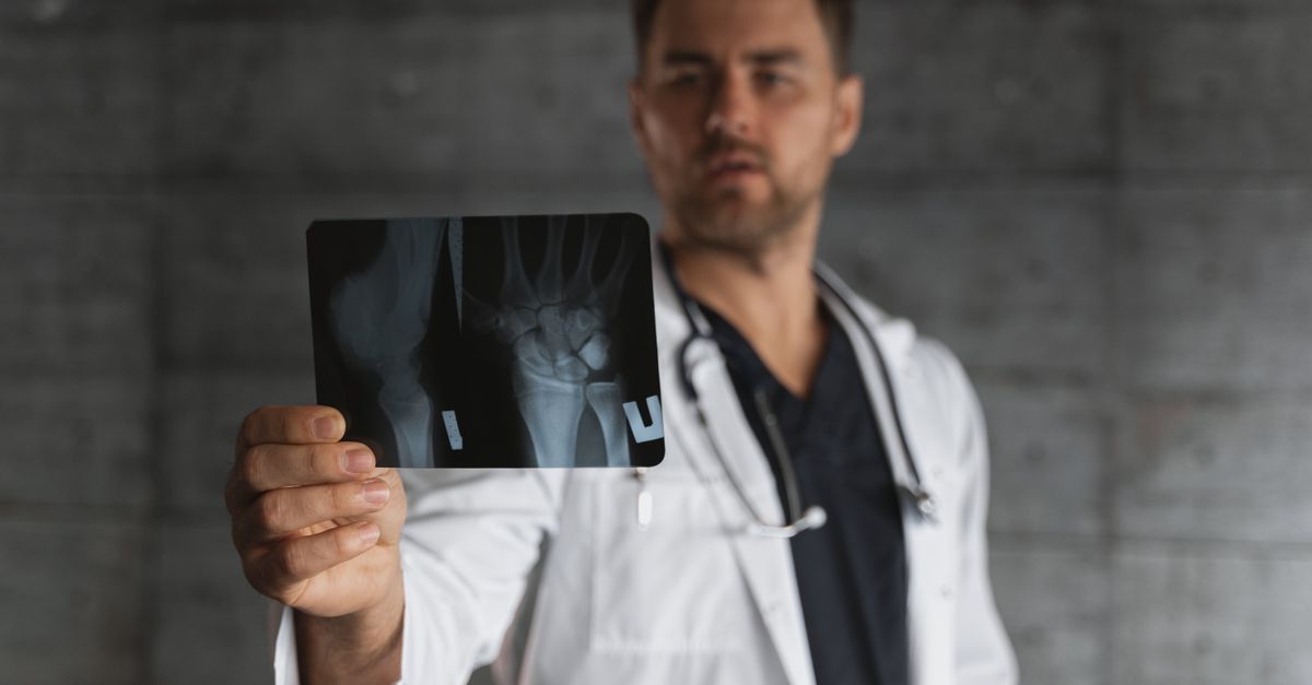
Radiographic evaluation is the first step in assessing structural integrity. High-quality X-rays evaluate joint spacing, detect osteoarthritic changes, and rule out fractures. In cases of suspected subtle fractures or complex carpal instability patterns, a computed tomography scan offers precise bony detail.
Magnetic resonance imaging is superior for evaluating soft tissue pathology. It clearly visualizes tendon tears, ligamentous ruptures, occult bone bruises, and the integrity of the triangular fibrocartilage complex. MRI provides a definitive roadmap when surgical intervention is being considered.
Electromyography and nerve conduction studies are the gold standard for diagnosing compressive neuropathies. These tests measure the speed and amplitude of electrical signals traveling through the peripheral nerves. They quantify the exact severity of nerve damage, guiding the decision between continued splinting or surgical decompression.
11. Differential Diagnosis
Neck pathology must always be considered when evaluating wrist and hand pain. Cervical radiculopathy, caused by a herniated disc in the neck, can compress a nerve root and send shooting pain, numbness, and tingling down the arm into the wrist. This mimics distal nerve entrapment syndromes.
Peripheral neuropathy, frequently a complication of diabetes mellitus, causes bilateral, stocking-glove sensory loss. Unlike carpal tunnel syndrome, which affects specific nerve territories, systemic neuropathy affects the entire hand and foot symmetrically and is accompanied by systemic metabolic derangements.
Infectious tenosynovitis is a rare but critical differential. A small puncture wound can introduce bacteria directly into the tendon sheath. This causes rapid, profound swelling, localized heat, and excruciating pain upon any passive extension of the fingers, requiring emergency surgical washout.
12. When to See a Doctor
Medical assessment is necessary if wrist pain is the result of high-velocity trauma, such as a fall onto an outstretched hand, and is accompanied by visible deformity or the inability to move the joint. These signs strongly indicate a displaced fracture or acute dislocation.
Immediate clinical evaluation is required if there are signs of infection. Redness, severe localized heat, rapid swelling, and systemic fever dictate urgent surgical consultation. Infections in the closed anatomical spaces of the hand can rapidly destroy tendons and articular surfaces.
Progressive neurological symptoms warrant prompt investigation. If numbness transitions into a constant loss of sensation, or if noticeable weakness develops in the grip, nerve compression is reaching a critical stage. Delaying treatment in these scenarios leads to permanent muscle atrophy.
13. Conservative Treatment and Ergonomics
Restoring function begins with strict activity modification and mechanical support. Rigid splints immobilize the joint in a neutral anatomical position. This maximizes the volume of the carpal tunnel and relieves tension on inflamed tendons. Splinting is particularly critical at night when unconscious wrist flexion exacerbates symptoms.
Ergonomic adjustments in the workplace form a foundational pillar of therapy. Ensuring proper keyboard height, utilizing ergonomic mouse designs, and taking frequent micro-breaks to stretch the flexor tendons significantly reduce daily biomechanical stress. Postural correction of the shoulders and neck also improves distal arm biomechanics.
Cryotherapy is highly beneficial during acute flare-ups. Cold therapy reduces vasodilation and local tissue edema. Once the acute inflammatory phase has passed, alternating heat and cold therapy improves local blood flow and relaxes surrounding muscle tension.
14. Physical Therapy and Injection Therapies
Hand therapy, conducted by specialized occupational therapists, focuses on restoring pain-free mobility. Tendon gliding exercises systematically move the flexor tendons through their full anatomical range, preventing the formation of restrictive scar tissue within the synovial sheaths.
Strengthening exercises are introduced gradually once inflammation subsides. Isometric contractions build muscle endurance without placing shear stress on the healing joint surfaces. Proprioceptive training improves the neuromuscular control of the wrist stabilizers, preventing recurrent microtrauma.
When conservative measures plateau, localized corticosteroid injections provide potent anti-inflammatory relief. Delivered directly into the carpal tunnel or the affected tendon sheath under ultrasound guidance, these injections rapidly suppress the immune response, shrinking swollen tissues and relieving mechanical pressure.
15. Surgical Interventions
Surgical management is indicated for structural failures or neuropathies refractory to conservative care. Carpal tunnel release involves surgically severing the transverse carpal ligament. This permanently increases the volume of the tunnel, providing immediate and profound relief of ischemic nerve pressure.
Tendon repairs or releases are performed for advanced tenosynovitis or traumatic ruptures. For De Quervain tenosynovitis, releasing the restrictive extensor retinaculum allows the trapped tendons to glide freely. For complex ligament tears, arthroscopic repair utilizes tiny anchors to reattach torn tissues to the bone.
In advanced osteoarthritis or severe rheumatoid joint destruction, salvage procedures are necessary. Proximal row carpectomy or partial wrist fusions maintain a functional arc of motion while eliminating painful bone-on-bone contact. Total wrist arthrodesis is the final option, completely eliminating motion to guarantee structural stability and pain relief.
16. Frequently Asked Questions (FAQ)
1. Can typing cause carpal tunnel syndrome?
While typing does not directly cause the anatomical structural changes of carpal tunnel syndrome, poor wrist positioning during prolonged computer use significantly exacerbates the pressure within the tunnel, triggering severe symptoms in susceptible individuals.
2. Why is my wrist pain worse at night?
During sleep, most people naturally curl their hands inward, adopting a flexed wrist posture. This position dramatically increases the pressure within the carpal tunnel and stretches inflamed dorsal tendons, leading to painful awakenings.
3. Will a wrist ganglion cyst go away on its own?
Yes, approximately half of all ganglion cysts resolve spontaneously without medical intervention. If a cyst is painless and does not restrict motion, simple observation is the standard clinical recommendation.
4. How can I tell if my wrist is sprained or broken?
While severe sprains and fractures both present with pain, swelling, and bruising, a fracture often involves deeper, sharper pain specifically upon bone loading. An X-ray is the only definitive method to distinguish a severe sprain from a non-displaced fracture.
5. What is the recovery time for De Quervain tenosynovitis?
With strict splinting and activity modification, mild cases resolve in four to six weeks. If a corticosteroid injection is administered, profound relief is often experienced within a few days, though complete tissue healing still requires several weeks of protected use.
17. Bibliography
Disclaimer: The content is for informational purposes only and does not replace medical advice. Always consult your doctor for personalized treatment.