1. Introduction
Pain provoked by exertion is a critical clinical symptom indicating that the body’s demand for oxygen or mechanical stability is exceeding its current physiological capacity. The most frequent causes include cardiovascular ischemia, peripheral arterial insufficiency, and musculoskeletal strain. Treatment is strictly dictated by the specific underlying etiology, ranging from rest and physical therapy to pharmacological intervention and surgical revascularization.
When physical activity initiates discomfort that reliably resolves upon resting, medical professionals immediately investigate potential vascular blockages or structural mechanical limits. The predictability of the pain the fact that it occurs after a specific distance walked or a certain number of stairs climbed is a hallmark diagnostic feature. Identifying the exact anatomical region affected provides the first major clue to the diagnosis.
Understanding the mechanisms behind exertional pain is essential for preventing severe complications. While skeletal muscle fatigue is a normal response to heavy exercise, sharp, restrictive, or crushing pain is a pathological warning sign. A comprehensive diagnostic workup ensures that potentially life-threatening cardiovascular conditions are not mistaken for benign muscular aches.
2. Physiology of Physical Exertion
During physical exertion, the metabolic demands of the working musculature increase exponentially. To meet this demand, the heart rate accelerates, and blood vessels supplying the active muscles dilate. This physiological adaptation ensures a steady delivery of oxygen and nutrients, alongside the rapid removal of metabolic byproducts like lactic acid.
If the arterial system is compromised, this increased blood flow cannot be achieved. The working tissues quickly deplete their available oxygen, leading to a state of localized ischemia. In the absence of oxygen, cells shift to anaerobic metabolism, which rapidly produces acidic byproducts. The accumulation of these byproducts stimulates local nociceptors, creating the sensation of ischemic pain.
Beyond metabolic demands, exertion places significant mechanical stress on the musculoskeletal system. Tendons stretch, joints bear increased compressive loads, and muscle fibers endure micro-tears. If these structures are already compromised by inflammation or degeneration, the mechanical loading of exercise will immediately provoke localized nociceptive signaling.
3. Cardiovascular Etiology: Stable Angina Pectoris
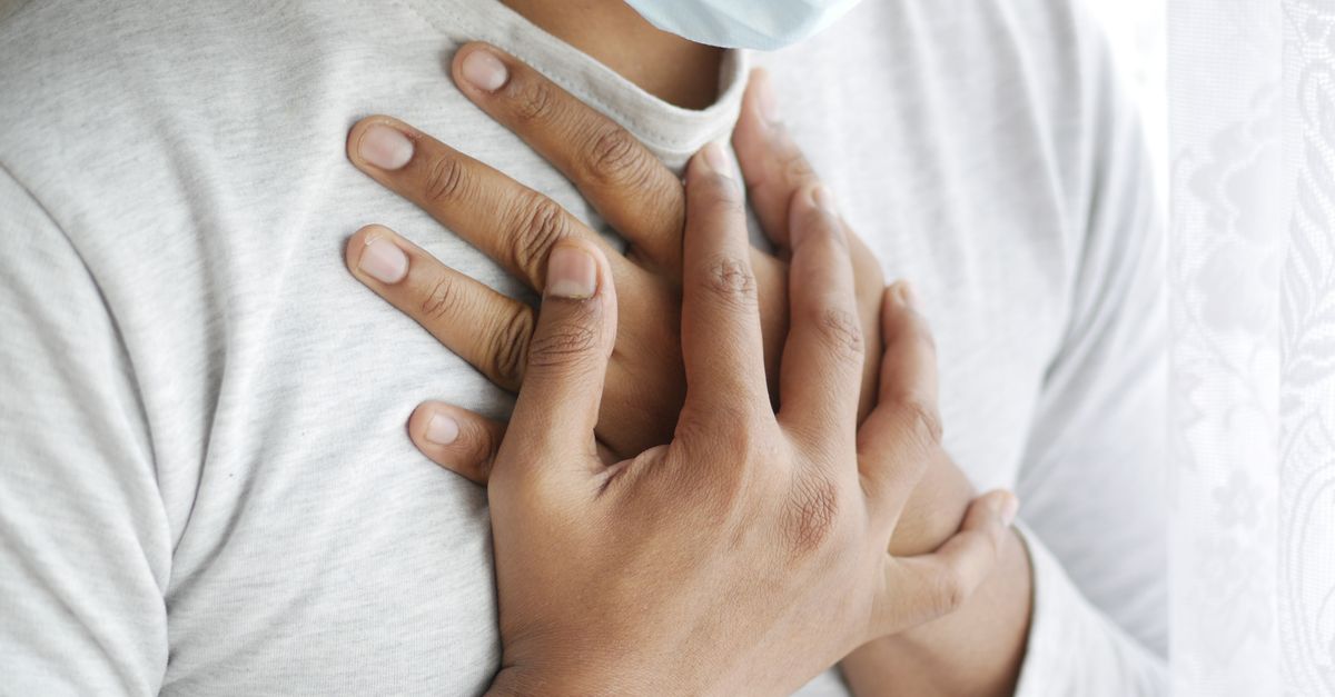
Stable angina pectoris is the classic manifestation of cardiovascular exertional pain. It occurs when atherosclerotic plaques narrow the coronary arteries, restricting blood flow to the heart muscle. At rest, the narrowed vessels provide adequate oxygen. However, during exertion, the myocardial oxygen demand outpaces the restricted supply, inducing cardiac ischemia.
Patients typically describe angina as a heavy, squeezing, or crushing pressure deep within the chest. This discomfort frequently radiates to the left arm, the jaw, or the upper back. The pain is highly predictable, triggered by specific levels of physical activity, and dependably subsides within minutes of resting or upon the administration of sublingual nitroglycerin.
Recognizing stable angina is crucial for preventing progression to an acute myocardial infarction. The ischemic pain serves as a critical warning that the coronary circulation is dangerously compromised. Early cardiological evaluation and risk factor modification can stabilize the plaque and improve myocardial perfusion.
4. Peripheral Vascular Etiology: Intermittent Claudication
Intermittent claudication is the peripheral equivalent of angina, typically affecting the lower extremities. It is a hallmark symptom of peripheral arterial disease, where atherosclerotic narrowing affects the arteries of the legs, such as the femoral or popliteal arteries. The calf muscles are most frequently involved due to their high metabolic demand during ambulation.
The pain is classically described as a deep, severe cramping or aching in the calf, thigh, or buttock that begins after walking a specific distance. This distance, known as the claudication distance, remains relatively constant for the patient. Crucially, the cramping completely resolves after a few minutes of standing still, allowing the patient to resume walking until the cycle repeats.
As peripheral arterial disease progresses, the claudication distance shortens. In severe stages, the ischemia becomes so profound that pain occurs even at rest, particularly at night when cardiac output naturally decreases. This stage, critical limb ischemia, represents a significant threat to tissue viability.
5. Musculoskeletal Causes: Strains and Tendinopathies
Musculoskeletal structures frequently generate pain strictly during physical exertion. A muscle strain occurs when muscle fibers are stretched beyond their mechanical limits, leading to partial tearing. During rest, the injured muscle remains relatively silent, but any active contraction or passive stretching during movement instantly provokes sharp, localized pain.
Tendinopathy involves the chronic degeneration and inflammation of a tendon, typically due to repetitive mechanical overload. Conditions such as Achilles tendinopathy or patellar tendinopathy present with pain that is remarkably specific to exertion. The tendon may feel stiff initially, warm up and feel better during light activity, but ache severely following strenuous loading.
Joint pathology, particularly osteoarthritis, also presents with exertion-provoked pain. As the protective cartilage degrades, the increased compressive forces of walking or running cause painful bone-on-bone friction. This mechanical joint pain is distinctly different from ischemic cramping, often presenting with associated joint swelling and crepitus.
| System Involved | Pathological Condition | Characteristics of Exertional Pain |
|---|---|---|
| Cardiovascular | Stable Angina | Crushing chest pressure, radiates to jaw/arm, relieved by rest. |
| Vascular | Intermittent Claudication | Calf or thigh cramping after a set walking distance. |
| Neurological | Spinal Stenosis | Leg heaviness and pain, relieved by leaning forward. |
| Musculoskeletal | Tendinopathy | Localized tendon pain that worsens with mechanical loading. |
6. Respiratory Causes: Exercise-Induced Asthma
The respiratory system can indirectly cause exertion-related discomfort. Exercise-induced bronchoconstriction, commonly known as exercise-induced asthma, occurs when the rapid inhalation of cool, dry air during physical exertion triggers the smooth muscles of the airways to spasm and contract.
Patients experience a tight, restrictive sensation in the chest, accompanied by a noticeable wheeze, shortness of breath, and a dry cough. This respiratory distress typically peaks a few minutes after the exertion ceases. The sensation is often described as chest tightness rather than the crushing pain of cardiac angina, though the two can be easily confused.
Management involves using inhaled bronchodilators shortly before initiating physical activity. By preemptively relaxing the airway smooth muscles, patients can safely engage in vigorous exercise without experiencing restrictive chest discomfort or oxygen deprivation.
7. Neurological Claudication and Spinal Stenosis
Neurogenic claudication is a symptom complex caused by lumbar spinal stenosis, which is the anatomical narrowing of the spinal canal. This condition compresses the descending nerve roots of the cauda equina. While it mimics the leg pain of peripheral vascular disease, the underlying mechanism is neurological compression rather than arterial ischemia.
Patients with neurogenic claudication experience bilateral leg pain, heaviness, and numbness that develops during walking or prolonged standing. However, unlike vascular claudication, simply stopping and standing still does not relieve the pain. The patient must flex their spine forward, such as sitting down or leaning over a shopping cart, to open the spinal canal and decompress the nerves.
This mechanical distinction is the primary diagnostic differentiator. A thorough clinical history focusing on exactly what posture relieves the exertional pain rapidly steers the clinician toward either a vascular or a neurological diagnostic pathway.
8. Metabolic and Myopathic Origins
Rarely, exertional pain is the primary symptom of an underlying metabolic myopathy. These genetic disorders impair the muscle cells’ ability to produce or utilize energy. McArdle disease, a glycogen storage disorder, prevents the breakdown of glycogen into glucose within the muscle, severely limiting anaerobic energy production.
Patients with metabolic myopathies experience profound muscle fatigue, severe cramping, and pain within minutes of initiating strenuous exercise. A hallmark of some metabolic myopathies is the “second wind” phenomenon, where a brief rest allows the body to switch to alternative energy pathways, subsequently permitting exercise to continue with less pain.
In severe cases, pushing through this metabolic pain can lead to rhabdomyolysis. This is a dangerous condition where starved muscle fibers rapidly break down, releasing large amounts of myoglobin into the bloodstream, which can precipitate acute kidney failure.
9. Differentiating Ischemic from Mechanical Pain
The fundamental step in evaluating exertional pain is distinguishing ischemic origins from mechanical ones. Ischemic pain, whether cardiac or peripheral, is deeply aching, cramping, or crushing. It is highly predictable based on the metabolic workload and rapidly resolves when the workload is removed. It is rarely affected by palpation or specific body movements.
Mechanical pain, stemming from muscles, tendons, or joints, is usually sharp, localized, and highly dependent on biomechanical positioning. It can often be reproduced in the clinic by applying direct pressure to the affected tissue or by moving the joint through a specific range of motion against resistance.
For example, chest pain that is provoked by pressing on the sternum or by taking a deep breath is highly indicative of musculoskeletal costochondritis, whereas true angina pectoris is unaffected by local pressure or respiratory mechanics.
10. Clinical Assessment and Patient History
The clinical assessment of exertional pain requires a meticulous history. The physician must determine the exact onset, duration, and character of the pain. Identifying the specific threshold of exertion that triggers the discomfort such as walking exactly two blocks or climbing one flight of stairs establishes the severity of the physiological limitation.
The medical history evaluates systemic risk factors. A history of smoking, hypertension, diabetes mellitus, or hyperlipidemia drastically increases the pre-test probability of atherosclerotic cardiovascular or peripheral arterial disease. Conversely, a history of recent heavy lifting or a change in sports training points toward musculoskeletal strain.
Physical examination focuses on the affected system. The clinician auscultates the heart and lungs, palpates peripheral pulses in the extremities, and evaluates the neurological function of the limbs. Diminished pedal pulses or the presence of an arterial bruit strongly suggest a vascular etiology.
11. Diagnostic Testing and Stress Tests
When cardiovascular or peripheral ischemic pain is suspected, stress testing is the diagnostic gold standard. An electrocardiogram exercise stress test monitors the heart’s electrical activity while the patient walks on a treadmill. Ischemic changes on the ECG during exertion provide definitive evidence of coronary artery disease.
For peripheral arterial disease, the ankle-brachial index is measured before and immediately after exercise. A significant drop in blood pressure at the ankle following exertion confirms that arterial narrowing is restricting distal blood flow when metabolic demand increases.
If neurological or musculoskeletal causes are suspected, diagnostic imaging is employed. Magnetic resonance imaging provides exquisite detail of the lumbar spine to confirm spinal stenosis or high-resolution imaging of specific tendons and joints to identify structural mechanical failure.
12. Differential Diagnosis
The differential diagnosis for exertional pain is extensive due to the variety of systems stressed during exercise. Exertional compartment syndrome must be differentiated from vascular claudication. In this condition, the muscle swells during exercise within a rigid fascial compartment, restricting its own microvascular blood supply and causing severe tightness and pain that resolves slowly after stopping.
Referred pain from intra-abdominal organs can occasionally manifest during exertion. A hiatal hernia or gastroesophageal reflux disease can cause burning chest pain during heavy exertion, particularly if the exercise involves bending or increased intra-abdominal pressure. This mimics angina but typically responds to antacid therapy.
Venous claudication, caused by deep vein thrombosis or chronic venous insufficiency, presents with a bursting, heavy pain in the leg during walking. Unlike arterial claudication, venous pain is often accompanied by significant leg swelling and is relieved more effectively by elevating the leg rather than simply resting.
13. When to See a Doctor
Any new onset of pain provoked by exertion requires prompt medical evaluation to rule out life-threatening ischemia. Pain in the chest, jaw, or left arm that reliably occurs during physical activity and stops with rest is a classic presentation of angina and demands an urgent cardiological workup.
Immediate emergency care is required if exertional chest pain does not resolve after resting. If the crushing pressure persists for more than a few minutes after stopping the activity, it strongly suggests that the ischemic event has transitioned into an acute myocardial infarction.
Patients should also seek specialized care if leg pain during walking is accompanied by cold, pale, or blue toes. These are signs of critical limb ischemia, indicating that the baseline blood flow is dangerously low and tissue death is imminent without rapid vascular intervention.
14. Pharmacological Interventions
Pharmacological therapy targets the underlying systemic disease. For stable angina, medications such as beta-blockers decrease the heart rate and contractility, thereby reducing the myocardial oxygen demand. Nitrates are used to rapidly vasodilate the coronary arteries, acutely relieving ischemic pain.
Management of peripheral arterial disease involves antiplatelet agents, such as aspirin or clopidogrel, to prevent clot formation on atherosclerotic plaques. Medications like cilostazol directly improve walking distance by promoting vasodilation and preventing platelet aggregation within the microcirculation of the legs.
For musculoskeletal pain, nonsteroidal anti-inflammatory drugs are utilized to blunt the acute inflammatory response following mechanical injury. However, masking exertional pain with analgesics to continue high-level exercise is strongly discouraged, as it rapidly leads to progressive tissue tearing and structural failure.
15. Physical Therapy and Rehabilitation
Supervised exercise therapy is a paradoxically effective treatment for intermittent vascular claudication. Patients are guided to walk on a treadmill until moderate pain develops, rest until it resolves, and repeat the cycle. Over several months, this ischemic preconditioning stimulates angiogenesis, the formation of new collateral blood vessels that bypass the arterial blockages.
For musculoskeletal etiologies, physical therapy focuses on biomechanical correction. Therapists design targeted strengthening programs to correct muscular imbalances and improve load distribution across injured tendons and joints. Eccentric strengthening protocols are specifically utilized to remodel chronically degenerated tendons.
In cases of neurogenic claudication, physical therapy focuses on lumbar flexion exercises and core stabilization. By strengthening the abdominal musculature and stretching the hip flexors, the resting posture of the pelvis is altered, which helps to slightly open the narrowed spinal canal and reduce nerve compression during ambulation.
16. Surgical and Interventional Options
When severe arterial blockages fail to respond to medication and exercise, revascularization is necessary. Percutaneous transluminal angioplasty involves threading a balloon catheter to the site of the narrowing, inflating it to compress the plaque, and often leaving a metallic stent to hold the artery open. This procedure is widely used for both coronary and peripheral arteries.
If the blockages are extensive or heavily calcified, surgical bypass grafting is performed. A healthy vein from the patient’s leg or a synthetic conduit is surgically attached to detour blood flow around the occluded arterial segment, restoring distal perfusion and eliminating exertional ischemia.
For structural mechanical failures, surgical options include tendon repairs, joint arthroplasty for severe osteoarthritis, or surgical decompression. In lumbar spinal stenosis, a laminectomy removes the posterior portion of the vertebra to permanently expand the spinal canal, definitively relieving the exertional neurological compression.
17. Frequently Asked Questions (FAQ)
1. Why does my chest only hurt when I walk up hills?
Walking uphill requires significantly more energy than walking on a flat surface. This drastically increases your heart’s oxygen demand. If a coronary artery is partially blocked, it cannot deliver the extra blood required, leading to temporary cardiac ischemia and chest pain.
2. Can dehydration cause muscle pain during exercise?
Yes. Severe dehydration alters the electrolyte balance within the muscle tissue. This disruption impairs normal muscle contraction and relaxation cycles, frequently leading to intense, painful muscle cramps during exertion.
3. Is it safe to push through the pain of intermittent claudication?
In a supervised medical exercise program, walking into moderate pain is encouraged to stimulate new blood vessel growth. However, you should never push through severe, agonizing pain, as this can cause localized muscle damage.
4. How is neurogenic claudication different from a herniated disc?
A herniated disc typically causes constant, sharp, shooting nerve pain down one leg, even at rest. Neurogenic claudication causes heavy, cramping pain in both legs that specifically occurs only after prolonged walking or standing.
5. What is the fastest way to relieve angina pain?
Stopping all physical exertion immediately reduces the heart’s workload. If prescribed, taking sublingual nitroglycerin rapidly dilates the blood vessels, restoring blood flow and relieving the ischemic chest pressure within minutes.
18. Bibliography
Disclaimer: The content is for informational purposes only and does not replace medical advice. Always consult your doctor for personalized treatment.