1. Introduction
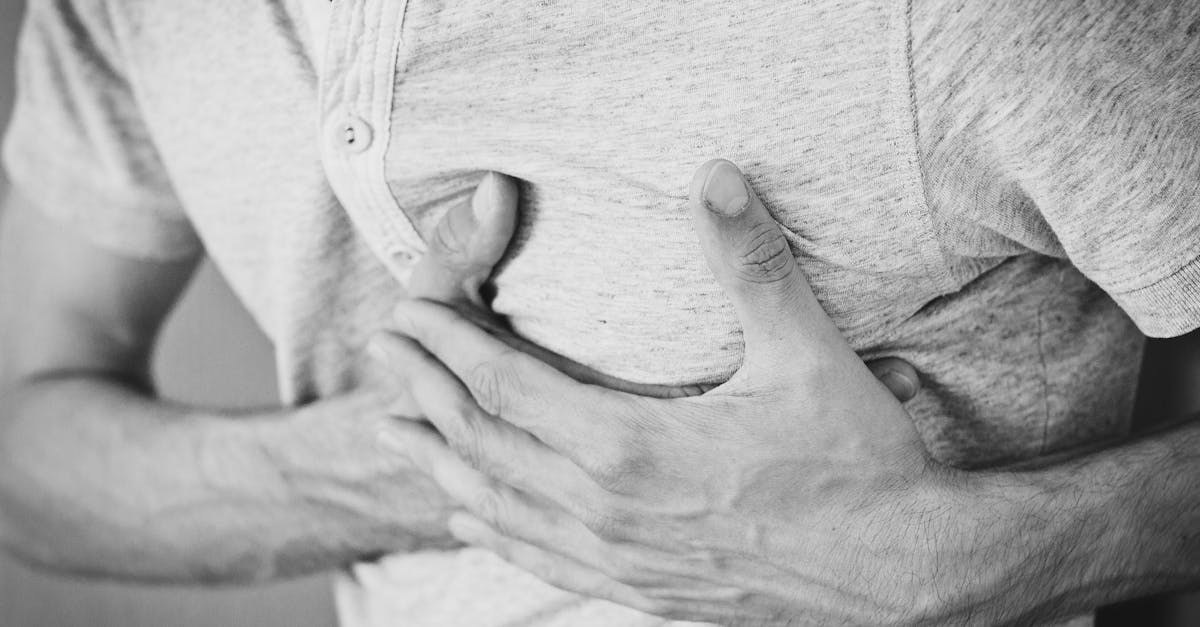
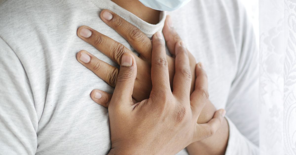
The sudden onset of a heavy, squeezing pressure in the center of the chest is a formidable medical symptom that mandates immediate clinical suspicion of acute cardiac ischemia. This specific sensation—often described by patients as an elephant sitting on their chest or a tight band constricting their ribs—is the classic hallmark of angina pectoris. It occurs when the heart muscle (myocardium) does not receive enough oxygen-rich blood to meet its immediate metabolic demands, typically due to a mechanical obstruction in the coronary arteries.
The chest cavity houses several vital organs, including the heart, lungs, esophagus, and major blood vessels. Because these organs share overlapping sensory nerve pathways, the brain often struggles to pinpoint the exact source of deep visceral pain. Therefore, a heavy pressure sensation can originate not only from the heart but also from a pulmonary embolism, a severe esophageal spasm, or an acute aortic dissection.
Given the potentially fatal consequences of cardiac and vascular emergencies, clinical protocols dictate treating sudden, heavy chest pressure as acute coronary syndrome until proven otherwise through objective diagnostic testing. Differentiating between a life-threatening myocardial infarction and a benign gastrointestinal or musculoskeletal etiology is the absolute priority of emergency medical triage.
2. Pathophysiology of Myocardial Ischemia
The heart is a highly aerobic muscle that requires a continuous, uninterrupted supply of oxygen to function. This oxygen is delivered via the coronary arteries, which branch off the base of the aorta and encircle the heart muscle. If the demand for oxygen exceeds the supply, the myocardial cells enter a state of ischemia.
During ischemia, the lack of oxygen forces the heart muscle cells to switch from efficient aerobic metabolism to inefficient anaerobic metabolism. This rapidly produces lactic acid and other metabolic byproducts. These chemicals accumulate in the local tissue and directly stimulate the visceral sensory nerve endings located within the myocardium.
These activated nerve fibers transmit distress signals to the central nervous system. However, visceral pain is notoriously diffuse and poorly localized. The brain interprets this distress not as a sharp, specific pain, but rather as a profound, heavy ache, tightness, or burning pressure deep within the retrosternal area (behind the breastbone).
3. Coronary Artery Disease and Angina Pectoris
The most common structural cause of myocardial ischemia is coronary artery disease. Over decades, cholesterol, inflammatory cells, and fibrous tissue accumulate within the walls of the coronary arteries, forming atherosclerotic plaques. These plaques physically narrow the lumen of the artery, restricting the maximum potential blood flow.
Stable angina occurs when this narrowing is significant but predictable. The patient may feel chest pressure only during physical exertion or acute emotional stress—situations where the heart beats faster and demands more oxygen. When the patient rests, the oxygen demand drops, the available blood flow is once again sufficient, and the pressure resolves within a few minutes.
Unstable angina, however, is a medical emergency. It occurs when a previously stable atherosclerotic plaque suddenly ruptures or erodes. The body’s clotting system immediately rushes to heal the ruptured plaque, forming a blood clot (thrombus) that rapidly obstructs the artery. Unstable angina presents as severe chest pressure that occurs at rest, lasts longer than typical angina, and is not relieved by rest or nitroglycerin.
4. Acute Coronary Syndromes (Heart Attack)
If the blood clot completely occludes the coronary artery, cutting off all blood flow to a segment of the heart muscle, an acute myocardial infarction (heart attack) occurs. Without oxygen, the affected myocardial cells begin to die (necrosis) within minutes to hours.
The chest pressure experienced during a myocardial infarction is typically severe, unrelenting, and often radiates to adjacent anatomical areas that share nerve pathways with the heart. Classic radiation patterns include the inner left arm, the jaw, the neck, and the interscapular region (between the shoulder blades).
Accompanying autonomic symptoms are highly predictive of a severe cardiac event. Patients frequently present with diaphoresis (profuse, cold sweating), severe nausea, vomiting, and a profound sense of impending doom. These systemic reactions are caused by a massive surge of adrenaline as the body reacts to the acute cardiac crisis.
5. Pulmonary Embolism and Vascular Obstructions
Not all heavy chest pressure originates from the coronary arteries. The pulmonary vasculature can also be the source of life-threatening emergencies. A pulmonary embolism occurs when a blood clot, typically originating from a deep vein thrombosis in the leg, breaks free, travels through the right side of the heart, and becomes lodged in a pulmonary artery.
This sudden blockage drastically increases the pressure in the right ventricle of the heart and prevents proper oxygenation of the blood. Patients experience sudden, severe chest pressure or sharp pain that worsens significantly with a deep breath (pleuritic pain).
Pulmonary embolism is almost universally accompanied by acute dyspnea (sudden shortness of breath) and resting tachycardia. The rapid onset of breathing difficulty alongside central chest pressure requires immediate imaging to visualize the pulmonary vascular tree.
6. Aortic Dissection as a Vascular Emergency
An acute aortic dissection is a catastrophic vascular event involving the aorta, the body’s main artery. It occurs when a tear develops in the innermost layer of the aortic wall (the intima). High-pressure blood surges through the tear, splitting the layers of the aortic wall apart.
While classic textbook descriptions characterize the pain of an aortic dissection as “tearing” or “ripping” radiating to the back, it frequently presents simply as sudden, agonizing, and heavy chest pressure. It is often distinguished by its abrupt onset—reaching maximal intensity immediately, unlike the crescendo nature of a heart attack.
Aortic dissections can compromise blood flow to the coronary arteries, the brain, or the spinal cord, depending on where the tear propagates. Rapid control of blood pressure and emergent surgical intervention are required for survival.
7. Gastroesophageal Reflux and Esophageal Spasm
Because the esophagus sits directly behind the heart and shares the exact same sensory nerve plexus, severe esophageal conditions can perfectly mimic the symptoms of acute angina. Gastroesophageal reflux disease allows highly acidic stomach contents to bathe the lower esophagus, causing severe chemical irritation.
This acid exposure can trigger an esophageal spasm, a condition where the smooth muscles of the esophagus contract violently and erratically. The resulting sensation is an intense, crushing chest pressure that radiates to the back and jaw, virtually indistinguishable from a myocardial infarction based on symptoms alone.
However, esophageal pressure often occurs after consuming large, acidic, or spicy meals, or upon lying flat. It may be accompanied by a sour taste in the mouth or difficulty swallowing (dysphagia). A trial of high-dose antacids or a “GI cocktail” in the emergency room sometimes relieves the pain, helping to differentiate it from cardiac ischemia.
8. Musculoskeletal Causes (Costochondritis)
The chest wall is composed of ribs, cartilage, and heavy musculature. Inflammation of the cartilage connecting the ribs to the sternum, known as costochondritis, is a frequent, benign cause of severe chest pain. It can mimic the pressure of a heart attack, especially if the inflammation is widespread.
Unlike cardiac ischemia, musculoskeletal pain is typically reproducible by mechanical stress. Taking a very deep breath, coughing, or twisting the torso often exacerbates the pain.
Crucially, in costochondritis, a clinician can usually reproduce the exact pain by pressing firmly on the costochondral junctions (where the ribs meet the breastbone). If the heavy pressure sensation can be localized and worsened by physical palpation, the likelihood of a primary cardiac event is significantly lower, though it does not entirely rule it out.
9. Panic Attacks and Psychosomatic Manifestations
The brain and the cardiovascular system are inextricably linked. Severe psychological distress, specifically panic disorder, can induce profound physical symptoms that mimic acute medical emergencies. During a panic attack, the sympathetic nervous system initiates a maximum “fight or flight” response.
Patients experience sudden, heavy chest tightness, a racing heartbeat, shortness of breath, trembling, and a genuine fear of dying. The hyperventilation associated with panic attacks can also cause respiratory alkalosis, leading to numbness and tingling in the extremities, further terrifying the patient.
While panic attacks are benign in terms of physical tissue damage, they are profoundly disabling. It is a critical tenet of emergency medicine that a panic attack is a diagnosis of exclusion. A clinician must never assume chest pressure is psychological without first definitively ruling out cardiac and pulmonary pathology.
10. Table: Differentiating Cardiac vs Non-Cardiac Chest Pain
| Condition | Nature of Sensation | Aggravating / Provocative Factors |
|---|---|---|
| Myocardial Ischemia | Heavy, squeezing, central pressure | Physical exertion, emotional stress |
| Pulmonary Embolism | Pressure with sharp pleuritic component | Taking a deep breath, lying flat |
| Esophageal Spasm | Squeezing, burning retrosternal pressure | Swallowing, lying down after large meals |
| Costochondritis | Aching, sharp pressure over breastbone | Direct palpation, twisting the torso |
11. Immediate Emergency Triage
When a patient presents to an emergency department with sudden chest pressure, triage protocols are initiated instantaneously. The primary goal is to identify a ST-segment Elevation Myocardial Infarction (STEMI), the most severe type of heart attack requiring immediate reopening of the blocked artery.
A twelve-lead electrocardiogram (ECG) is performed and interpreted within ten minutes of arrival. The ECG analyzes the electrical vectors of the heart. If a major coronary artery is completely blocked, specific electrical changes, such as ST-segment elevation or new bundle branch blocks, will be immediately apparent.
Intravenous access is established, and the patient is placed on continuous cardiac monitoring. Oxygen is administered if oxygen saturation is low, and aspirin is typically given immediately to inhibit further platelet aggregation and clot expansion.
12. Electrocardiography and Cardiac Biomarkers
While the ECG is vital, it can appear perfectly normal during unstable angina or a non-STEMI heart attack. Therefore, laboratory testing for cardiac biomarkers is the necessary second step in the diagnostic pathway.
When myocardial cells are damaged or die due to ischemia, they release specific intracellular proteins into the bloodstream. High-sensitivity cardiac troponin is the gold standard biomarker. An elevated troponin level definitively confirms that myocardial necrosis (heart muscle damage) has occurred.
Troponin levels are typically drawn upon arrival and then serially every few hours. Observing the trend of the troponin levels—whether they are rising or falling—helps clinicians determine the precise timing and severity of the ischemic event, guiding subsequent interventional decisions.
13. Advanced Cardiac Imaging
If the initial ECG and troponin markers are inconclusive, or if the patient requires further risk stratification, advanced cardiac imaging is utilized. Echocardiography uses ultrasound to visualize the mechanical pumping action of the heart in real-time. If a segment of the heart muscle is starved of blood, that specific wall will appear sluggish or completely immobile (a regional wall motion abnormality).
Coronary Computed Tomography Angiography (CCTA) is an advanced non-invasive scan that creates highly detailed, three-dimensional images of the coronary arteries. It can visualize the presence and extent of atherosclerotic plaques and rule out life-threatening conditions like aortic dissection or pulmonary embolism simultaneously.
If there is strong evidence of an acute blockage, an invasive coronary angiogram is performed in the cardiac catheterization laboratory. A catheter is threaded from the wrist or groin to the heart, and contrast dye is injected to visualize the exact location of the arterial obstruction under X-ray guidance.
14. Acute Medical and Interventional Management
If a blocked coronary artery is identified, the immediate goal is reperfusion—restoring blood flow to the dying muscle. Percutaneous Coronary Intervention (PCI) is the preferred treatment. A deflated balloon is advanced across the blockage and inflated to crush the plaque against the artery walls. A metallic mesh tube, called a stent, is then deployed to keep the artery permanently open.
Pharmacological management is equally aggressive. Dual antiplatelet therapy, utilizing aspirin alongside drugs like clopidogrel or ticagrelor, is mandatory to prevent blood clots from forming inside the newly placed stent. High-dose statins are initiated to stabilize existing cholesterol plaques and reduce vascular inflammation.
Nitroglycerin may be administered sublingually or intravenously. It is a potent vasodilator that expands the coronary arteries to increase blood flow and expands the peripheral veins to reduce the mechanical workload of the heart, rapidly alleviating the sensation of heavy chest pressure.
15. Frequently Asked Questions (FAQ)
1. How do I know if my chest pressure is a heart attack or just severe anxiety?
You cannot reliably tell the difference at home. Both can cause severe pressure, sweating, and shortness of breath. Because a heart attack is life-threatening, you must seek emergency medical care immediately to have an ECG and blood tests performed.
2. Does chest pressure from a heart attack always hurt?
No. Many patients, particularly women, the elderly, and those with diabetes, may not experience sharp pain. They often describe a heavy, dull ache, extreme tightness, or profound fatigue and shortness of breath.
3. Will taking antacids stop the pressure if it is a heart attack?
No. Antacids only neutralize stomach acid. If antacids relieve the pressure completely within a few minutes, it points toward a gastrointestinal cause. However, relying on this to delay seeking emergency care is highly dangerous.
4. Why did the pressure spread to my jaw and left arm?
The heart and the nerves in your arm and jaw share the same neural pathways traveling to the spinal cord. When the heart is in distress, the brain gets confused and perceives the pain as originating in the arm or jaw, a phenomenon called referred pain.
5. What should I do immediately if I feel this sudden, heavy pressure?
Call emergency services (911) immediately. Do not attempt to drive yourself to the hospital. Chew and swallow a 325 mg aspirin (unless you are allergic or instructed otherwise by a doctor) while waiting for the paramedics to arrive.
16. Bibliography
Disclaimer: The content is for informational purposes only and does not replace medical advice. Always consult your doctor for personalized treatment.