1. Introduction
Prolonged sitting can directly cause severe pain in the tailbone by placing sustained mechanical pressure on the coccyx and its surrounding sacrococcygeal ligaments. This constant structural compression restricts localized blood flow and initiates a profound inflammatory response, leading to a clinical condition diagnosed as coccydynia. When the human body remains seated for extended periods, the natural biomechanics of the pelvis are altered, shifting the primary weight-bearing burden away from the robust ischial tuberosities directly onto the fragile distal segments of the spine.
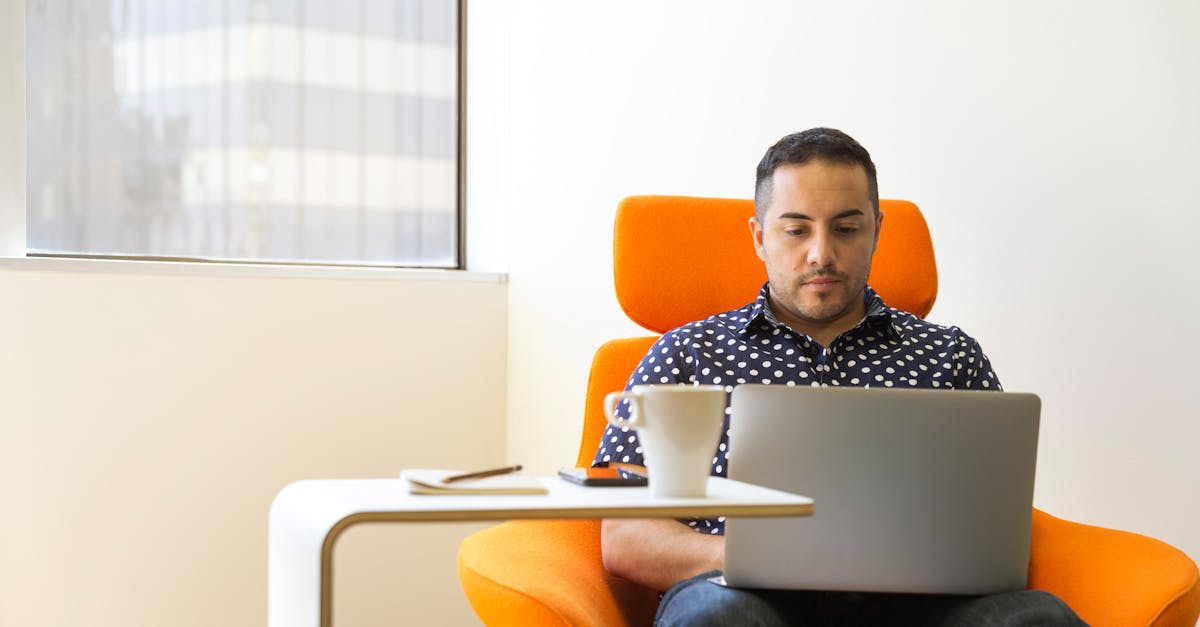
Coccydynia is a frequent and debilitating complaint in modern occupational environments where sedentary behavior is the norm. The pain is typically described as a sharp, localized ache that intensifies immediately upon transitioning from a seated to a standing position. Understanding the precise anatomical and environmental factors driving this discomfort is necessary for effective rehabilitation.
Clinical intervention focuses on removing the mechanical stressor, restoring proper pelvic alignment, and reducing the localized tissue inflammation. Without modifying seating ergonomics and postural habits, the continuous microtrauma outpaces the body’s natural healing capabilities, resulting in chronic, intractable pelvic pain.
2. Anatomy of the Coccyx and Sacrococcygeal Joint
The coccyx, commonly referred to as the tailbone, is the terminal segment of the human vertebral column. It typically consists of three to five fused or semi-fused rudimentary vertebrae. Superiorly, it articulates with the sacrum at the sacrococcygeal joint, a symphysis held together by a dense network of fibrocartilage and strong ligamentous bands.
Despite being a vestigial structure from an evolutionary standpoint, the coccyx serves a vital biomechanical purpose. It functions as a critical insertion point for numerous pelvic floor muscles, including the levator ani and the coccygeus, as well as the robust gluteus maximus.
The sacrococcygeal joint possesses a small degree of natural mobility. During sitting or leaning back, the coccyx flexes slightly forward to act as a functional shock absorber. This subtle movement is essential for dissipating the vertical loads transmitted through the spine.
3. Biomechanics of Sitting Posture
The human pelvis is designed to bear the weight of the seated body on the ischial tuberosities, colloquially known as the sit bones. When an individual sits with a neutral spine and an upright posture, the pelvis rocks forward slightly into an anterior tilt. In this optimal position, the ischial tuberosities support the torso, and the coccyx remains elevated and free from direct surface contact.
However, as sitting time increases, postural fatigue inevitably sets in. The core musculature relaxes, causing the pelvis to rock backward into a posterior tilt. This slouched position fundamentally alters the distribution of mechanical load across the pelvic floor.
As the pelvis tilts posteriorly, the ischial tuberosities roll forward, and the point of maximum contact shifts directly onto the sacrum and the coccyx. The tailbone is forced to bear a substantial percentage of the upper body’s weight, a task for which its delicate structure is entirely unsuited.
4. Mechanism of Mechanical Compression
When the coccyx is subjected to the direct weight of the torso, it is mechanically compressed against the seating surface. This compression forces the sacrococcygeal joint beyond its normal physiological range of flexion. The distal segments of the tailbone are pushed sharply inward toward the pelvic cavity.
This sustained compression has immediate physiological consequences. The soft tissues overlying the coccyx, which possess minimal subcutaneous fat for cushioning, become crushed between the rigid bone and the unyielding chair. This creates a localized area of immense mechanical stress.
Furthermore, the continuous pressure compresses the microvasculature supplying the periosteum of the bone and the surrounding connective tissue. This reduction in blood flow induces tissue hypoxia, preventing the delivery of oxygen and nutrients required to maintain cellular integrity during load-bearing activities.
5. Ligamentous Strain and Instability
The stability of the sacrococcygeal joint depends entirely on the integrity of the sacrococcygeal ligaments. When a posterior pelvic tilt forces the coccyx into extreme flexion for hours at a time, these ligaments are stretched tightly across the joint space.
Ligaments are composed of dense, inelastic collagen fibers designed to resist sudden, forceful movements, not sustained, static stretching. Prolonged elongation leads to a phenomenon known as ligamentous creep, where the fibers slowly deform and lose their structural resilience.
This continuous stretching causes microscopic tears within the ligamentous matrix. The resulting microtrauma triggers a robust inflammatory cascade. The ligaments become swollen and exquisitely tender, producing the deep, localized ache that characterizes early-stage coccydynia.
6. Ischemia and Localized Inflammation
The mechanical crushing of tissues and the tearing of ligaments initiate a profound localized inflammatory response. The body attempts to repair the microtrauma by flooding the area with pro-inflammatory cytokines, prostaglandins, and fluid, resulting in tissue edema.
However, because the patient remains seated, the inflamed tissues continue to be subjected to compressive forces. The ongoing pressure prevents the lymphatic system from effectively draining the inflammatory fluid. The area becomes a stagnant pool of inflammatory mediators, which constantly stimulate the local nociceptors, or pain-sensing nerves.
This localized inflammation makes the periosteum of the coccyx hypersensitive. Even minor mechanical inputs, such as shifting weight in a chair or wearing tight clothing, trigger a disproportionately severe pain signal to the central nervous system.
7. The Role of the Pelvic Floor Musculature
The coccyx serves as an anchor for the pelvic floor musculature. When the tailbone is inflamed and painful, the body initiates a protective reflex known as muscle guarding. The muscles attaching to the coccyx, particularly the levator ani, contract involuntarily in an attempt to splint the joint and prevent further painful movement.
This constant, involuntary isometric contraction leads to severe muscle fatigue and the development of myofascial trigger points within the pelvic floor. The pain radiates outward from the coccyx, frequently causing a deep, cramping sensation in the perineum and the bilateral gluteal regions.
Consequently, patients with chronic coccydynia often develop secondary pelvic floor dysfunction. This can manifest as pain during bowel movements or a deep aching sensation in the pelvis that persists even when the patient is lying down and free from coccygeal compression.
8. Ergonomic Factors and Seating Surfaces
The physical properties of the seating surface interact directly with pelvic anatomy to either mitigate or exacerbate coccygeal pressure.
| Seating Surface Characteristic | Biomechanical Impact | Risk of Coccydynia |
|---|---|---|
| Rigid, Unyielding (Wood, Metal) | Zero pressure redistribution, direct bone impact | Considerable risk, rapid onset of pain |
| Overly Soft, Sagging (Old Couch) | Promotes severe posterior pelvic tilt | Considerable risk, insidious onset of pain |
| Firm with Coccyx Cut-out | Suspends the tailbone, loads the ischial tuberosities | Low risk, therapeutic for active pain |
| Forward-Tilting Ergonomic Chair | Promotes anterior pelvic tilt, maintains lumbar curve | Low risk, optimal for prevention |
9. Anthropometric Variations and Susceptibility
Individual anatomical variations significantly influence a person’s susceptibility to sitting-induced coccydynia. Body mass index plays a complex role. Both rapid weight loss and clinical obesity increase the risk, albeit through different mechanisms.
In rapid weight loss, the depletion of the gluteal and ischiorectal fat pads removes the natural cushioning surrounding the coccyx. Without this adipose buffer, the bone is subjected to direct, unmitigated friction against the seating surface.
Conversely, in patients with a higher body mass index, the sheer magnitude of the gravitational load increases the total compressive force on the sacrococcygeal joint. Additionally, the specific anatomical curvature of the coccyx—whether it points straight down or curves sharply forward—dictates how much direct contact it makes with a chair during a posterior pelvic tilt.
10. Differentiating Coccydynia from Lumbar Pathology
Pain in the lowest region of the spine requires careful clinical differentiation. It is critical to distinguish isolated coccydynia from referred pain originating in the lumbar spine, such as nerve pain caused by sciatica or a herniated lumbar disc.
Coccydynia is precisely localized. A clinician can typically reproduce the patient’s exact pain by applying direct, external pressure to the tip of the tailbone. The pain does not radiate down the back of the legs, and neurological examinations of the lower extremities remain entirely normal.
Lumbar radiculopathy, on the other hand, frequently presents with shooting, electrical pain that travels from the lower back through the buttocks and down the leg, often accompanied by numbness or motor weakness in the foot. Accurate differentiation is mandatory, as the treatment protocols for the two conditions are entirely different.
11. Clinical Presentation and Pain Patterns
The pain pattern of mechanically induced coccydynia is highly specific. Patients universally report that the pain is absent or minimal upon waking and gradually intensifies in direct proportion to the duration of sitting.
The hallmark clinical symptom is a sharp, agonizing stab of pain experienced exactly at the moment the patient transitions from a seated to a standing position. This occurs because the sacrococcygeal joint, which was forced into flexion during sitting, must rapidly snap back into its resting alignment. The inflamed ligaments are abruptly stretched during this transition, eliciting a severe pain reflex.
Once the patient is fully standing and has taken several steps, the acute pain typically subsides into a dull, throbbing ache as the mechanical compression is relieved and normal blood flow begins to return to the ischemic tissues.
12. Diagnostic Imaging and Evaluation
While the diagnosis of coccydynia is primarily clinical, based on patient history and physical palpation, diagnostic imaging is frequently utilized to rule out structural anomalies or traumatic fractures, particularly if the pain is unrelenting.
Standard static X-rays (taken while the patient is standing) are often unrevealing. The gold standard for evaluating sitting-induced coccydynia is dynamic radiography. This involves taking an X-ray while the patient is standing, and a second X-ray while the patient is seated in their painful posture.
Comparing these two images allows the physician to measure the exact degree of coccygeal mobility. A joint that flexes more than twenty-five degrees during sitting is deemed hypermobile or structurally unstable, confirming that the ligaments have failed and the bone is undergoing pathological subluxation under load.
13. Conservative Management and Postural Correction
The foundation of treating sitting-induced coccydynia is strict adherence to conservative mechanical management. The primary objective is absolute pressure relief. If the coccyx continues to bear weight, the inflammation will never resolve.
Patients must utilize a specialized coccyx cushion whenever seated. These cushions, often U-shaped or wedge-shaped with a distinct cut-out at the posterior border, allow the tailbone to hang suspended in the air while transferring the body’s weight onto the thighs and ischial tuberosities. Standard donut pillows should be avoided, as they increase pressure on the central pelvic floor and can exacerbate venous congestion.
Postural correction is equally critical. Patients must actively practice sitting with an anterior pelvic tilt, keeping the spine neutral and the weight centered over the thighs. Implementing standing desks and taking mandatory ambulation breaks every forty-five minutes prevents the onset of postural fatigue and subsequent posterior pelvic collapse.
14. Physical Therapy and Pelvic Floor Rehabilitation
When secondary muscle guarding and pelvic floor tension complicate coccydynia, targeted physical therapy is required. A specialized pelvic floor physical therapist can perform internal and external myofascial release techniques to break the cycle of involuntary muscle spasms.
Therapy focuses on lengthening the chronically contracted levator ani and coccygeus muscles. Stretching the surrounding hip musculature, particularly the piriformis and the hamstrings, helps correct the mechanical imbalances that force the pelvis into a posterior tilt during sitting.
Patients are instructed in deep diaphragmatic breathing exercises, which naturally relax the pelvic floor and improve local vascular circulation, accelerating the clearance of stagnant inflammatory fluid from the sacrococcygeal region.
15. Pharmacological and Interventional Treatments
If mechanical offloading and physical therapy fail to provide adequate relief, pharmacological intervention is utilized to suppress the localized inflammatory cascade. Nonsteroidal anti-inflammatory drugs taken consistently can reduce the baseline edema within the joint capsule.
For recalcitrant pain, a physician may perform a localized corticosteroid injection directly into the sacrococcygeal joint. This powerful anti-inflammatory medication bathes the inflamed ligaments and periosteum, frequently providing rapid and profound pain relief that lasts for several months, allowing the patient to participate comfortably in physical rehabilitation.
In exceptionally rare cases where dynamic imaging confirms severe structural instability and all conservative measures have failed for over a year, a surgical coccygectomy (removal of the tailbone) may be considered, though this remains an absolute last resort due to the extended and complicated recovery profile.
16. Frequently Asked Questions (FAQ)
1. How long does it take for coccyx pain to go away?
With strict use of a coccyx cut-out cushion and postural correction, mild acute coccydynia typically resolves within four to six weeks. Chronic cases, where the ligaments have undergone significant microtrauma, may take three to six months of dedicated conservative management.
2. Can an MRI show why my tailbone hurts from sitting?
While dynamic X-rays are better for showing bone movement, an MRI is highly effective at displaying soft tissue inflammation, bone marrow edema, or a sacrococcygeal joint effusion, confirming the exact location of the inflammatory process.
3. Is it safe to use a heating pad on a sore tailbone?
During the initial acute flare-up, ice is preferred to constrict blood vessels and reduce swelling. After the first week, alternating with a heating pad can promote blood flow and relax the surrounding muscle spasms in the pelvic floor.
4. Why do donut pillows make tailbone pain worse?
A circular donut pillow places circumferential pressure on the gluteal muscles and the perimeter of the pelvic floor. This pressure pulls the tissues outward, which can actually increase tension on the coccyx and trap inflammatory fluid in the center of the ring.
5. Can tight hamstrings contribute to tailbone pain?
Yes. The hamstrings attach to the bottom of the pelvis. When they are chronically tight, they constantly pull the bottom of the pelvis forward, forcing the top of the pelvis backward into a posterior tilt. This posture guarantees that you sit directly on your tailbone.
17. Bibliography
Disclaimer: The content is for informational purposes only and does not replace medical advice. Always consult your doctor for personalized treatment.