
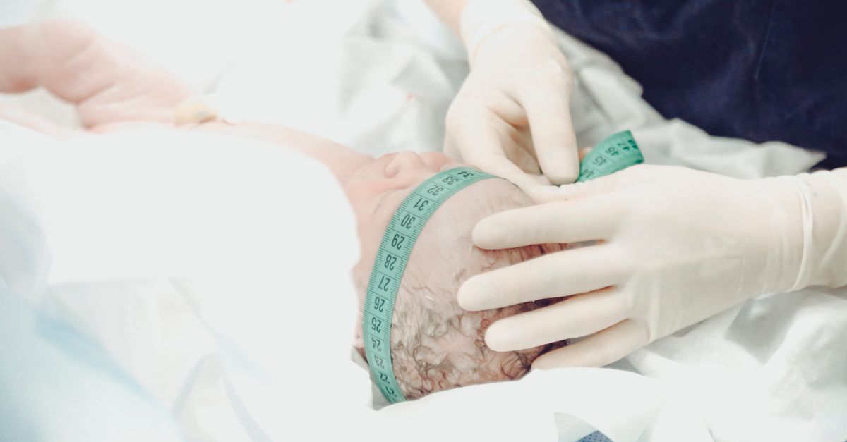
1. Introduction to Postmature Infancy
Postmature infancy refers to the clinical condition of a neonate born after completing forty-two weeks of gestation, exhibiting specific physical and metabolic characteristics related to a prolonged intrauterine environment. When a pregnancy extends significantly beyond the optimal due date, the placenta gradually loses its physiological efficiency. This decline in placental function deprives the fetus of optimal oxygen and nutrient delivery during the final weeks of gestation, leading to a distinct clinical syndrome upon delivery.
Caring for a postmature infant requires specialized neonatal assessment and proactive management. These infants frequently present with a unique set of physical traits, such as dry, peeling skin and an unusually alert demeanor. More importantly, they are at an elevated risk for acute metabolic complications, including hypoglycemia and respiratory distress. Prompt clinical recognition and rigorous monitoring in the immediate postnatal period are essential to stabilize the infant and prevent long-term neurological or physical deficits.
2. Defining Postmaturity vs. Macrosomia
It is essential to distinguish between a prolonged pregnancy and the specific syndrome of postmaturity. A prolonged pregnancy simply defines a gestational age exceeding forty-two weeks. Many infants born at this stage are healthy but large for gestational age, a condition known as macrosomia. Macrosomic infants continue to receive adequate placental nourishment and grow continuously until delivery.
In contrast, postmature infancy, also known as dysmaturity syndrome, occurs when the aging placenta begins to fail. The infant stops growing and may even lose weight in utero as they metabolize their own subcutaneous fat stores for energy. The resulting neonate appears characteristically thin and malnourished, rather than large and robust.
3. The Pathophysiology of Placental Senescence
The placenta is a temporary organ programmed for a lifespan of approximately forty weeks. As gestation extends beyond this physiological limit, the placental tissues undergo progressive senescence, characterized by micro-infarctions, calcifications, and decreased villous blood flow. This vascular deterioration impairs the vital exchange of oxygen, carbon dioxide, and nutrients between the maternal and fetal circulations.
This state of chronic uteroplacental insufficiency creates a hostile intrauterine environment. The fetus responds by redirecting its limited blood flow to essential organs, primarily the brain and heart, at the expense of peripheral tissues and the kidneys. The decreased renal perfusion leads to a critical drop in fetal urine production, causing severe oligohydramnios, a dangerous reduction in amniotic fluid volume.
4. Physical Characteristics of the Postmature Infant
Upon delivery, the physical appearance of a dysmature infant is highly distinctive and easily recognizable by the neonatal care team. Because the infant has lost subcutaneous fat, the skin appears loose, wrinkled, and lacking the typical plumpness of a healthy full-term newborn. The absence of vernix caseosa, the protective white biofilm that coats the fetus, leaves the epidermis exposed to amniotic fluid for an extended period.
Consequently, the skin is extremely dry, cracked, and actively peeling, particularly on the palms of the hands and the soles of the feet. Postmature infants also exhibit remarkably long fingernails and an abundance of scalp hair. Behaviorally, these infants often present with a wide-eyed, hyper-alert expression, reflecting the chronic physiological stress experienced in utero.
5. Meconium Aspiration Syndrome
One of the most severe respiratory complications associated with postmature infancy is meconium aspiration syndrome. The physiological stress of placental hypoxia frequently triggers an involuntary relaxation of the fetal anal sphincter, causing the passage of meconium into the limited amniotic fluid. The thick, tarry substance mixes with the fluid, heavily staining the infant’s skin, nails, and umbilical cord a yellowish-green hue.
If the distressed fetus gasps while in utero or takes a deep breath immediately upon delivery, the meconium-laden fluid is inhaled deep into the pulmonary tree. Meconium is highly toxic to lung tissue; it physically obstructs the airways, causes severe chemical pneumonitis, and inactivates pulmonary surfactant. This cascade leads to profound respiratory failure requiring immediate and aggressive neonatal resuscitation.
6. Managing Neonatal Hypoglycemia
Hypoglycemia is a ubiquitous risk in the postmature neonate. The brain demands a constant, robust supply of glucose to function. Because the failing placenta restricted nutrient transfer, the postmature infant is born with severely depleted hepatic glycogen stores. Furthermore, the loss of subcutaneous fat removes the infant’s secondary energy reserves.
Without these vital energy buffers, the infant’s blood glucose levels can plummet rapidly within the first few hours of life. Routine and rigorous glucose monitoring is mandatory. Hypoglycemia manifests clinically as jitteriness, lethargy, poor feeding, or even seizures. Immediate intervention with early feeding, or intravenous dextrose solutions in severe cases, is critical to prevent irreversible neurodevelopmental damage.
7. Thermoregulation Challenges
Maintaining a stable core body temperature is incredibly difficult for the postmature infant. Normal thermoregulation in neonates relies heavily on the metabolism of brown fat, specialized adipose tissue deposited during the third trimester. Due to intrauterine starvation, the dysmature infant has consumed the majority of these brown fat stores before birth.
The lack of insulating subcutaneous fat, combined with a high surface-area-to-mass ratio, makes the infant highly susceptible to rapid heat loss. Hypothermia exacerbates other metabolic issues, accelerating glycogen depletion and increasing cellular oxygen consumption. Neonatal care focuses on strict environmental temperature control, utilizing radiant warmers and promoting skin-to-skin contact with the mother to stabilize the infant’s temperature.
8. Polycythemia and Hyperviscosity
Chronic intrauterine hypoxia stimulates the fetal kidneys to release erythropoietin, a hormone that drives the excessive production of red blood cells. This physiological attempt to maximize oxygen-carrying capacity results in polycythemia, an abnormally high hematocrit level in the newborn’s blood.
While advantageous in a hypoxic environment, polycythemia becomes detrimental after birth. The elevated red blood cell count significantly increases blood viscosity, making the blood thick and sluggish. Hyperviscosity impairs capillary blood flow to vital organs, increasing the risk of microthrombi, neurological impairment, and severe neonatal jaundice as the excessive red blood cells eventually break down.
9. Delivery Room Resuscitation Protocols
The anticipation of a postmature delivery necessitates the presence of a specialized neonatal resuscitation team. The immediate focus is evaluating the infant’s respiratory effort and heart rate. If the amniotic fluid is meconium-stained and the infant is depressed, non-vigorous, or apneic, immediate intervention is required.
Current protocols dictate rapid clearing of the airway and the initiation of positive pressure ventilation. Intubation and deep tracheal suctioning may be indicated if the airway is visibly obstructed by thick meconium. The swift establishment of adequate oxygenation and ventilation is the paramount priority in preventing hypoxic-ischemic encephalopathy.
10. Neonatal Intensive Care Interventions
Infants demonstrating severe dysmaturity or respiratory distress are promptly transferred to the neonatal intensive care unit. Management is highly systematic and focuses on stabilizing the infant’s fragile metabolic state.
| Clinical Complication | Physiological Cause | NICU Management |
|---|---|---|
| Hypoglycemia | Depleted glycogen and fat stores. | Frequent feeding, IV dextrose infusion. |
| Polycythemia | Chronic hypoxia increasing RBC production. | Hydration, partial exchange transfusion if severe. |
| Hypothermia | Lack of insulating brown and white fat. | Incubator care, radiant warmers. |
11. Neurological Assessment and Monitoring
The brain of a postmature infant has often endured subtle, chronic hypoxia during the final weeks of gestation. Consequently, meticulous neurological monitoring is an ongoing priority. The clinical team continuously assesses the infant’s tone, reflexes, and states of arousal.
An exaggerated startle reflex, persistent hypotonia, or abnormal feeding behaviors can be early indicators of hypoxic brain injury. In high-risk cases, cerebral function monitoring via continuous electroencephalography helps detect subclinical seizures, ensuring that any neurological insult is treated rapidly with specific neuroprotective protocols.
12. Nutritional Support and Feeding Strategies
Feeding the postmature infant presents a delicate balance. While rapid caloric intake is vital to correct hypoglycemia and reverse the state of starvation, the infant’s gastrointestinal tract may be compromised. Hypoxia redirects blood away from the intestines, increasing the vulnerability of the gut mucosa to injury, particularly necrotizing enterocolitis.
Enteral feeding is introduced cautiously. Maternal breast milk is the absolute preferred nutrition due to its optimal digestibility and rich immunological properties. Feeds are initiated in small, frequent volumes and advanced slowly, with continuous assessment of the infant’s tolerance and bowel function.
13. Long-Term Developmental Outcomes
With modern obstetric surveillance, severe cases of postmature infancy are increasingly rare. When caught early and managed properly in the neonatal period, the vast majority of these infants achieve normal catch-up growth and demonstrate excellent long-term neurological outcomes. The lost subcutaneous fat is rapidly replaced within the first few weeks of life, and the dry, peeling skin resolves completely without intervention.
However, infants who suffered severe meconium aspiration syndrome or profound, prolonged hypoglycemia face higher risks of chronic reactive airway disease and subtle neurodevelopmental delays. These infants require enrollment in early intervention programs and long-term pediatric follow-up to monitor developmental milestones.
14. Frequently Asked Questions FAQ
1. Why does a postmature baby lose weight before birth?
When a pregnancy goes too far past the due date, the placenta begins to age and fails to deliver adequate nutrients. To survive, the fetus starts burning its own fat and energy stores, leading to weight loss and a thin appearance at birth.
2. What does a dysmature newborn look like?
They typically appear very thin with loose skin because they lack normal newborn body fat. Their skin is very dry, cracked, and peeling, and they often have unusually long fingernails and abundant scalp hair.
3. Why is meconium dangerous for the baby?
If a stressed baby passes meconium in the womb, it mixes with the amniotic fluid. If inhaled into the lungs, this thick substance physically blocks the airways and causes severe inflammation, making it very difficult for the baby to breathe after birth.
4. How do doctors treat low blood sugar in these infants?
Doctors monitor blood sugar closely immediately after birth. If it drops, they encourage early, frequent feeding. If the sugar level remains dangerously low, the baby is given an intravenous drip containing glucose to protect the brain.
5. Will a postmature baby have long-term health problems?
Most postmature infants recover completely and catch up in weight quickly with no long-term issues. However, if they experienced severe oxygen deprivation or severe meconium inhalation, they may require long-term developmental monitoring.
15. Bibliography
Disclaimer: The content is for informational purposes only and does not replace medical advice. Always consult your doctor for personalized treatment.

