
1. Introduction
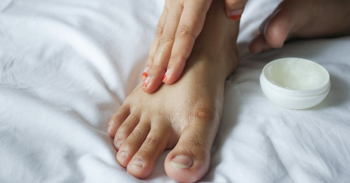
To get rid of deep cracked heels that cause pain when walking, you must implement a rigorous daily regimen utilizing keratolytic creams containing high concentrations of urea or salicylic acid to chemically break down the thickened, dead skin. Following chemical exfoliation, gently use a pumice stone on damp skin to reduce the mechanical thickness of the callus. To repair the severe epidermal micro-fractures, generously apply an occlusive emollient, such as petroleum jelly, and wear cotton socks overnight to trap the moisture, allowing the stratum corneum to heal and regain its natural elasticity.
2. Anatomy and Physiology of the Plantar Skin
The skin on the sole of the foot, specifically the plantar aspect of the heel, is anatomically unique. Because it must bear the entire weight of the human body and withstand immense frictional forces during locomotion, the epidermis here is vastly thicker than anywhere else on the body.
The outermost layer of the epidermis, the stratum corneum, is heavily fortified with dense keratin proteins. Additionally, plantar skin completely lacks sebaceous glands, the microscopic glands responsible for secreting the natural oils (sebum) that keep skin lubricated and flexible.
Because the heel relies entirely on sweat glands for moisture, it is inherently prone to severe desiccation. When the moisture content of the stratum corneum drops below ten percent, the keratinized tissue loses its pliability. It becomes rigid, brittle, and unable to stretch in response to mechanical pressure.
3. Pathophysiology of Heel Fissures
Deep cracked heels are clinically termed heel fissures. The pathology begins with the development of a dense, thick callus around the rim of the heel, a condition known as hyperkeratosis. This callus is a biological defense mechanism, forming in response to excessive, repetitive mechanical stress and friction.
When a person stands or walks, the downward gravitational force of the body expands the fat pad situated directly beneath the heel bone. This fat pad naturally spreads outward, exerting immense lateral tension on the overlying skin.
If the skin is healthy and highly moisturized, it simply stretches to accommodate this lateral expansion. However, if the perimeter of the heel is encased in a thick, dry, and brittle hyperkeratotic callus, the skin cannot expand. The mechanical tension exceeds the tensile strength of the dry tissue, causing it to snap and tear, creating deep, painful fissures.
4. Mechanical Stress and Footwear
The primary environmental catalyst for heel fissures is inappropriate footwear combined with hard, unyielding walking surfaces. Shoes that feature an open back, such as sandals, flip-flops, or backless mules, provide absolutely no lateral support to the heel pad.
Without a rigid shoe counter to contain the fat pad, the tissue is allowed to expand outward maximally with every step, dramatically increasing the lateral stretching force on the skin. Furthermore, the constant slapping motion of a loose sandal physically strikes the heel, inducing micro-trauma that accelerates the formation of a rigid callus.
Prolonged barefoot walking on hard surfaces, such as hardwood floors or concrete, subjects the heel to direct, unmitigated impact, rapidly advancing the cycle of hyperkeratosis and subsequent fissuring. Transitioning to closed-heel, well-cushioned footwear is a mandatory step for mechanical offloading and fissure healing.
5. Systemic Causes of Plantar Xerosis
While environmental factors are significant, severe dry skin (xerosis) on the feet is frequently a manifestation of an underlying systemic or metabolic condition.
| Systemic Condition | Mechanism of Heel Xerosis and Fissuring |
|---|---|
| Diabetes Mellitus | Autonomic neuropathy severely impairs sweat gland function, leading to profound dryness. High blood sugar impedes cellular healing. |
| Hypothyroidism | A depressed metabolic rate globally reduces skin cell turnover and lipid production, creating dry, thick, inflexible skin. |
| Sjogren’s Syndrome | An autoimmune disorder that attacks moisture-producing glands, resulting in systemic, severe cutaneous desiccation. |
| Nutritional Deficiencies | Lack of Vitamin A, zinc, or essential omega-3 fatty acids compromises the structural integrity of the epidermal lipid barrier. |
Identifying and managing these underlying systemic issues is critical, as topical treatments will only provide temporary relief if the biological environment causing the dryness remains uncorrected.
6. Inflammatory and Fungal Etiologies
Not all thick, cracking skin is simply a benign callus; inflammatory dermatological conditions and pathogenic infections frequently masquerade as standard dry heels.
Tinea pedis, commonly known as athlete’s foot, often presents as a chronic, moccasin-type fungal infection. The dermatophyte fungus actively invades the stratum corneum, producing an enzyme called keratinase that digests the keratin. This infection results in a widespread, powdery, thick scaling covering the entire sole and heel, which cracks deeply when stressed.
Similarly, plantar psoriasis or severe atopic dermatitis causes rapid, inflammatory hyperkeratosis. The immune system mistakenly forces the skin cells to multiply at an accelerated rate, creating thick, red, scaly plaques that form rigid fissures. If basic moisturizing protocols fail, a dermatologist must evaluate the heel to rule out these active pathologies.
7. Immediate Intervention: Keratolytic Therapy
The foundational clinical treatment for heel fissures is the breakdown and removal of the inflexible callus, a process achieved through keratolytic therapy. Merely applying standard lotion to a thick callus is completely ineffective, as the dense keratin acts as an impenetrable shield.
Keratolytic agents are specialized chemical exfoliants designed to dissolve the intracellular bonds holding the dead skin cells together. Urea is the gold standard ingredient. At low concentrations, urea is a powerful humectant, drawing water into the skin. At higher concentrations (twenty to forty percent), it becomes a potent keratolytic, melting away the thick, rigid layers of the callus.
Salicylic acid and lactic acid are also highly effective keratolytic options. These acidic creams should be applied strictly to the thickened callus borders once or twice daily. They systematically soften the hard edges of the fissure, restoring pliability to the skin and preventing the crack from tearing deeper during ambulation.
8. Proper Debridement Techniques
Once the keratolytic cream has chemically softened the hyperkeratotic tissue, gentle mechanical debridement is required to physically reduce the thickness of the callus.
After bathing or soaking the feet in lukewarm water for ten minutes to fully hydrate the stratum corneum, the patient should use a pumice stone or a fine-grit foot file. The objective is to gently file away the top layers of the softened, dead skin.
This procedure requires strict caution. Aggressive filing, or the dangerous use of razor blades and sharp calluses shavers, can easily remove too much tissue, exposing the raw dermis, causing profuse bleeding, and creating an open wound highly susceptible to bacterial infection. Debridement should be a slow, gradual process performed two to three times a week.
9. Epidermal Barrier Repair and Occlusion
Following chemical and mechanical debridement, the exposed, compromised skin barrier must be aggressively repaired and sealed. This requires the application of heavy, occlusive emollients.
Standard water-based lotions evaporate too quickly to provide meaningful hydration to plantar skin. Instead, patients must utilize thick, greasy ointments, such as pure petroleum jelly, anhydrous lanolin, or specialized heel balms containing high levels of ceramides and dimethicone.
These occlusive agents create a non-permeable, artificial lipid barrier on the surface of the heel. This seal traps the body’s natural moisture inside the epidermis, forcing it to rehydrate the damaged tissue from the inside out. Applying this thick ointment immediately before bed and covering the foot with a 100% cotton sock ensures maximum absorption and prevents the product from rubbing off onto the bedding.
10. Pharmacological Management of Complicated Fissures
When heel fissures become so deep that they breach the dermis, they expose the delicate sensory nerve endings and capillary beds. This results in agonizing, sharp pain when bearing weight and frequent bleeding.
For these deep, painful cracks, a physician or podiatrist may utilize specialized medical glues, such as liquid cyanoacrylate skin protectants. The glue is applied directly into the clean fissure, physically holding the edges of the wound together, instantly blocking the pain receptors, and providing a sterile, waterproof environment for the dermis to heal.
If the deep fissure shows signs of significant inflammation, a short course of a topical corticosteroid ointment may be prescribed to suppress the localized immune response, accelerating the closure of the wound.
11. Biomechanical Support and Orthotics
If mechanical stress is the root cause of the hyperkeratosis, topical treatments must be paired with biomechanical correction to prevent recurrence. The physical load placed on the heel pad must be evenly distributed.
Patients must wear supportive, closed-heel shoes with a deep heel cup. The firm heel cup physically surrounds the fat pad, preventing it from expanding laterally under the weight of the body, thereby completely eliminating the stretching force that tears the skin.
For individuals with structural foot abnormalities or those who spend long hours standing on hard surfaces, custom orthotic inserts or specialized silicone heel cups provide essential shock absorption. This cushioning minimizes the micro-trauma to the plantar skin, drastically slowing the biological formation of the protective callus.
12. Recognizing Signs of Secondary Infection
A deep heel fissure is an open portal of entry for environmental pathogens. The feet are constantly exposed to dark, damp, and highly populated bacterial environments, particularly inside shoes.
Patients must meticulously monitor deep cracks for signs of secondary bacterial colonization. A localized infection presents with a sudden, significant increase in pain, spreading redness, localized heat, and significant swelling around the heel perimeter. The presence of yellow or green purulent discharge (pus) emanating from the fissure is a definitive sign of an active infection.
Infectious complications are exceptionally dangerous for diabetic patients or those with peripheral arterial disease, as a simple infected fissure can rapidly escalate into a limb-threatening ulceration. Any sign of infection demands immediate clinical evaluation for oral or topical antibiotic therapy.
13. Frequently Asked Questions (FAQ)
1. Is it safe to use a motorized foot grinder to remove the hard skin?
While motorized callus removers are highly effective, they must be used with extreme caution. Because they remove tissue very rapidly, it is easy to accidentally grind down to the sensitive, living tissue, causing severe pain and open wounds. They should only be used on completely dry skin using a light touch.
2. Why do my cracked heels seem to get worse during the winter?
During winter, ambient humidity drops significantly, and indoor heating systems severely desiccate the air. This extremely dry environment rapidly pulls moisture out of the stratum corneum, exacerbating the brittleness of the heel callus and making it much more likely to fissure.
3. Does soaking my feet in Epsom salts help heal the cracks?
Epsom salt (magnesium sulfate) is highly osmotic; it draws moisture out of tissues. While a brief soak can soften the dead skin for filing, prolonged soaking in Epsom salts will severely dry out the healthy tissue, making the cracking problem worse. Always follow a soak with heavy moisturization.
4. Can walking barefoot on the beach cause heel fissures?
Yes. Sand is highly abrasive and acts like a giant, uncontrolled pumice stone, stripping away the protective oils and upper layers of the skin. Combined with the drying effect of salt water, a day at the beach can rapidly trigger severe plantar xerosis.
5. How long does it take for a deep, painful heel crack to heal completely?
With aggressive, twice-daily keratolytic and occlusive therapy, a deep, painful fissure typically closes and becomes pain-free within seven to fourteen days. However, completely removing the surrounding hyperkeratosis to prevent the next crack requires several weeks of consistent maintenance.
14. Bibliography
Disclaimer: The content is for informational purposes only and does not replace medical advice. Always consult your doctor for personalized treatment.


