
1. Introduction
Cracked and bleeding heels during the hot, dry summer months are clinically known as heel fissures. This painful dermatological condition occurs when the thick skin on the bottom of the heel loses its natural elasticity and moisture, causing it to split under the mechanical pressure of walking. The combination of arid environmental conditions, open-backed footwear, and repetitive physical stress compromises the structural integrity of the skin barrier.
The human heel is designed to withstand massive compressive forces. To accomplish this, the stratum corneum, the outermost layer of the epidermis, thickens to form a protective callous. Under normal physiological conditions, this thick skin remains supple and flexible due to adequate internal hydration and natural lipid production.
However, summer environments strip the skin of its essential moisture. When this dehydrated, rigid callous is forced to expand outward as body weight bears down on the heel, the inelastic tissue simply tears. These tears can penetrate deeply into the vascularized dermis, leading to sharp pain and active bleeding.
Addressing heel fissures requires a dual approach of intense rehydration and mechanical pressure management. Understanding the environmental triggers and proper dermatological care empowers individuals to restore the skin barrier and prevent recurrent fissuring.
2. Dermatological Anatomy of the Heel
The skin on the plantar aspect, or bottom, of the foot is anatomically unique. It lacks hair follicles and sebaceous glands, which are the primary producers of sebum, the natural oil that lubricates and waterproofs the rest of the human skin.
Because the heel cannot secrete its own moisturizing oils, it relies entirely on the sweat glands for surface hydration and the internal vascular system to deliver moisture from within. The outermost layer of this skin is exceptionally thick, consisting of tightly packed, dead keratinocytes.
This thickened pad acts as a biological shock absorber. When a person steps down, the fat pad beneath the skin flattens and expands outward laterally. The overlying skin must stretch to accommodate this rapid expansion.
If the stratum corneum becomes overly thick and brittle, it loses its tensile strength. When the fat pad expands, the brittle epidermis cannot stretch, resulting in a mechanical fracture of the skin layer.
3. The Impact of Summer Environments
The summer season introduces a combination of environmental factors that actively degrade skin hydration. Hot, arid climates cause rapid trans-epidermal water loss. The moisture within the superficial layers of the skin evaporates directly into the dry air.
Air conditioning further exacerbates this issue. Indoor cooling systems remove humidity from the air, creating an artificially dry environment that continuously draws moisture out from the skin surface.
Additionally, summer activities often involve prolonged exposure to harsh environmental elements. Walking barefoot on hot sand or concrete pulls moisture out of the feet and causes microscopic thermal damage to the skin barrier.
Chlorinated swimming pools are particularly harsh. Chlorine is a powerful chemical that strips away any remaining natural lipids on the skin surface, leaving the heels profoundly dehydrated and vulnerable to cracking.
4. Open-Backed Footwear and Mechanical Stress
The transition to summer footwear is a primary catalyst for heel fissures. Sandals, flip-flops, and open-backed shoes provide zero structural support for the heel pad.
In a closed shoe, the rigid heel counter encapsulates the fat pad. When weight is applied, the shoe prevents the fat pad from expanding outward, thereby minimizing the stretching force applied to the skin.
In a backless sandal, there is no physical barrier to contain the fat pad. With every step, the heel strikes the firm sole of the sandal, and the fat pad splay laterally without restriction. This maximized lateral expansion places immense shearing stress on the rim of the heel.
Furthermore, flip-flops require a modified, heavier heel-strike gait to keep the shoe on the foot. This repetitive, heavy slapping against the sole of the shoe accelerates the build-up of defensive callous tissue, compounding the problem.
5. Hyperkeratosis and Callous Formation
Hyperkeratosis is the physiological process where the body rapidly produces extra layers of skin cells in response to friction or pressure. It is a protective mechanism designed to prevent blister formation.
Walking in unsupportive summer shoes stimulates rapid hyperkeratosis around the rim of the heel. While this thick callous prevents friction burns, it creates a significant structural problem. A thick callous is inherently rigid and devoid of moisture.
As the callous grows thicker, it becomes a distinct, inflexible ring of tissue. It acts much like a piece of dried clay. When pressure is applied to dried clay, it shatters.
Managing hyperkeratosis is a delicate balance. The goal is to gently reduce the extreme thickness of the callous without removing it entirely, as the heel still requires a baseline layer of protection for walking.
6. The Physiology of a Heel Fissure
A heel fissure begins as a microscopic, superficial crack in the dead epidermal layer. At this stage, it is painless and appears as a fine white line within the callous.
If the mechanical stress and dehydration continue, the crack deepens. It propagates downward through the epidermis, driven apart by the constant lateral splay of the fat pad during walking.
When the fissure finally breaches the epidermal-dermal junction, it enters the living tissue of the dermis. The dermis is heavily vascularized and packed with sensory nerve endings.
Once the dermis is compromised, the fissure bleeds freely. The pain becomes sharp and stinging, particularly when standing up after a period of rest. The edges of the fissure pull apart with every step, preventing the delicate dermal tissue from bridging the gap and healing.
7. Infection Risks and Complications
A bleeding heel fissure represents a complete breach in the body’s primary immunological barrier. The foot is in constant contact with the ground, making it highly susceptible to environmental pathogens.
Bacteria, such as Staphylococcus aureus, and various fungal organisms can easily infiltrate the open wound. An infected fissure becomes increasingly painful, red, and swollen. It may discharge a purulent fluid and emit a foul odor.
For individuals with compromised immune systems, particularly those with diabetes mellitus, a simple heel fissure can rapidly escalate into a limb-threatening diabetic foot ulcer. Elevated blood sugar impairs localized circulation and blunts the neurological pain response, allowing the infection to spread unchecked.
Immediate attention to wound hygiene is paramount to prevent these serious clinical complications. Readers seeking to understand related immune responses may review our article on swollen lymph nodes.
8. Analyzing Clinical Risk Factors
Certain systemic conditions predispose individuals to severe heel fissuring beyond simple environmental exposure.
| Systemic Condition | Physiological Impact on the Foot | Resulting Complication |
|---|---|---|
| Diabetes Mellitus | Autonomic neuropathy reduces sweating | Profoundly dry skin, delayed wound healing |
| Hypothyroidism | Slowed metabolic rate affects skin turnover | Thick, coarse, extremely dry epidermis |
| Obesity | Increased vertical mechanical load | Maximal lateral splay of the heel fat pad |
| Eczema / Psoriasis | Altered structural integrity of the skin | Chronic inflammation and rapid scaling |
9. Immediate First Aid for Bleeding Fissures
When a heel fissure begins to bleed, it must be treated clinically as an open laceration. Stop all abrasive treatments immediately. Do not attempt to file or scrape a bleeding heel, as this will further traumatize the living dermis.
Cleanse the fissure gently with mild soap and warm water to flush out environmental debris. Apply a topical antibacterial ointment directly into the crack to establish a barrier against pathogens.
Draw the edges of the fissure together. This can be accomplished using specialized liquid skin adhesives or medical closure strips. Holding the edges together minimizes the pain caused by the tissue stretching during walking and allows the dermal layer to begin cellular repair.
Cover the heel with a sterile, non-stick dressing and transition immediately to closed-back, supportive footwear to eliminate lateral fat pad expansion.
10. Keratolytic Agents and Exfoliation
Once the active bleeding has ceased and the dermal layer has sealed, the focus shifts to removing the rigid hyperkeratotic tissue. This requires the use of keratolytic agents.
Keratolytics are specialized chemical compounds that break the intercellular bonds holding the dead skin cells together. Urea is the gold standard for treating heel fissures. Creams containing twenty to forty percent urea not only dissolve the thick callous but also actively draw moisture into the skin.
Salicylic acid and lactic acid are also effective keratolytic ingredients. They provide gentle, continuous chemical exfoliation, slowly softening the rigid rim of the heel without the need for aggressive physical scraping.
Consistent application of a high-percentage urea cream twice daily can transform a hardened callous into pliable tissue within a matter of weeks.
11. Emollients and Moisture Trapping
While keratolytics remove the dead skin, emollients are required to restore flexibility and trap moisture within the newly exposed tissues. Emollients contain lipids that fill the microscopic spaces between skin cells, creating a smooth, supple surface.
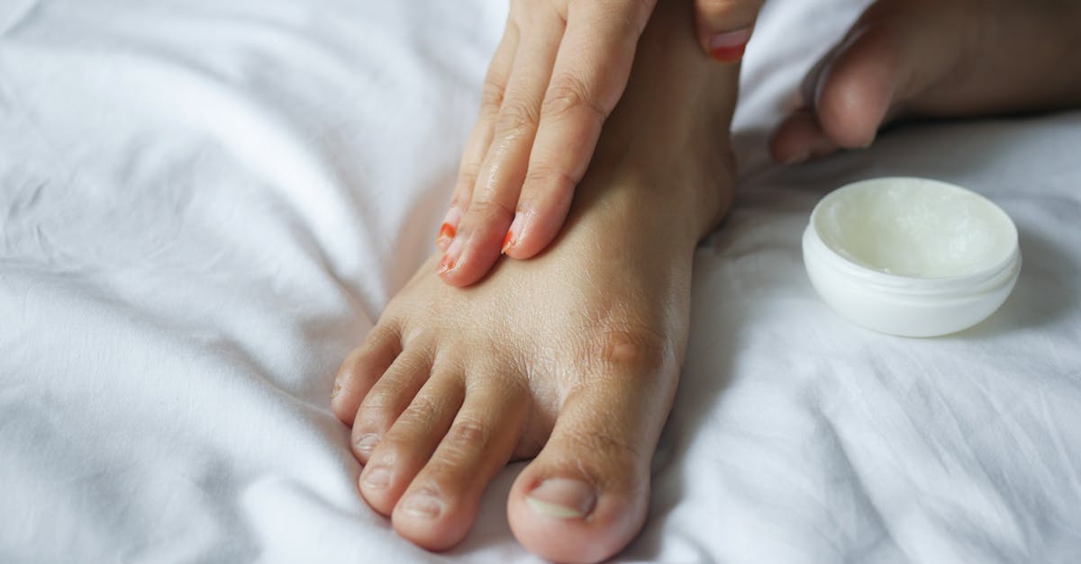
Thick, occlusive ointments containing petroleum jelly or lanolin are highly effective. Because the heel lacks sebaceous glands, these ointments artificially replicate the skin’s natural oil barrier.
To maximize their effectiveness, emollients must be applied immediately after bathing while the skin is still damp. Applying a thick layer of ointment and covering the foot with a cotton sock before bed creates a localized greenhouse effect. The sock traps the body heat, opens the pores, and forces the emollient deep into the epidermis overnight.
12. Safe Physical Debridement Techniques
Physical debridement is the mechanical removal of dead skin. It must be performed safely and correctly to avoid worsening the fissures.
Never use sharp blades or razor-style callous shavers at home. These tools easily slip and slice into the healthy dermis, causing severe injury and massive infection risk.
Instead, utilize a pumice stone or a fine-grit foot file. Debridement should only be performed on damp skin, ideally at the end of a warm shower when the callous is softened.
File gently in a single direction, following the natural contour of the heel. The goal is to reduce the thickness of the callous gradually over several sessions, not to aggressively scrub the skin down to a pink, sensitive layer in one day.
13. Modifying Summer Footwear
Preventing the recurrence of fissures requires a compromise in summer footwear fashion. Constant reliance on flat, backless sandals will inevitably lead to a return of hyperkeratosis and cracking.
Select sandals that feature a deep heel cup. A contoured heel cup cradles the fat pad, providing a physical boundary that prevents the tissue from splaying outward laterally when standing.
Incorporate supportive athletic shoes or closed-back canvas shoes into the weekly rotation. Wearing closed shoes for part of the week gives the heels a necessary break from the intense mechanical shearing forces of flip-flops.
If open-backed shoes must be worn for formal occasions, preemptively apply a thick layer of emollient to the heels to maintain flexibility during use.
14. Nutritional Support for Skin Health
The structural integrity of the epidermis is heavily influenced by systemic nutrition. The skin requires specific vitamins and fatty acids to manufacture its lipid barrier and maintain cellular turnover.
Omega-3 fatty acids, found in fatty fish, walnuts, and flaxseeds, are crucial components of the cell membrane. Adequate dietary intake ensures that the newly formed skin cells are structurally robust and capable of retaining moisture.
Vitamin C is essential for collagen synthesis, the protein that provides the dermis with its strength and elasticity. Zinc plays a vital role in wound healing and epidermal repair. Ensuring a balanced diet rich in these nutrients provides the necessary building blocks to heal deep fissures from the inside out.
15. When to Consult a Podiatrist or Dermatologist
While most heel fissures respond well to diligent home care, professional medical evaluation is necessary for recalcitrant or complicated cases.
If a fissure shows signs of clinical infection, such as expanding redness, severe heat, throbbing pain at rest, or pus drainage, seek immediate medical attention. Oral antibiotics are frequently required to clear a dermal infection.
Individuals with diabetes, severe peripheral neuropathy, or compromised circulation must not attempt to self-treat bleeding fissures. These patients should visit a podiatrist for professional, sterile debridement and specialized wound care to prevent ulceration.
A dermatologist can also evaluate chronic, non-healing cracks to ensure they are not a manifestation of a deeper dermatological condition, such as palmoplantar psoriasis or a severe fungal infection requiring prescription-strength topical therapies.
16. Frequently Asked Questions FAQ
1. Why does walking barefoot on the beach make my heels crack faster?
Sand is highly abrasive and acts like a natural file, stripping away the outer layers of skin. Furthermore, salt water is extremely hypertonic; it rapidly draws moisture out of the cellular tissues through osmosis, leaving the skin profoundly dehydrated and prone to splitting.
2. Is it safe to use superglue to close a bleeding heel crack?
While cyanoacrylate adhesives, similar to superglue, are used in medicine, standard household superglue contains toxic industrial solvents that can irritate the wound and delay healing. It is much safer to use a medically approved liquid skin protectant available at pharmacies.
3. Will drinking more water cure my dry heels?
Systemic hydration is important, but the heel lacks the sebaceous glands needed to lock that moisture in. You can drink gallons of water, but if you do not apply an external occlusive emollient, the environmental air will simply evaporate the moisture away from the skin surface.
4. How long does it take for a deep, bleeding fissure to fully heal?
Once the edges are brought together and protected from mechanical stretching, the dermal layer usually seals within three to seven days. However, completely restoring the epidermal barrier and returning the callous to a healthy, flexible state typically requires a strict four-week regimen of moisturizing and filing.
5. Can a fungal infection cause heel cracks?
Yes. A condition known as moccasin-type tinea pedis is a fungal infection that presents as widespread, dry, powdery scaling on the soles and heels. It degrades the structural integrity of the skin, making it highly susceptible to fissuring. It requires antifungal medication, not just moisturizers.
17. Bibliography
Disclaimer: The content is for informational purposes only and does not replace medical advice. Always consult your doctor for personalized treatment.
