1. Introduction
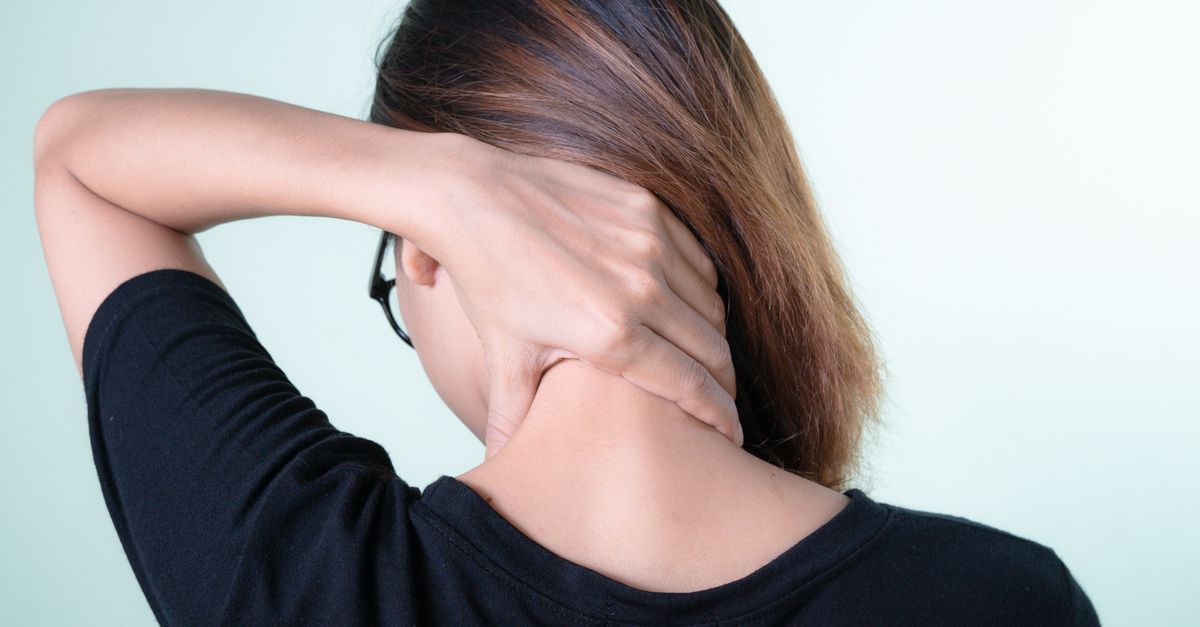
Experiencing a sharp, profound pain in the neck when looking up at the ceiling is typically caused by the mechanical compression of inflamed facet joints or the pinching of cervical nerve roots. Tilting the head backward places the cervical spine into extension. This specific movement narrows the anatomical spaces at the back of the neck, forcing the bony structures closer together. If the cartilage is worn or if a spinal disc is bulging, this compression triggers immediate and severe pain signals.
The human neck consists of seven cervical vertebrae designed to support the weight of the head while providing a substantial range of motion. This flexibility relies on a delicate balance between the intervertebral discs in the front and the facet joints in the back. Maintaining an upright posture requires these structures to absorb and distribute mechanical loads evenly.
When structural degradation occurs due to age, poor posture, or injury, the mechanics of the cervical spine alter. Extension becomes a provocative movement. Understanding the specific anatomy involved in cervical extension helps clarify why looking upward acts as a direct trigger for structural joint pain and neural irritation.
2. Biomechanics of Cervical Extension
Cervical extension is the physiological movement of tilting the head backward. During this motion, the anterior portion of the cervical spine stretches, while the posterior portion compresses. The intervertebral discs at the front of the spinal column decompress slightly, shifting their internal load.
Simultaneously, the facet joints located at the rear of the vertebrae glide over one another to accommodate the backward tilt. These joints bear an increased mechanical load during extension compared to neutral standing or forward flexion. The space between the posterior bony elements physically decreases to allow the head to angle upward.
In a healthy spine, the cartilage and joint capsules absorb this compressive force smoothly. However, if the functional space is already compromised, the physiological compression of normal extension turns into pathological impingement, immediately activating local pain receptors.
3. The Role of the Facet Joints
The facet joints connect each vertebra to the one above and below it, forming a continuous chain down the back of the spine. They are synovial joints, completely encased in a fibrous capsule and lubricated by synovial fluid. These joints guide the direction of spinal movement and prevent excessive twisting.
When you look up, the facet joints in the neck bear the brunt of the compressive force. If a patient suffers from cervical osteoarthritis, the smooth cartilage lining these joints is degraded or entirely absent. The underlying bone, which is densely packed with sensory nerves, becomes exposed.
Forcing these worn, arthritic joints together during cervical extension causes the exposed bone surfaces to grind against each other. This mechanical friction generates a sharp, localized ache in the back of the neck, often accompanied by a sensation of stiffness or structural blockage.
4. Neuroforaminal Narrowing
Nerves branch off from the spinal cord and exit the cervical spine through small bony tunnels called neural foramina. These openings must remain clear to allow the nerve roots to travel freely down into the shoulders, arms, and hands.
The act of looking up naturally decreases the diameter of these neural foramina by up to twenty percent. For a healthy individual, this temporary narrowing is harmless. The nerve root has ample room to slide and accommodate the changing bony boundaries.
However, if a patient has bone spurs or a thickened ligament occupying space in the foramen, the baseline diameter is already critically reduced. When extension further narrows this compromised space, the bone physically crushes the exiting nerve root, resulting in sharp, electrical pain.
5. Cervical Spondylosis and Degeneration
Cervical spondylosis is the medical term for age-related wear and tear affecting the spinal disks and joints in the neck. Over decades of supporting the heavy skull, the cervical intervertebral discs gradually lose hydration. They flatten and shrink, reducing the vertical height between the vertebrae.
As the discs lose height, the facet joints at the back of the spine are forced to overlap more than they should. This abnormal overlap alters the entire mechanical alignment of the neck. The body attempts to stabilize this wobbly alignment by growing extra bone, known as osteophytes or bone spurs.
These bone spurs form around the edges of the facet joints and inside the neural foramina. When the patient looks up at the ceiling, these rigid bone spurs crash into adjacent structures, causing deep, unyielding pain and severely restricting the backward range of motion.
6. Muscular Spasms and Tension
The muscles at the back of the neck, particularly the suboccipital muscles and the upper trapezius, are responsible for pulling the head backward into extension. These muscles constantly work to keep the head balanced over the center of gravity.
Chronic poor posture, such as staring down at a smartphone for hours, forces these muscles into a constant state of elongation and strain. When they are finally asked to contract concentrically to tilt the head upward, they often spasm due to fatigue and poor conditioning.
A muscle spasm is a sudden, involuntary contraction. When the cervical muscles spasm during extension, they lock the neck in place. This creates a severe, pulling ache that feels distinctly different from the sharp, electrical pain of a pinched nerve.
7. Disc Herniation Mechanics
While forward flexion typically exacerbates intervertebral disc issues, posterior disc herniations can cause intense pain during extension. A herniated disc occurs when the soft inner gel of the disc pushes through a tear in the tough outer ring.
If the disc herniates directly backward toward the spinal cord, tilting the head up can compress the spinal canal. The ligamentum flavum, a thick ligament running down the inside of the spinal canal, buckles slightly during extension.
This buckling pushes against the herniated disc material, creating a pincer effect on the delicate neural tissues. This specific mechanism generates a deep, severe pain that often radiates down the center of the back or into both arms simultaneously.
8. Identifying the Source of Neck Pain
Distinguishing between joint pain and nerve pain dictates the appropriate clinical approach.
| Clinical Symptom | Facet Joint Origin | Nerve Root Origin |
|---|---|---|
| Location of Pain | Localized to the back of the neck | Radiates down the shoulder or arm |
| Type of Sensation | Dull ache or sharp mechanical catch | Electrical, burning, or shooting pain |
| Associated Deficits | Stiffness and restricted motion | Numbness, tingling, or muscle weakness |
| Response to Extension | Immediate localized sharp pain | Immediate shooting pain down the limb |
9. Whiplash and Ligamentous Sprain
A previous history of whiplash, often from a motor vehicle accident, can leave the neck structurally vulnerable. Whiplash involves a rapid, forceful hyperflexion followed by hyperextension of the cervical spine. This trauma tears the ligaments supporting the front and back of the vertebrae.
When ligaments heal, they often form dense, inelastic scar tissue. This scar tissue fails to stretch smoothly during normal movements. Furthermore, damaged anterior ligaments allow the vertebrae to slide backward excessively during extension.
This abnormal backward sliding, known as segmental instability, causes the facet joints to jam together violently when looking up. The resulting pain is a combination of joint impaction and the pulling of stiff, scarred connective tissue.
10. The Impact of Upper Crossed Syndrome
Upper crossed syndrome is a postural imbalance characterized by forward head posture, rounded shoulders, and a pronounced curve in the upper back. This alignment drastically alters the resting position of the cervical spine.
In a forward head posture, the lower cervical spine is hyper-flexed, while the upper cervical spine near the skull is constantly hyper-extended to keep the eyes looking straight ahead. The facet joints at the top of the neck are perpetually compressed.
When a patient with this posture attempts to look up at the ceiling, they have no available extension left in the upper neck. They must force the lower neck to hinge abruptly, placing concentrated, profound stress on one or two specific vertebrae rather than distributing the movement evenly.
11. Diagnostic Imaging and Evaluation
When cervical extension consistently produces severe pain, a thorough clinical evaluation is mandatory. A physician will begin with a neurological exam, testing reflexes, muscle strength, and sensory perception in the arms to check for nerve root compression.
Standard X-rays are utilized to evaluate the overall alignment of the cervical spine, the height of the intervertebral discs, and the presence of significant bone spurs. X-rays are excellent for diagnosing advanced cervical spondylosis.
For a detailed view of the soft tissues, a Magnetic Resonance Imaging scan is required. This advanced imaging clearly visualizes the spinal cord, the exiting nerve roots, and the condition of the facet joint cartilage, pinpointing the exact anatomical structure causing the impingement.
12. Conservative Management and Physical Therapy
The first line of defense against extension-based neck pain involves conservative therapy. Physical therapy focuses on restoring proper cervical and thoracic alignment. Strengthening the deep neck flexor muscles at the front of the throat provides better muscular support for the vertebrae.
Therapists utilize manual traction to gently decompress the cervical spine. This passive stretching pulls the vertebrae slightly apart, opening up the neural foramina and providing immediate relief to compressed nerve roots and jammed facet joints.
Postural re-education is critical. Teaching the patient to retract their chin and sit with a neutral spine prevents the chronic facet joint compression associated with forward head posture, creating a healthier mechanical baseline for movement.
13. Pharmacological Interventions
To manage the acute inflammation driving the pain, physicians often recommend nonsteroidal anti-inflammatory drugs. These medications reduce the chemical swelling inside the facet joint capsules and around irritated nerve roots, creating more physical space in the narrowed canals.
If muscle spasms are severely limiting movement, a short course of muscle relaxants may be prescribed. Breaking the spasm cycle allows the patient to participate comfortably in physical therapy and perform necessary stretching exercises.
For persistent facet joint pain, a pain management specialist might perform a targeted corticosteroid injection. Using fluoroscopic guidance, the physician injects potent anti-inflammatory medication directly into the inflamed joint, providing profound relief that can last for several months.
14. Ergonomic Adjustments
Modifying the daily environment significantly reduces the strain placed on the cervical spine. Computer monitors should be elevated so the top third of the screen is at eye level. This prevents the constant forward leaning and upward tilting that jams the posterior neck structures.
When painting a ceiling or performing tasks that require sustained upward gazing, using a ladder to bring the work closer to eye level eliminates the need for extreme cervical extension.
Sleeping posture also plays a vital role. Utilizing a contoured cervical pillow supports the natural curve of the neck during sleep, preventing the joints from collapsing into painful extension or side-bending throughout the night.
15. When to Seek Urgent Medical Attention
While most extension-based neck pain is mechanical and non-emergent, certain red flag symptoms demand immediate medical evaluation. If looking up causes sudden dizziness, blurred vision, or fainting, it may indicate compression of the vertebral arteries, which supply blood to the brain.
Pain that shoots down both arms simultaneously, or is accompanied by a sudden loss of bowel or bladder control, suggests severe spinal cord compression, known as cervical myelopathy. This is a surgical emergency.
Additionally, if the severe neck pain is accompanied by an unexplained fever, night sweats, or unexpected weight loss, it requires urgent investigation to rule out spinal infections or underlying malignancies.
16. Frequently Asked Questions (FAQ)
1. Why does my neck crack when I look up?
The cracking sound is usually caused by the release of nitrogen gas bubbles from within the synovial fluid of the facet joints as they glide over one another. It is generally harmless unless accompanied by sharp pain.
2. Will a neck brace help stop the pain when I look up?
A soft cervical collar can provide temporary support and remind you not to move your head to extreme angles. However, wearing one long-term weakens the neck muscles, which ultimately worsens structural instability and pain.
3. Can stress make the pain in the back of my neck worse?
Yes. Psychological stress causes involuntary tensing of the upper trapezius and cervical muscles. This constant muscular tension increases the baseline compression on your joints, making movements like looking up much more painful.
4. Is it safe to do neck rolls to stretch out the stiffness?
Full circular neck rolls are generally not recommended. Rolling the head backward into deep extension grinds the facet joints together. It is safer to stretch by gently tilting the ear toward the shoulder or tucking the chin forward.
5. How do I know if the pain is from a muscle or a nerve?
Muscle pain typically feels like a dull, widespread ache or a stiff pulling sensation restricted to the neck. Nerve pain is distinctly sharp, electrical, and usually travels away from the neck down into your shoulder blade, arm, or hand.
17. Bibliography
Disclaimer: The content is for informational purposes only and does not replace medical advice. Always consult your doctor for personalized treatment.