1. Introduction
A solitary fibrous tumour is a rare and unique type of mesenchymal neoplasm that originates from fibroblastic cells. Historically, it was believed to occur exclusively within the pleura, the protective lining of the lungs. However, modern medicine now recognizes that these tumors can develop almost anywhere in the body, including the abdomen, pelvis, head, neck, and soft tissues of the extremities. They typically present as a slow-growing, solid mass that may remain asymptomatic until it grows large enough to compress surrounding organs.
Receiving a diagnosis of this nature can prompt significant questions, especially given the variable clinical behavior of the condition. The majority of these tumors are biologically benign and can be completely cured with surgery. Nevertheless, a small percentage exhibit malignant traits, displaying a more aggressive growth pattern and a risk of spreading to other tissues. Precise pathological grading is essential to determine the exact nature of the tumor.
Advanced diagnostic testing and a multidisciplinary medical approach are necessary to manage this disease effectively. Through careful surgical planning and rigorous pathological analysis, healthcare teams can provide definitive care and establish appropriate long-term monitoring plans to safeguard patient health.
2. Understanding the Pathology
The cellular landscape of a solitary fibrous tumour is highly characteristic and requires expert histological interpretation. Under a microscope, pathologists observe a proliferation of spindle-shaped cells arranged in a haphazard, patternless architecture. These cells are embedded within a dense, collagen-rich stroma, creating alternating areas of high and low cellularity.
A defining microscopic feature is the prominent network of branching, thin-walled blood vessels that resemble staghorns. This specific vascular pattern is termed hemangiopericytoma-like, a historical term once used to describe a separate disease entity now unified under the solitary fibrous tumour classification.
Pathologists meticulously evaluate the tissue for specific signs of aggression. They assess the rate of cellular division, the presence of dead tissue areas known as necrosis, and the degree of cellular abnormality. These factors dictate whether the tumor is classified as benign, which is the most common presentation, or as a malignant variant requiring more intensive therapy.
3. Prevalence and Demographics
This condition is classified as a rare neoplasm, accounting for a very small fraction of all diagnosed soft tissue and pleural tumors worldwide. It most frequently occurs in adults during their fifth to seventh decades of life, though cases have been occasionally reported in young adults and children.
Epidemiological data shows an equal distribution between men and women, with no discernible gender predilection. Furthermore, the disease appears across all global populations without any specific racial or ethnic clustering.
Because of the rarity and varied anatomical locations of these tumors, patients are typically referred to specialized tertiary care centers. Treatment at these specialized institutions ensures access to experienced sarcoma surgeons and dedicated soft tissue pathologists who are familiar with the nuances of the disease.
4. Primary Clinical Symptoms
The clinical symptoms produced by a solitary fibrous tumour depend entirely on its anatomical location and size. Because the tumor generally grows very slowly, it can reach a substantial size before causing any noticeable issues.
When located in the chest cavity, patients may experience vague respiratory symptoms. A chronic cough, shortness of breath, or a dull aching pain in the chest are common complaints as the mass compresses the lung tissue. In some instances, the tumor may cause fluid to accumulate in the pleural space, further restricting breathing capacity.
When the tumor arises in extrapleural sites, such as the abdomen or deep soft tissues, the presentation is typically a painless, palpable mass. Systemic symptoms are generally absent, though a rare paraneoplastic condition known as Doege-Potter syndrome can occur. In this syndrome, the tumor secretes an insulin-like growth factor, leading to episodes of severe hypoglycemia, or low blood sugar.
5. Underlying Causes and Risk Factors
The precise medical causes triggering the development of this specific tumor remain largely unknown. Unlike lung cancers that are heavily linked to tobacco smoke or mesotheliomas tied to asbestos exposure, solitary fibrous tumours have no proven connection to environmental toxins, lifestyle choices, or occupational hazards.
The medical consensus points toward sporadic genetic mutations occurring randomly within a single connective tissue cell. These intrinsic errors allow the cell to bypass normal growth restrictions, leading to the gradual formation of a tumor mass. The condition is not considered hereditary, meaning patients do not pass an increased risk to their children.
Currently, there are no preventive strategies or screening protocols recommended for the general population. Diagnosis relies primarily on the clinical investigation of new, unexplained symptoms or the incidental discovery of a mass during imaging for an unrelated issue.
6. Cellular and Molecular Mechanisms
The most significant advancement in understanding this disease has been the discovery of a defining molecular signature. In almost all cases, these tumors harbor a specific genetic mutation involving the inversion of a section of chromosome twelve. This structural rearrangement fuses two genes together: NAB2 and STAT6.
The resulting NAB2-STAT6 fusion gene acts as a powerful driver of tumor growth. It forces the continuous activation of pathways that promote cellular proliferation and survival. This specific genetic anomaly is unique to this condition, setting it apart from all other spindle cell neoplasms.
The discovery of this fusion gene has revolutionized diagnostic pathology. By testing the tumor tissue for the presence of the STAT6 protein in the cell nucleus, pathologists can now definitively confirm the diagnosis, ensuring exceptional accuracy even in histologically challenging cases.
7. Differential Diagnosis
Because these tumors can occur in virtually any organ system and share morphological features with other connective tissue growths, the differential diagnosis is extensive. A precise diagnosis is crucial, as the treatment protocols for mimicking conditions differ dramatically.
The medical team must utilize clinical, radiological, and advanced pathological data to rule out alternative diagnoses. The table below outlines key conditions that mimic this disease.
| Condition | Differentiating Characteristics |
|---|---|
| Malignant Mesothelioma | Strongly linked to asbestos, primarily grows along pleural surfaces rather than as a discrete mass. |
| Gastrointestinal Stromal Tumor | Specific to the digestive tract, driven by different genetic mutations, requires targeted drug therapy. |
| Synovial Sarcoma | Highly aggressive, distinct chromosomal translocation, occurs near large joints. |
| Schwannoma | Benign nerve sheath tumor, distinct neural markers present on pathology. |
Accurate differentiation, primarily driven by STAT6 immunohistochemistry, directs the patient to the correct surgical and oncological pathways.
8. Diagnostic Imaging Modalities
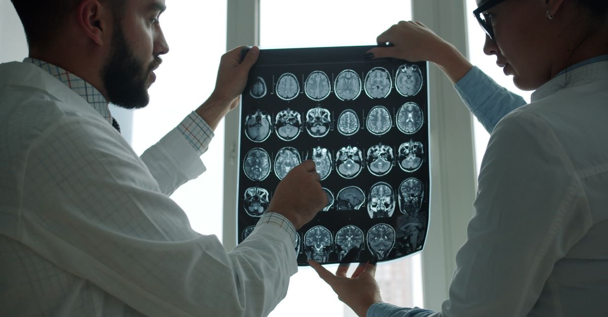
Imaging studies are critical for identifying the tumor, assessing its size, and planning the surgical approach. Computed tomography is the most frequently utilized initial imaging modality, especially for tumors located in the chest or abdomen.
On a CT scan, a solitary fibrous tumour generally appears as a well-defined, solid, and often highly vascular mass. Because of the extensive internal blood vessel network, these tumors typically exhibit intense enhancement when intravenous contrast dye is administered. Large tumors may display areas of central fluid accumulation or calcification.
Magnetic resonance imaging is particularly valuable for tumors located in the pelvis, head, neck, or extremities. MRI provides superior soft tissue contrast, allowing surgeons to precisely map the tumor’s relationship to adjacent nerves, major blood vessels, and muscular structures.
9. Biopsy and Histological Analysis
Before committing to major surgery, a tissue biopsy is usually performed to establish a definitive diagnosis. A core needle biopsy, guided by ultrasound or CT imaging, is the preferred method to obtain a representative sample of the solid tumor tissue.
During histological analysis, the pathologist relies on immunohistochemical staining. The hallmark of a solitary fibrous tumour is strong, diffuse nuclear staining for the STAT6 protein, which acts as a direct surrogate marker for the underlying genetic fusion. The tissue is also typically positive for CD34, a marker of vascular and connective tissues.
This rigorous pathological review is the most critical step in the diagnostic process. It not only confirms the identity of the tumor but also provides the preliminary grading that will dictate the surgical urgency and scope.
10. Staging and Tumor Classification
Risk stratification is a more accurate concept than traditional staging for this specific disease. Clinicians utilize comprehensive scoring systems to predict the biological behavior of the tumor, focusing on the risk of local recurrence and distant metastasis.
The evaluation criteria include the patient’s age, the anatomical size of the tumor, the mitotic rate observed under the microscope, and the presence of tissue necrosis. Tumors that are large, divide rapidly, and contain dead tissue areas are classified as malignant and carry a higher risk of spreading, most commonly to the lungs, liver, or bones.
Most tumors, however, fall into the low-risk or benign category. Thorough preoperative evaluation ensures that the treatment plan is appropriately scaled to match the specific risk profile of the individual patient’s tumor.
11. Surgical Treatment Options
Complete surgical resection is the primary, most effective, and often curative treatment for this condition. The overarching goal of the surgical team is to remove the tumor in its entirety with negative microscopic margins, ensuring no tumor cells remain in the adjacent healthy tissue.
The surgical approach depends entirely on the tumor’s location. For pleural tumors, a thoracic surgeon may perform a video-assisted thoracoscopic surgery or an open thoracotomy, carefully peeling the mass away from the lung and chest wall. For abdominal or extremity tumors, surgical oncologists perform wide local excisions.
Because these tumors are highly vascular and prone to bleeding, surgeons must operate with meticulous care. In cases involving massive tumors, a preoperative procedure called embolization may be performed to deliberately block the blood vessels feeding the mass, thereby reducing surgical blood loss.
12. Role of Radiation Therapy
While surgery is the definitive treatment, radiation therapy plays a strategic role in comprehensive disease management, particularly for tumors displaying malignant characteristics. Radiation utilizes focused energy beams to eradicate localized cancer cells.
Adjuvant radiation, given after surgery, is strongly considered if the tumor is deemed malignant or if achieving clear surgical margins was physically impossible due to proximity to vital organs. This localized treatment significantly decreases the likelihood of the tumor recurring in the surgical bed.
In some rare instances, radiation may be employed as the primary treatment if a patient is medically unfit for surgery or if the tumor is located in an inoperable anatomical position.
13. Systemic Therapies and Targeted Treatments
Systemic chemotherapy is generally reserved for advanced, malignant cases that are surgically unresectable or have metastasized to distant organs. Traditional cytotoxic drugs have historically shown limited efficacy, but they remain an option in aggressive disease scenarios.
Modern oncology is intensely focused on targeted therapies. Anti-angiogenic drugs, which specifically inhibit the formation of the abnormal blood vessels that feed the tumor, have shown promise in controlling advanced disease.
The presence of the NAB2-STAT6 fusion gene provides a clear target for future pharmacological research. Clinical trials are continually exploring novel medications designed to block the specific signaling pathways driven by this genetic mutation, offering hope for more effective systemic treatments in the future.
14. Post-Treatment Monitoring and Prognosis
The clinical prognosis is highly favorable for patients with benign, completely resected tumors, with most experiencing a normal life expectancy. However, because even histologically benign tumors possess a small potential for late recurrence, long-term surveillance is mandatory.
The follow-up protocol typically involves routine physical examinations and scheduled imaging studies, such as CT or MRI scans, of the original tumor site. In the first few years following surgery, these check-ups occur frequently, usually every three to six months.
Patients diagnosed with the malignant variant require more intensive monitoring, including systemic imaging to check for distant spread. Adhering to the prescribed surveillance schedule is critical for the early detection and successful management of any recurrent disease.
15. Living with Soft Tissue Neoplasms
Navigating the diagnosis, treatment, and recovery from a rare tumor involves significant physical and emotional adjustment. Postoperative recovery varies widely based on the surgical site; thoracic surgery requires respiratory rehabilitation, while extremity surgery necessitates physical therapy to restore mobility.
Patients often face psychological stress related to the uncertainty of tumor recurrence. Establishing a strong support network, including family, friends, and professional counselors, provides essential emotional stability. Open communication with the oncology team ensures that patients remain informed and actively involved in their survivorship care plan.
Embracing a healthy lifestyle, focusing on balanced nutrition and appropriate physical activity, supports the body’s healing processes and improves overall well-being during the long-term follow-up phase.
16. When to See a Doctor
It is crucial to consult a healthcare professional if you develop persistent, unexplained symptoms or discover a new mass anywhere on your body. Symptoms such as a chronic, worsening cough, unprovoked shortness of breath, or a painless, growing lump require prompt clinical evaluation.
Seek immediate medical attention if you experience sudden episodes of severe dizziness, confusion, or fainting, which could indicate the rare hypoglycemia associated with this condition. Early detection through proper medical imaging and specialist consultation provides the highest probability for a complete surgical cure.
17. Frequently Asked Questions FAQ
1. Is a solitary fibrous tumour a type of cancer?
Most of these tumors are benign and non-cancerous, but a small percentage are malignant and can behave aggressively. Pathology testing determines the specific grade of the tumor.
2. What causes this tumor to grow?
It is caused by a random genetic mutation within a single cell, specifically a fusion of the NAB2 and STAT6 genes. It is not caused by lifestyle factors or environmental exposures.
3. Will I need chemotherapy?
Chemotherapy is rarely used for this disease. It is only considered in advanced, malignant cases where surgery is not possible or the disease has spread significantly.
4. Can this condition cause low blood sugar?
Yes, in very rare cases, the tumor secretes a substance that lowers blood sugar levels, causing a condition known as Doege-Potter syndrome, which resolves once the tumor is removed.
5. How is this tumor permanently cured?
Complete surgical removal with clear margins is the most effective way to cure the disease and prevent it from returning.
18. Bibliography
Disclaimer: The content is for informational purposes only and does not replace medical advice. Always consult your doctor for personalized treatment.