1. Introduction
A grade 2 solitary fibrous tumour, historically and sometimes still referred to as a haemangiopericytoma, is a rare soft tissue neoplasm originating from specialized connective tissue cells. This condition typically presents as a painless, slow-growing mass that can arise almost anywhere in the body, most commonly within the deep soft tissues, the pelvis, or the protective lining of the brain and lungs. The grade 2 classification indicates an intermediate biological behavior, meaning the tumor cells display more active growth than completely benign lesions but lack the highly aggressive features of high-grade sarcomas.
Patients receiving this diagnosis often experience uncertainty due to the complex medical terminology and the historical shifting of the disease names. The medical community now understands that haemangiopericytoma and solitary fibrous tumour represent the same biological spectrum of disease, driven by identical genetic alterations. A grade 2 designation requires careful clinical attention because these tumors possess a distinct potential to recur locally or, in uncommon instances, spread to other organs.
Providing accurate, evidence-based care is the primary focus of modern oncology teams. Through advanced diagnostic imaging, precise tissue analysis, and targeted surgical interventions, healthcare professionals can effectively manage this condition. A comprehensive understanding of the disease process empowers patients and ensures a structured, vigilant approach to long-term health monitoring.
2. Understanding the Pathology
The cellular landscape of this neoplasm is highly distinctive and requires expert pathological evaluation. Under a microscope, pathologists observe a proliferation of spindle-shaped cells that are arranged in a random, patternless architecture. These cells grow within a collagen-rich framework, interspersed with a prominent network of thin-walled, branching blood vessels.
This specific vascular pattern resembles the antlers of a deer and was the original reason these lesions were termed haemangiopericytomas. Pathologists meticulously evaluate the tissue to determine the grade of the tumor. For a grade 2 classification, the medical professional looks for an increased number of dividing cells, increased cellular density, and occasionally small areas of tissue necrosis.
Accurate grading is fundamental because it directly predicts how the tumor might behave over time. A grade 2 tumor occupies the middle ground between a completely indolent growth and a rapidly advancing cancer, necessitating a customized treatment protocol tailored to this intermediate risk profile.
3. The Shift in Medical Terminology
For decades, the medical literature treated solitary fibrous tumours and haemangiopericytomas as two separate and distinct diseases. Haemangiopericytomas were thought to originate specifically from pericytes, which are cells that wrap around the outside of tiny blood vessels. Meanwhile, solitary fibrous tumours were believed to arise solely from the pleura lining the lungs.
Advancements in molecular biology have revolutionized this understanding. Researchers discovered that both conditions share the exact same genetic mutation and display overlapping physical characteristics under the microscope. Consequently, the World Health Organization revised its classification system to merge these entities into a single diagnostic category.
Today, the term solitary fibrous tumour is the preferred and most accurate nomenclature. However, many pathology reports and legacy medical documents still utilize the term haemangiopericytoma, particularly when describing tumors located within the central nervous system or the meninges.
4. Prevalence and Demographics
This intermediate-grade neoplasm is considered exceptionally rare, representing a very small percentage of all diagnosed soft tissue tumors. It typically affects adults in their fourth to sixth decades of life, although cases have been documented in young adults and, very rarely, in children.
There is no significant disparity in occurrence between genders, as both men and women are affected at relatively equal rates. The condition has been identified globally across all ethnic and racial populations without any specific demographic clustering.
Because of its rarity, general practitioners often refer suspected cases to specialized medical centers. These tertiary care facilities possess the multidisciplinary expertise required to navigate the diagnostic nuances and coordinate the complex surgical care often required for these rare growths.
5. Primary Clinical Symptoms
The clinical presentation of a grade 2 solitary fibrous tumour is entirely dependent on its anatomical location and size. Because these tumors often grow slowly in their early stages, they can become quite large before producing any noticeable physical symptoms. A painless, palpable lump beneath the skin is the most frequent initial sign for extrapleural tumors.
When the tumor develops within the chest cavity, it can compress lung tissue or the airways. This mechanical pressure may lead to a chronic cough, unprovoked shortness of breath, or a dull aching sensation in the chest. Similarly, tumors located in the abdomen or pelvis may cause feelings of fullness, alterations in bowel habits, or urinary frequency due to pressure on adjacent organs.
In very rare instances, the tumor can produce an insulin-like growth factor. This hormonal activity causes severe, unexplainable drops in blood sugar levels, leading to episodes of dizziness, confusion, or fainting. This specific clinical scenario resolves completely once the tumor is surgically removed.
6. Underlying Causes and Genetic Factors
The precise environmental or lifestyle triggers for this condition remain completely unknown. Medical science has found no credible links between the development of these tumors and external factors such as tobacco use, radiation exposure, dietary habits, or occupational hazards. The condition is considered sporadic, arising from spontaneous errors during cellular division.
The defining characteristic of this disease is a specific genetic rearrangement occurring within the connective tissue cells. A structural inversion on chromosome twelve causes two separate genes, NAB2 and STAT6, to fuse together. This abnormal genetic fusion acts as a continuous biological switch, instructing the cells to multiply without standard regulatory controls.
This genetic mutation is not inherited from parents and cannot be passed down to children. It is a localized error confined strictly to the tumor cells, rendering routine genetic screening for family members entirely unnecessary.
7. Cellular and Molecular Mechanisms
The NAB2 and STAT6 fusion gene is the primary molecular driver of the disease. The resulting fusion protein migrates into the nucleus of the cell, where it actively alters the expression of numerous other genes responsible for cellular growth and survival.
By disrupting normal cellular pathways, the fusion protein prevents the cells from undergoing programmed cell death. This immortalization allows the spindle cells to accumulate slowly over time, forming a solid mass. Furthermore, the tumor cells secrete factors that stimulate the creation of the extensive, staghorn-like blood vessel network that supplies the growing mass with oxygen and nutrients.
Understanding these exact molecular mechanisms has profoundly impacted diagnostic pathology. The presence of the STAT6 protein inside the cell nucleus is a reliable, universal marker that pathologists use to confirm the diagnosis definitively, separating it from similar soft tissue growths.
8. Differential Diagnosis
Because these spindle cell neoplasms can develop anywhere in the body, they mimic a wide variety of other medical conditions. Establishing a precise differential diagnosis is a critical step to ensure the patient receives the appropriate oncological care rather than treatments intended for entirely different diseases.
Clinicians must carefully evaluate the radiological findings alongside the tissue biopsy. The table below outlines several conditions that may present similarly.
| Differential Diagnosis | Distinguishing Features |
|---|---|
| Malignant Mesothelioma | Linked to asbestos exposure, typically spreads diffusely along pleural surfaces rather than forming a distinct solitary mass. |
| Synovial Sarcoma | Highly aggressive soft tissue cancer driven by a different genetic translocation, frequently occurring near major joints. |
| Gastrointestinal Stromal Tumor | Develops strictly within the digestive tract, responds to targeted oral therapies, lacks the STAT6 mutation. |
| Schwannoma | A completely benign tumor arising from nerve sheaths, demonstrating specific neural markers during pathological testing. |
Thorough differentiation relies on molecular testing to identify the specific genetic signature of the grade 2 solitary fibrous tumour.
9. Diagnostic Imaging Modalities
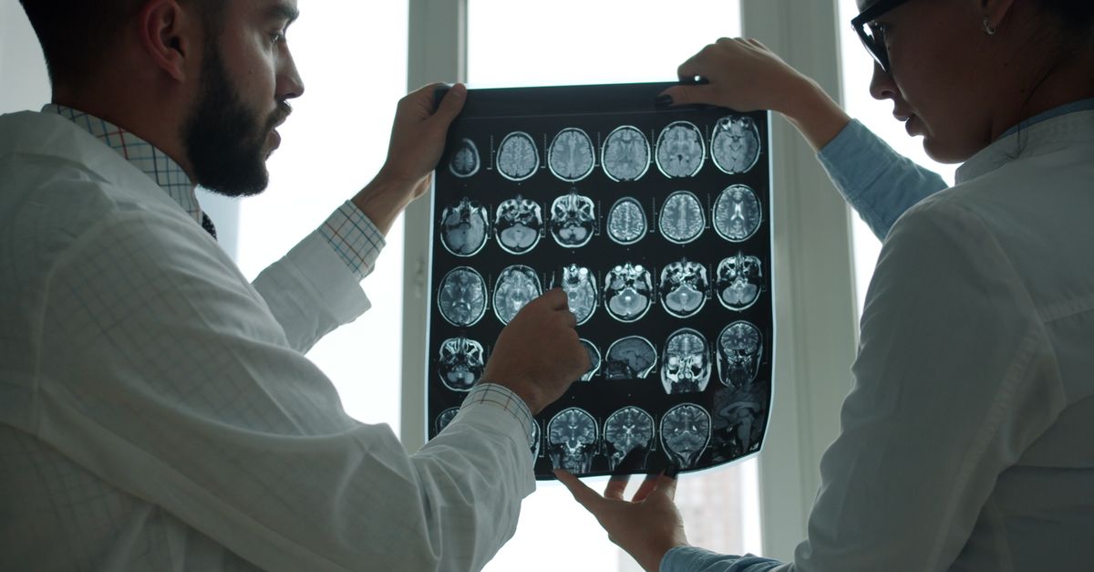
Medical imaging is essential for mapping the exact anatomical extent of the tumor before any surgical intervention is attempted. Computed tomography scans are widely used for tumors located in the chest, abdomen, or pelvis. The scan provides excellent detail of the tumor boundaries and helps detect any potential spread to the lungs or liver.
Because of their rich internal blood supply, these tumors typically enhance significantly when intravenous contrast dye is administered during a CT scan. This bright, prominent appearance on the scan is a key radiological feature. Large tumors may also show dark central areas, indicating cystic changes or tissue necrosis.
Magnetic resonance imaging provides superior contrast for soft tissues and is the preferred modality for tumors located in the brain, spine, head, neck, or extremities. An MRI allows the surgical team to visualize the intricate relationship between the tumor mass and essential nerves, major arteries, and joint structures.
10. Biopsy and Histological Analysis
A definitive diagnosis requires a tissue biopsy. A core needle biopsy is typically performed, utilizing advanced imaging guidance to safely extract a small cylinder of tissue from the solid mass. This procedure preserves the delicate architecture of the cells, which is necessary for an accurate pathological assessment.
Once the tissue reaches the laboratory, the pathologist applies specialized immunohistochemical stains. The tumor cells consistently demonstrate positive nuclear staining for the STAT6 protein, which serves as direct evidence of the underlying genetic mutation. The tissue also typically expresses CD34, a marker common to connective and vascular tissues.
The pathologist evaluates the specific number of dividing cells and the degree of cellular abnormality to assign the grade 2 classification. This formal pathology report acts as the foundational blueprint for the multidisciplinary treatment plan.
11. Surgical Treatment Options
Complete surgical resection is the primary and most effective treatment strategy. The primary goal for the surgeon is to remove the entire mass with clear, negative margins, ensuring no microscopic tumor cells are left behind in the adjacent healthy tissue.
The complexity of the surgery depends heavily on the tumor location. Tumors in the extremities may require wide local excision, while tumors in the chest cavity may necessitate the removal of a portion of the lung or chest wall. Given the dense network of fragile blood vessels within the tumor, surgical bleeding is a recognized risk requiring meticulous surgical technique.
In cases involving massive or highly vascular tumors, a specialized interventional radiologist may perform a preoperative embolization. This procedure involves deliberately blocking the specific arteries feeding the tumor, significantly reducing the risk of heavy bleeding during the subsequent operation.
12. Role of Radiation Therapy
While complete surgical removal is the optimal goal, radiation therapy serves a vital supplementary role in comprehensive disease management. Radiation utilizes highly focused energy beams to destroy abnormal cells and is frequently employed in the postoperative setting for grade 2 tumors.
If the surgical margins are exceptionally narrow or if tiny fragments of the tumor could not be safely removed due to proximity to critical organs, adjuvant radiation therapy is strongly recommended. The targeted radiation sterilizes the surgical bed, profoundly reducing the statistical risk of the tumor growing back in the same location.
In rare scenarios where a patient is medically unfit for major surgery, or if the tumor is located in an inoperable area, radiation therapy might be utilized as the primary standalone treatment to halt tumor progression and alleviate pressure symptoms.
13. Systemic Therapies and Targeted Treatments
Systemic therapy involves medications that travel throughout the entire body to treat disease. For grade 2 solitary fibrous tumours, traditional cytotoxic chemotherapy is rarely effective and is generally not utilized as a first-line treatment. The relatively slow division rate of these cells renders them largely resistant to standard chemotherapy drugs.
Systemic treatments are generally reserved for advanced cases where the disease has spread to distant organs or recurred aggressively despite surgery and radiation. In such instances, oncologists may explore anti-angiogenic medications. These specific drugs are designed to block the formation of new blood vessels, effectively starving the highly vascular tumor of its essential blood supply.
Clinical trials are continuously evaluating new targeted biological therapies that specifically inhibit the cellular pathways driven by the NAB2 and STAT6 fusion gene, offering potential future avenues for medical management.
14. Post-Treatment Monitoring and Prognosis
The clinical prognosis for individuals with a grade 2 solitary fibrous tumour is generally favorable following a complete surgical excision. However, the grade 2 designation indicates an intermediate risk level, meaning the tumor possesses a tangible capacity to recur locally or spread distantly over time.
Due to this recognized risk, lifelong medical surveillance is considered mandatory. The post-treatment protocol typically involves regular physical examinations combined with scheduled CT or MRI scans of the primary tumor site and the chest. During the first few years following surgery, these check-ups are scheduled frequently, typically every four to six months.
Strict adherence to this monitoring schedule allows the medical team to detect any potential recurrence at its earliest microscopic stage, facilitating prompt and highly effective salvage interventions.
15. Living with Connective Tissue Neoplasms
Managing a diagnosis of a rare soft tissue tumor involves navigating significant physical and emotional transitions. Postoperative recovery varies widely based on the surgical location. Patients recovering from chest surgery often engage in specific respiratory therapy, while those undergoing limb surgery benefit immensely from structured physical rehabilitation to restore full strength and joint mobility.
The emotional impact of carrying an intermediate-grade diagnosis can lead to feelings of anxiety regarding future health. Establishing an open, continuous dialogue with the oncology team provides reassurance and clarity. Patients are encouraged to utilize professional counseling services and connect with specialized support networks.
Maintaining a balanced diet, engaging in regular appropriate physical activity, and avoiding tobacco products support the immune system and enhance overall physical resilience during the long-term recovery phase.
16. When to See a Doctor
It is critical to seek a comprehensive medical evaluation if you notice a new, unexplained lump or mass anywhere on your body, regardless of whether it causes pain. Furthermore, persistent internal symptoms such as a chronic cough, unprovoked shortness of breath, or unexplained abdominal fullness require prompt investigation.
Seek immediate emergency medical care if you experience sudden, severe episodes of dizziness, profound confusion, or fainting. While rare, these symptoms could indicate a dangerous drop in blood sugar levels associated with specific hormone-secreting tumors. Early clinical assessment and precise imaging are the most reliable methods for securing an accurate diagnosis and implementing curative treatment.
17. Frequently Asked Questions FAQ
1. Is a grade 2 solitary fibrous tumour considered cancer?
It is classified as an intermediate-grade neoplasm. While it is not a highly aggressive cancer, it has a recognized potential to grow back locally or spread, requiring careful surgical removal and long-term monitoring.
2. What is the difference between this and a haemangiopericytoma?
Medically, they are now considered the exact same disease caused by the same genetic mutation. The medical community has shifted to using the term solitary fibrous tumour to describe all of these lesions.
3. Will I need chemotherapy after surgery?
Standard chemotherapy is generally ineffective for this condition and is rarely used unless the disease reaches an advanced, widely spread stage that cannot be treated with surgery or radiation.
4. Can this tumor be prevented?
There are no known environmental risks or lifestyle choices that cause this tumor. It arises from a spontaneous genetic error, meaning there is no way to prevent its occurrence.
5. What happens if the tumor comes back?
If routine imaging detects a recurrence, the primary treatment approach is typically another surgical procedure to remove the new growth, often supplemented by targeted radiation therapy.
18. Bibliography
Disclaimer: The content is for informational purposes only and does not replace medical advice. Always consult your doctor for personalized treatment.