1. Introduction
Heart valve disorders are a diverse group of cardiovascular conditions characterized by the structural or functional impairment of one or more of the heart’s four valves. These delicate structures are responsible for maintaining the unidirectional flow of blood through the cardiac chambers and into the systemic and pulmonary circulations. When a valve fails to operate correctly, the heart must work substantially harder to maintain adequate circulation.
The pathology of valvular disease falls into two primary categories: stenosis, where the valve opening narrows and restricts forward flow, and regurgitation, where the valve fails to close completely, allowing blood to leak backward. These mechanical defects can occur in isolation or in combination, and they may affect multiple valves simultaneously.
The clinical progression of a heart valve disorder is often insidious, developing over many years before overt symptoms arise. Proper diagnosis relies on specialized imaging techniques to quantify the severity of the mechanical defect. Once identified, modern cardiology offers an array of pharmacological and surgical interventions to alleviate symptoms, prevent permanent myocardial damage, and restore a normal quality of life.
2. Anatomy and Function of Cardiac Valves
The human heart contains four valves divided into two functional pairs. The atrioventricular valves, known as the tricuspid and mitral valves, separate the upper atria from the lower ventricles. The semilunar valves, consisting of the pulmonary and aortic valves, separate the ventricles from the major arteries exiting the heart.
Each valve is composed of thin, strong flaps of tissue called leaflets or cusps. The mitral valve uniquely possesses two leaflets, while the other three valves possess three. The leaflets of the atrioventricular valves are tethered to the ventricular walls by strong, string-like structures called chordae tendineae, which prevent the valves from inverting during forceful ventricular contractions.
In a healthy heart, these valves open fully to allow blood to pass with minimal resistance and snap shut tightly to prevent any backward flow. This precise, synchronized opening and closing is entirely driven by pressure gradients created as the heart muscle contracts and relaxes during the cardiac cycle.
3. Pathophysiology of Valvular Dysfunction
Valvular dysfunction creates a significant mechanical disadvantage for the myocardium. The specific pathophysiological consequences depend on whether the defect is primarily stenotic or regurgitant. Both mechanisms force the heart to employ compensatory strategies to maintain cardiac output, which ultimately leads to detrimental structural remodeling.
When the heart is faced with a mechanical burden, the cardiac muscle responds much like skeletal muscle. It attempts to adapt. Over time, the affected chamber may undergo hypertrophy, where the muscle walls become abnormally thick to generate higher pressures. Alternatively, the chamber may dilate, stretching outward to accommodate an increased volume of blood.
While these adaptations initially preserve blood flow, they are not sustainable. Hypertrophied muscle becomes stiff and requires excessive oxygen, while dilated muscle eventually loses its contractile strength. If the mechanical valve defect is not corrected, this continuous strain inexorably leads to progressive heart disease and failure.
4. Mechanisms of Stenosis
Valvular stenosis is the narrowing of the valve orifice. This narrowing is typically the result of the valve leaflets becoming thickened, fused together, or heavily calcified. As the opening shrinks, the heart chamber immediately preceding the stenotic valve must generate significantly higher pressure to force blood through the restricted space.
Aortic stenosis is the most common and clinically profound stenotic lesion in developed nations. As the aortic valve narrows, the left ventricle must pump with immense force to overcome the obstruction. This continuous pressure overload leads to severe left ventricular hypertrophy.
Mitral stenosis, frequently a late consequence of rheumatic fever, obstructs blood flowing from the left atrium into the left ventricle. This causes pressure to build rapidly in the left atrium, leading to atrial enlargement and increasing the pressure backward into the pulmonary veins, which forces fluid into the lungs.
5. Mechanisms of Regurgitation
Valvular regurgitation, also known as insufficiency or incompetence, occurs when the valve leaflets fail to coapt, or seal completely. This allows a portion of the ejected blood to flow backward into the preceding chamber during the resting phase of the cardiac cycle.
This backward flow creates a volume overload. The heart must now pump both the normal amount of incoming blood plus the extra volume that leaked backward. To accommodate this excess volume, the affected heart chamber progressively dilates.
Mitral regurgitation allows blood to leak backward into the left atrium during ventricular contraction. Aortic regurgitation allows blood that was just pumped into the aorta to fall back into the left ventricle. Both conditions force the left ventricle to work tirelessly to process the excess volume, leading to eventual myocardial exhaustion.
6. Congenital Valvular Abnormalities
Many heart valve disorders are present at birth, resulting from errors during embryological development. The most frequent congenital anomaly is a bicuspid aortic valve. In this condition, the aortic valve develops with only two functional leaflets instead of the normal three.
A bicuspid valve may function perfectly for decades. However, because the two leaflets must bear the entire mechanical stress of opening and closing, they are highly prone to premature wear and tear. Individuals with a bicuspid valve frequently develop severe aortic stenosis or regurgitation much earlier in life than those with normal anatomy.
Other congenital defects include Ebstein anomaly, where the tricuspid valve is malformed and displaced downward into the right ventricle, and pulmonary valve stenosis, which restricts blood flow to the lungs and places a heavy burden on the right side of the heart.
7. Degenerative and Age-Related Changes
In developed countries, degenerative changes associated with aging are the leading cause of heart valve disorders. The continuous mechanical stress placed on the valves over a lifetime causes cumulative damage to the collagen matrix of the leaflets.
Senile calcific aortic stenosis occurs when calcium deposits accumulate on the aortic valve leaflets over decades, causing them to become rigid and immobile. This process shares many pathophysiological similarities with atherosclerosis, involving lipid deposition, chronic inflammation, and active tissue calcification.
Myxomatous degeneration primarily affects the mitral valve. In this condition, the structural connective tissue within the valve leaflets becomes abnormally weak and spongy. The leaflets stretch and prolapse backward into the atrium during contraction, a condition known as mitral valve prolapse, which frequently leads to progressive mitral regurgitation.
8. Infectious and Rheumatic Etiologies
Infectious agents can directly destroy heart valves. Infective endocarditis is a severe, life-threatening condition where circulating bacteria adhere to the valve surface, forming growths called vegetations. These bacteria literally digest the valve tissue, leading to rapid, acute regurgitation that often requires emergency surgical replacement.
Rheumatic heart disease is a profound cause of valvular damage, particularly in developing nations. It is a delayed autoimmune complication of untreated group A streptococcal pharyngitis (strep throat). The immune system produces antibodies to fight the bacteria, but these antibodies mistakenly attack the tissues of the heart valves.
The inflammatory process of rheumatic fever causes the valve leaflets to become scarred, thickened, and fused together over many years. This typically results in a combination of stenosis and regurgitation, most commonly affecting the mitral and aortic valves, creating complex, multi-valvular disease profiles.
9. Clinical Symptoms and Presentation
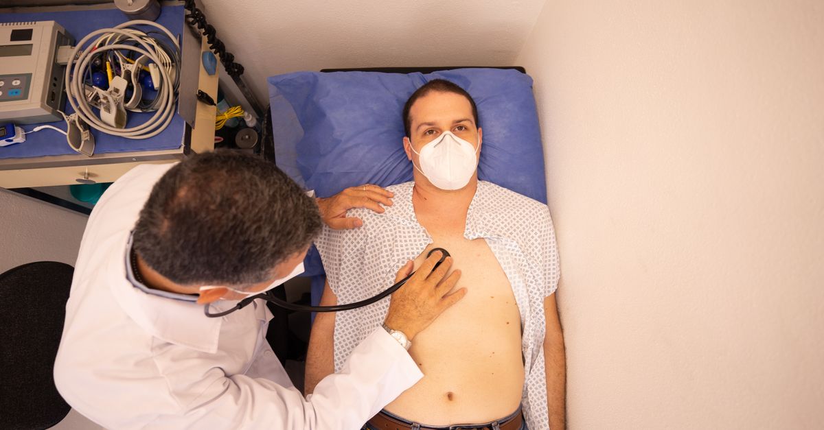
The clinical presentation of a heart valve disorder correlates directly with the severity of the mechanical defect and the heart’s ability to compensate. During the early stages, patients are frequently entirely asymptomatic. The disorder is often discovered incidentally when a physician hears a heart murmur during a routine stethoscope examination.
As the condition progresses, patients typically report a gradual decline in exercise tolerance. They may notice unusual fatigue, weakness, and shortness of breath during physical exertion. This exertional dyspnea indicates that the heart can no longer increase cardiac output sufficiently to meet the body’s demands.
In advanced stages, the symptoms become prominent even at rest. Patients may experience orthopnea (shortness of breath when lying flat), paroxysmal nocturnal dyspnea, palpitations resulting from associated arrhythmias, and significant swelling in the ankles and abdomen due to systemic fluid retention.
10. Hemodynamic Consequences
The hemodynamic consequences of severe valvular disease are profound. Stenotic lesions lead to severe pressure gradients across the affected valve, while regurgitant lesions lead to massive volume shifts. Both scenarios culminate in elevated pressures within the pulmonary or systemic venous systems.
When left-sided valves (mitral or aortic) fail, the elevated pressures transfer backward into the pulmonary circulation. This causes pulmonary hypertension, which forces fluid into the alveolar spaces of the lungs, resulting in pulmonary edema and severe respiratory distress.
When right-sided valves (tricuspid or pulmonary) fail, the elevated pressure transfers backward into the systemic venous circulation. This leads to hepatic congestion (an engorged liver), ascites (fluid accumulation in the abdomen), and profound peripheral edema in the lower extremities.
11. Diagnostic Evaluation
A thorough diagnostic evaluation is mandatory to determine the exact nature and severity of the valvular defect. The foundation of this evaluation is echocardiography. This non-invasive ultrasound utilizes high-frequency sound waves to generate real-time, dynamic images of the heart structures.
Transthoracic echocardiography allows the cardiologist to visualize the opening and closing of the valve leaflets, measure the thickness of the heart walls, and calculate the ejection fraction. Doppler ultrasound is utilized simultaneously to visualize the direction and velocity of blood flow, providing precise measurements of stenotic gradients and the volume of regurgitant jets.
If a more detailed view is required, a transesophageal echocardiogram is performed. A specialized ultrasound probe is passed down the esophagus, resting directly behind the heart, providing exceptionally clear images of the posterior cardiac structures, particularly the mitral valve.
12. Pharmacological Management
Currently, there are no medical therapies capable of reversing or repairing a mechanically damaged heart valve. Pharmacological management is utilized exclusively to optimize the heart’s function, alleviate symptoms, and prevent complications while awaiting definitive surgical intervention.
Diuretics are the cornerstone of symptom management. They promote the excretion of excess fluid through the kidneys, reducing the volume burden on the heart and alleviating pulmonary congestion and peripheral edema.
Other medications, such as beta-blockers, angiotensin-converting enzyme inhibitors, and calcium channel blockers, may be used to lower blood pressure, reduce the heart rate, and decrease the resistance the heart must pump against. Anticoagulants are frequently prescribed for patients who develop atrial fibrillation due to atrial enlargement, preventing the formation of systemic blood clots.
13. Surgical and Transcatheter Interventions
Definitive treatment for severe heart valve disorders requires mechanical correction. The traditional approach is open-heart surgery to either repair or replace the damaged valve. Valve repair is the preferred option, particularly for mitral valve disease, as it preserves the patient’s native tissue and avoids the need for long-term anticoagulation.
If the valve is irreparably calcified or damaged, it must be replaced. Mechanical valves are highly durable but require the patient to take lifelong blood-thinning medications to prevent clotting. Bioprosthetic valves, made from bovine or porcine tissue, do not require lifelong anticoagulation but tend to wear out over ten to fifteen years.
In recent years, transcatheter therapies have revolutionized the field. Procedures like transcatheter aortic valve replacement allow cardiologists to insert a new, fully collapsible replacement valve through a catheter in the groin, delivering it precisely over the old, diseased valve without the need for open-chest surgery.
14. Summary of Valvular Lesions
Differentiating between the specific mechanical failures is essential for determining the appropriate clinical response. The table below outlines common valvular pathologies.
| Valve Defect | Mechanism | Primary Consequence |
|---|---|---|
| Aortic Stenosis | Narrowing of outflow tract | Left ventricular pressure overload and hypertrophy |
| Mitral Regurgitation | Incomplete closure during systole | Left atrial dilation and ventricular volume overload |
| Mitral Stenosis | Narrowing of inflow tract | Left atrial pressure overload and pulmonary edema |
| Aortic Regurgitation | Backward leak during diastole | Left ventricular volume overload and dilation |
Early detection and precise surgical timing are the most critical factors in preventing irreversible myocardial damage and ensuring a favorable long-term prognosis.
15. Frequently Asked Questions (FAQ)
1. Can medications fix a leaking heart valve?
No. Medications can relieve the symptoms of a leaking valve, such as shortness of breath or fluid retention, by helping the heart pump more efficiently. However, they cannot physically repair the torn or stretched tissue of the valve itself.
2. What is a heart murmur?
A heart murmur is simply an extra sound the doctor hears through a stethoscope. It is the sound of turbulent blood flow. While some murmurs are innocent, a prominent murmur often indicates that blood is rushing forcefully through a narrowed valve or leaking backward.
3. Why do I need antibiotics before seeing the dentist if I have a valve disorder?
Certain structural valve abnormalities increase the risk of infective endocarditis. Routine dental work can release normal mouth bacteria into the bloodstream, where they might stick to a damaged valve and cause a severe infection. Prophylactic antibiotics prevent this.
4. Is open-heart surgery the only way to replace a valve?
No. Transcatheter procedures, such as TAVR for aortic valves, are now widely used. These minimally invasive techniques allow doctors to replace a valve using a thin tube inserted through an artery in the leg, avoiding open-chest surgery entirely.
5. How long do biological replacement valves last?
Bioprosthetic valves, typically made from cow or pig tissue, usually last between ten to fifteen years before they begin to degenerate and require replacement. Mechanical metal valves can last a lifetime but require the patient to take strong blood thinners forever.
16. Bibliography
Disclaimer: The content is for informational purposes only and does not replace medical advice. Always consult your doctor for personalized treatment.