1. Introduction to Joint Pain (Arthralgia)
Joint pain, clinically documented as arthralgia, is a highly prevalent medical symptom resulting from inflammation, acute structural injury, systemic disease, or progressive mechanical degeneration of the articular structures. Because joints form the dynamic connections between bones, allowing for all bodily movement, pathology within these spaces severely impairs physical function and diminishes the overall quality of life. Rapid and accurate clinical identification of the pain source is the foundation of effective medical intervention.
The diagnostic evaluation of joint pain is complex and requires distinguishing between a primary intra-articular process, involving the joint capsule and cartilage, and a periarticular process, involving the surrounding tendons, ligaments, and bursae. Furthermore, the clinician must ascertain whether the underlying etiology is mechanical and localized, or inflammatory and systemic.
Understanding the specific onset, duration, temporal pattern, and exact number of joints involved provides immediate, critical clues. A systematic medical approach ensures that severe destructive conditions, such as autoimmune arthritis or deep joint space infections, are recognized and treated aggressively, preventing irreversible structural deformity.
2. Anatomy and Physiology of Synovial Joints
Synovial joints are the most common and highly mobile type of joint in the human body. They are characterized by a distinct anatomical structure designed to minimize friction and absorb immense mechanical loads. The opposing bony surfaces within the joint are capped with a layer of smooth, resilient hyaline cartilage. This cartilage acts as a highly efficient shock absorber, distributing compressive forces safely across the underlying subchondral bone.
The entire articulation is enclosed within a tough, fibrous joint capsule that provides static structural stability. The inner surface of this capsule is lined with a highly vascularized synovial membrane. This membrane continually secretes a viscous, nutrient-rich synovial fluid into the joint cavity, which drastically reduces mechanical friction and nourishes the avascular articular cartilage.
Dynamic stability is provided by an intricate network of dense ligaments and surrounding musculotendinous units crossing the joint space. A rich network of sensory nerves densely innervates the joint capsule and ligaments, immediately transmitting pain signals in response to excessive mechanical stretch, structural damage, or localized chemical inflammation.
3. Osteoarthritis: Degenerative Joint Disease
Osteoarthritis represents the most frequent cause of chronic joint pain worldwide. It is a progressive, primarily non-inflammatory disease characterized by the gradual deterioration and ultimate loss of the protective articular cartilage. As the cartilage wears down over decades of continuous mechanical loading, the underlying subchondral bones grind against each other.
The biological response to this increased mechanical stress includes subchondral bone sclerosis and the formation of osteophytes (bone spurs) along the joint margins. Patients typically describe a deep, localized, aching pain that is reliably provoked by physical exertion and weight-bearing, but significantly improves with rest.
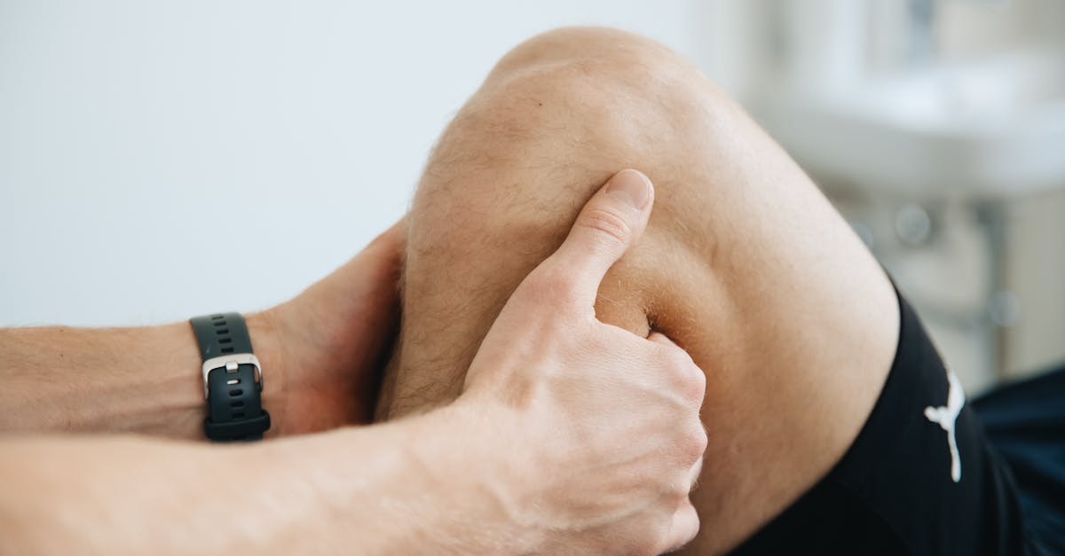
Osteoarthritis most commonly affects the major weight-bearing joints, including the knees and hips, as well as the small joints of the hands and spine. While it frequently causes brief morning stiffness, this stiffness typically dissipates within thirty minutes of waking. Management relies heavily on weight management, physical therapy to strengthen supporting muscles, and pharmacological analgesia.
4. Rheumatoid Arthritis: Autoimmune Inflammation
Rheumatoid arthritis is a profound systemic autoimmune disorder in which the body’s immune system erroneously targets the synovial membranes of the joints. This aggressive immune attack causes severe synovitis, resulting in immense joint swelling, pronounced warmth, and deep, throbbing pain.
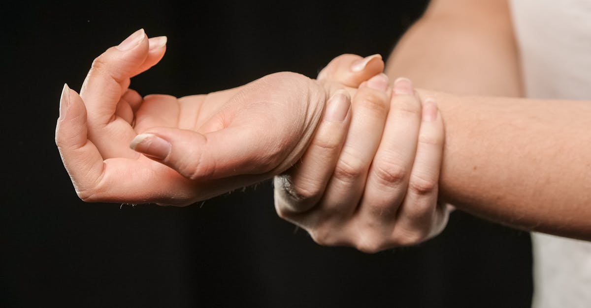
Unlike osteoarthritis, the joint pain of rheumatoid arthritis is highly symmetrical, typically affecting the small joints of the hands and wrists on both sides of the body simultaneously. A clinical hallmark of the disease is profound, debilitating morning stiffness that lasts for several hours and only slowly improves with physical movement.
The chronic inflammatory cascade releases powerful destructive enzymes that rapidly erode the articular cartilage and the underlying bone, leading to severe, irreversible structural deformities. Diagnosis requires sophisticated serological testing. Immediate initiation of targeted disease-modifying antirheumatic drugs is absolute paramount to suppress the immune response and preserve functional joint architecture.
5. Gout and Crystal Arthropathies
Gout is a severely painful, acute inflammatory arthritis caused by the systemic accumulation of uric acid in the bloodstream. When serum uric acid concentrations exceed their physiological saturation point, the excess molecules precipitate into microscopic, needle-like monosodium urate crystals. These crystals preferentially deposit within the synovial fluid of cooler peripheral joints.
The sudden shedding of these crystals into the joint space triggers an explosive localized immune response. Patients experience the rapid onset—often overnight—of agonizing pain, intense erythema, and profound swelling, most frequently involving the first metatarsophalangeal joint of the great toe, a condition known as podagra.
Pseudogout is a clinically similar crystal arthropathy caused by the deposition of calcium pyrophosphate crystals, typically affecting larger joints such as the knee. Diagnosis is definitively established by aspirating synovial fluid and identifying the specific crystals under a polarized microscope. Acute management utilizes potent anti-inflammatory agents, while chronic care requires systemic urate-lowering pharmacotherapy.
6. Infectious and Septic Arthritis
Septic arthritis constitutes a true orthopedic emergency. It occurs when a highly virulent bacterial pathogen, most commonly Staphylococcus aureus, gains entry into the sterile joint space. Bacteria can enter directly through a penetrating traumatic wound, spread from an adjacent bone infection (osteomyelitis), or travel hematogenously through the bloodstream from a distant infection site.
The bacterial proliferation triggers a massive influx of neutrophils, turning the synovial fluid into thick, purulent pus. This intense inflammatory reaction rapidly degrades the articular cartilage, destroying the joint within a matter of days. Patients present with sudden, excruciating pain, an acutely swollen, hot, and erythematous joint, and a severe absolute inability to bear weight or tolerate any passive movement.
Associated systemic symptoms, including high fever and rigors, are frequently present. Immediate medical intervention requires the urgent aspiration of the joint fluid for microbiological culture, followed by rapid surgical washout of the joint space and the initiation of broad-spectrum intravenous antibiotic therapy.
7. Traumatic Injuries and Ligament Damage
Acute physical trauma is a highly common etiology of sudden, severe joint pain. High-energy impacts, athletic injuries, or severe twisting forces can stretch or completely rupture the stabilizing ligaments surrounding the joint, resulting in a sprain. If the force exceeds the ultimate tensile strength of the capsule, a joint dislocation occurs, where the articular surfaces are completely separated.
Traumatic joint injuries are characterized by immediate, sharp pain, rapid and profound soft tissue swelling, and visible mechanical instability. Hemarthrosis, or the rapid bleeding directly into the joint space, frequently accompanies severe ligamentous tears or intra-articular fractures, causing immense capsular distension and intense pain.
Clinical evaluation necessitates immediate radiographic imaging to identify or rule out structural bone fractures. The acute management of traumatic joint injuries follows the strict protocols of rest, immobilization, and localized cryotherapy, with severe ligamentous ruptures frequently requiring delayed surgical reconstruction to restore mechanical stability.
8. Viral Arthralgia and Systemic Infections
A multitude of systemic viral infections can cause transient, severe joint pain, referred to as viral arthropathy. Viruses such as Parvovirus B19, Hepatitis B and C, Rubella, and the Epstein-Barr virus can provoke a generalized inflammatory response that affects the synovial tissues.
Viral joint pain typically presents as a rapid-onset, symmetrical polyarthralgia involving multiple joints simultaneously. The pain is frequently accompanied by classic systemic viral symptoms, including low-grade fever, extreme fatigue, generalized myalgia, and a diffuse maculopapular skin rash.
The joint pain associated with viral infections is usually self-limiting, resolving spontaneously over several weeks without causing any permanent structural degradation to the articular cartilage or bone. Management is entirely supportive, focusing on symptomatic relief utilizing standard oral nonsteroidal anti-inflammatory drugs and ensuring adequate hydration and rest.
9. Lupus and Connective Tissue Disorders
Systemic lupus erythematosus is a complex, multi-organ autoimmune disease that frequently presents with significant joint involvement. The immune system produces vast quantities of autoantibodies that deposit within various tissues, triggering chronic systemic inflammation.
The joint pain of lupus is typically migratory, moving unpredictably from one joint to another, and frequently involves the wrists and small joints of the hands. While the joints may be highly painful and visibly swollen, lupus arthritis rarely causes the severe, erosive bone destruction characteristic of rheumatoid arthritis.
The pain often fluctuates in severity and is frequently accompanied by other systemic manifestations, such as severe photosensitivity, the classic malar “butterfly” rash across the face, and profound fatigue. Thorough rheumatological evaluation and sophisticated serological profiling are necessary to establish the diagnosis and direct systemic immunosuppressive therapy.
10. Tendinopathy and Periarticular Pain
Frequently, pain perceived within the joint actually originates from the soft tissues immediately surrounding the articulation, a phenomenon known as periarticular pain. Tendons, which attach muscles to bone, and bursae, which provide frictionless gliding surfaces, are highly susceptible to acute inflammation and chronic degenerative wear due to repetitive mechanical overload.
Conditions such as lateral epicondylitis (tennis elbow), rotator cuff tendinopathy, or trochanteric bursitis of the hip generate severe, highly localized pain. Unlike true intra-articular joint disease, where pain is present throughout the entire range of motion, periarticular pain is reliably provoked only during specific active movements that load the affected tendon, while passive movement by the examiner is often painless.
Treatment for periarticular pain focuses intensely on activity modification, biomechanical correction, and highly targeted physical therapy to stimulate cellular repair and restore the structural integrity of the affected soft tissues.
11. Fibromyalgia and Non-Articular Pain Syndromes
Fibromyalgia is a complex, chronic central pain processing disorder characterized by widespread musculoskeletal pain, profound fatigue, and significant sleep disturbances. While patients frequently complain of severe, generalized joint pain, the underlying pathology does not involve active inflammation or structural tissue damage within the joints themselves.
Instead, the disorder involves a pathological amplification of sensory nerve signals within the central nervous system. The brain processes normal sensory input as severe pain. Clinical examination typically reveals multiple specific “tender points” located in the soft tissues and muscles, while the joints themselves appear structurally normal without any swelling or restricted range of motion.
Management of fibromyalgia requires a highly comprehensive, multidisciplinary approach. Pharmacological interventions utilize specific neuromodulating agents and antidepressants to alter central pain processing, combined with a graded, low-impact aerobic exercise program and cognitive behavioral therapy.
12. The Impact of Obesity and Biomechanical Stress
Obesity plays a massive and multifaceted role in the development and rapid progression of chronic joint pain, particularly in the lower extremities. The sheer mechanical load placed upon the knees and hips during normal ambulation is drastically amplified by excess body weight, accelerating the physical degradation of the articular cartilage.
Beyond pure mechanical wear, adipose tissue is highly metabolically active. It continuously secretes potent inflammatory cytokines, known as adipokines, directly into the systemic circulation. This creates a state of chronic, low-grade systemic inflammation that negatively impacts the biological integrity of all synovial joints, even those not subjected to weight-bearing stress, such as the hands.
Consequently, comprehensive weight loss is considered a primary, highly effective therapeutic intervention. Reducing body mass directly unloads the structural cartilage and mitigates the systemic inflammatory drive, providing profound relief of chronic joint pain.
13. Diagnostic Assessment and Blood Panels
The clinical evaluation of joint pain relies on establishing the exact pattern of involvement. The clinician notes whether the pain is monoarticular (one joint) or polyarticular (multiple joints), and whether the distribution is symmetrical or asymmetrical.
| Clinical Pattern | Associated Symptoms | Most Probable Etiology |
|---|---|---|
| Asymmetrical, large weight-bearing joints | Pain worsened by activity, brief morning stiffness | Osteoarthritis |
| Symmetrical, small joints of hands/wrists | Severe, prolonged morning stiffness, swelling | Rheumatoid Arthritis |
| Acute, extremely painful single swollen joint | Intense redness, overnight onset | Gout (Crystal Arthropathy) |
| Acute, hot, swollen joint with fever | Inability to bear weight, severe systemic illness | Septic Arthritis (Infection) |
Laboratory blood panels are critical for diagnosing systemic conditions. Erythrocyte sedimentation rate (ESR) and C-reactive protein (CRP) assess global inflammation, while specific autoantibodies (Rheumatoid Factor, Anti-CCP, ANA) precisely identify underlying autoimmune pathologies.
14. Radiographic Imaging and Fluid Analysis
Standard plain radiographs (X-rays) are the universally accepted first step in imaging painful joints. They clearly demonstrate bony anatomy, revealing acute fractures, the joint space narrowing indicative of cartilage loss in osteoarthritis, and the characteristic marginal bone erosions seen in advanced rheumatoid arthritis.
When complex soft tissue injury is suspected, such as meniscal tears in the knee or ligamentous ruptures, magnetic resonance imaging provides unparalleled anatomical resolution. Diagnostic ultrasound is highly effective for identifying joint effusions and active synovial inflammation.
In the presence of an acutely swollen joint without a history of trauma, joint aspiration (arthrocentesis) is mandatory. The clinician extracts a sample of synovial fluid utilizing a sterile needle. Analyzing the cell count, presence of crystals, and performing microbiological cultures definitively differentiates between an acute gout flare and a life-threatening septic joint infection.
15. Non-Pharmacological Management and Therapy
Conservative, non-pharmacological interventions are the absolute foundation of long-term joint pain management. Physical therapy is critically important. A structured, progressive exercise program prevents severe muscular deconditioning and enhances the dynamic stability provided by the muscles crossing the affected joint, effectively unloading the painful articular surfaces.
Occupational therapy assists patients in modifying their daily activities, instructing them in joint-protection techniques, and providing customized splints to rest inflamed joints in optimal anatomical alignment. Thermal modalities, including localized cryotherapy for acute inflammatory flares and deep heat for chronic mechanical stiffness, offer significant, medication-free symptomatic relief.
Dietary modifications, specifically adopting an anti-inflammatory diet rich in omega-3 fatty acids and antioxidants, support the systemic reduction of inflammatory mediators and promote overall joint health.
16. Pharmacological and Surgical Treatments
The pharmacological strategy is strictly dictated by the established underlying diagnosis. Simple analgesics and nonsteroidal anti-inflammatory drugs provide the first line of defense for osteoarthritis and acute periarticular injuries. For aggressive autoimmune arthritis, highly specialized disease-modifying antirheumatic drugs and modern biologic therapies specifically target and neutralize the inflammatory cytokines driving joint destruction.
Intra-articular corticosteroid injections provide rapid, potent suppression of severe localized inflammation, offering a critical window of pain relief that enables patient participation in physical rehabilitation. Viscosupplementation, the injection of hyaluronic acid into the joint, provides a temporary mechanical lubricant for arthritic knees.
When conservative and pharmacological measures fail to control intractable pain or when structural joint destruction severely impairs mobility, surgical intervention is required. Advanced surgical options range from arthroscopic debridement to complete joint arthroplasty (joint replacement), which offers dramatic pain relief and the full restoration of mechanical function.
17. Frequently Asked Questions (FAQ)
1. Does cracking your knuckles cause arthritis?
No. Comprehensive medical research demonstrates absolutely no link between cracking knuckles and the development of osteoarthritis. The “cracking” sound is simply the rapid release of gas bubbles forming within the synovial fluid of the joint capsule.
2. Does cold, damp weather make joint pain worse?
Many patients report increased joint pain during cold, damp weather. Clinicians theorize that a drop in barometric pressure causes the tissues surrounding the joint to expand slightly, placing increased mechanical pressure on the already sensitive, inflamed nerves within the joint capsule.
3. Can dietary supplements like glucosamine rebuild cartilage?
No currently available dietary supplement, including glucosamine or chondroitin, has been clinically proven to regenerate or rebuild lost articular cartilage. While some patients report mild symptomatic relief, they do not reverse the structural damage of osteoarthritis.
4. Is rest the best treatment for sore joints?
While brief rest is necessary during an acute inflammatory flare or after a traumatic injury, prolonged rest is highly detrimental. It causes rapid muscle wasting and increases joint stiffness. Gentle, low-impact movement is essential to nourish the cartilage and maintain function.
5. How do I know if my joint pain is serious?
Immediate medical evaluation is required if the joint pain is accompanied by severe swelling, intense redness, the joint feels hot to the touch, or if you develop an unexplained systemic fever, as these are critical signs of a severe bacterial joint infection.
Bibliography
Disclaimer: The content is for informational purposes only and does not replace medical advice. Always consult your doctor for personalized treatment.