
1. Introduction
Having persistently bloodshot eyes can indeed be a sign of an underlying health issue. While occasional eye redness is a common and harmless result of fatigue, minor irritation, or a poor night of sleep, a red eye that refuses to clear up signals a chronic state of localized inflammation. The visible redness occurs when the tiny, superficial blood vessels on the surface of the eye become engorged and dilated. Determining the exact cause requires evaluating the specific nature of the redness and identifying any concurrent symptoms, such as pain, visual disturbances, or systemic medical conditions.
The human eye is a highly sensitive and vulnerable organ, constantly exposed to environmental elements and deeply connected to the systemic vascular and immune networks of the body. When the eye remains perpetually red, it is an anatomical warning sign that the delicate surface tissues are being continuously assaulted by structural dryness, allergic reactions, active infections, or dangerous spikes in internal pressure.
Addressing chronic bloodshot eyes requires a meticulous clinical approach. Dismissing persistent redness as simple fatigue and relying heavily on over-the-counter cosmetic eye drops can mask severe, vision-threatening pathologies. A comprehensive ophthalmological examination is strictly necessary to differentiate benign surface irritation from sight-threatening intraocular diseases, ensuring the implementation of targeted therapies that preserve long-term visual acuity.
2. The Vascular Anatomy of the Eye
To comprehend why an eye appears red, one must look at the anatomy of the conjunctiva and the episclera. The sclera is the tough, fibrous white outer layer of the eyeball. Covering the sclera is a thin, transparent, and highly vascularized mucous membrane known as the conjunctiva, which also lines the inside of the eyelids.
Beneath the conjunctiva lies the episclera, another thin layer of tissue containing a rich network of blood vessels. In a healthy, resting state, the blood vessels in these superficial layers are incredibly narrow and virtually invisible to the naked eye, allowing the bright white of the underlying sclera to shine through.
However, these vessels are highly reactive. In response to physical irritation, chemical allergens, or internal inflammatory signals, the autonomic nervous system commands these vessels to dilate rapidly. This vasodilation increases blood flow to deliver immune cells and oxygen to the affected area. When hundreds of these tiny vessels enlarge simultaneously, the white of the eye is visually overpowered by the distinct red or pink hue of circulating blood.
3. Dry Eye Syndrome
Dry eye syndrome is arguably the most prevalent cause of persistently bloodshot eyes in the modern adult population. The surface of the eye relies on a continuous, stable tear film for lubrication, oxygen delivery, and protection against friction. This tear film is a complex mixture of water, oils, and mucus.
When the lacrimal glands fail to produce enough aqueous tears, or when the meibomian glands in the eyelids fail to produce sufficient oil, the tear film evaporates too rapidly. The exposed surface of the cornea and conjunctiva becomes structurally desiccated. This chronic dryness causes microscopic damage to the superficial cells, prompting a continuous, low-grade inflammatory response.
Patients with dry eye syndrome frequently complain of a gritty, burning, or sandy sensation in their eyes, accompanied by persistent redness that typically worsens toward the end of the day or after prolonged periods staring at a digital screen. Restoring the tear film with specialized lubricating drops is essential to break the inflammatory cycle.
4. Allergic Conjunctivitis
For individuals with highly reactive immune systems, allergic conjunctivitis is a primary driver of chronic eye redness. The conjunctiva is heavily populated with mast cells, which are immune sentinels packed with histamine. When the eye is exposed to environmental allergens like pollen, pet dander, or dust mites, the mast cells degranulate aggressively.
The sudden, massive release of histamine into the local tissues causes profound and immediate vasodilation of the conjunctival blood vessels, turning the eye bright red. It also causes the tiny capillaries to leak fluid, resulting in noticeable swelling of the clear conjunctival tissue, a symptom known as chemosis.
The absolute hallmark symptom of allergic conjunctivitis is intense, unyielding itching. Unlike viral or bacterial infections, allergic redness is almost always bilateral, affecting both eyes simultaneously. Managing this condition requires identifying and avoiding the specific environmental triggers, alongside the use of topical ophthalmic antihistamines to stabilize the hyperactive mast cells.
5. Viral and Bacterial Conjunctivitis
Infectious conjunctivitis, colloquially known as pink eye, is a highly contagious condition that causes severe eye redness. Viral conjunctivitis is most commonly caused by an adenovirus, the same virus responsible for the common cold. It typically begins in one eye and spreads rapidly to the other, causing a bright, watery red eye accompanied by enlarged, tender lymph nodes directly in front of the ear.
Bacterial conjunctivitis is characterized by the presence of a thick, purulent discharge. The eye becomes deeply red, and the patient frequently wakes up with their eyelids heavily crusted and physically glued shut by yellow or green pus.
While both infectious forms cause significant redness, they are typically acute processes that resolve within one to two weeks, distinguishing them from conditions that cause months of persistent bloodshot eyes. However, if a bacterial infection is suspected, topical antibiotic drops are required to accelerate clearance and prevent corneal damage.
6. Glaucoma and Intraocular Pressure
A persistently bloodshot eye can occasionally be a visible sign of a catastrophic internal crisis, specifically acute angle-closure glaucoma. The eye maintains a specific shape by continuously producing a clear fluid called aqueous humor, which flows through the front chamber of the eye and drains out through a complex anatomical meshwork.
If the drainage angle suddenly becomes blocked, the fluid has nowhere to go, and the intraocular pressure spikes astronomically within hours. This extreme internal pressure compresses the delicate optic nerve at the back of the eye and severely compromises blood flow to the ocular tissues.
Acute angle-closure glaucoma presents with a deeply red eye, excruciating orbital pain, a cloudy or hazy cornea, and profound visual disturbances such as seeing rainbow-colored halos around lights. This is a profound ophthalmological emergency. If the pressure is not lowered immediately through systemic medications or laser surgery, the patient faces permanent, irreversible blindness.
7. Uveitis and Autoimmune Conditions
Uveitis is the inflammation of the uvea, the deeply pigmented middle layer of the eye that includes the iris, ciliary body, and choroid. Unlike surface conjunctivitis, uveitis involves inflammation deep within the structural tissues of the eyeball. The redness associated with uveitis is distinct; it is often concentrated in a dark, purplish-red ring directly surrounding the colored iris, a pattern known as ciliary flush.
Uveitis is frequently an ocular manifestation of a systemic autoimmune disease. The immune system mistakenly attacks the internal structures of the eye, assuming them to be foreign tissue. This condition is strongly linked to systemic disorders such as ankylosing spondylitis, rheumatoid arthritis, and inflammatory bowel disease.
Patients with uveitis experience a deeply aching red eye, significant blurred vision, and extreme photophobia, where exposure to even normal room lighting causes severe, stabbing pain. Because the intense inflammation can rapidly cause permanent scarring and cataracts, urgent evaluation and potent topical corticosteroid therapy are mandatory.
8. Subconjunctival Hemorrhage
A sudden, terrifyingly bright red patch on the white of the eye is typically a subconjunctival hemorrhage. This occurs when a single, tiny blood vessel in the conjunctiva breaks and bleeds into the space between the conjunctiva and the underlying sclera. Because the tissue is transparent, the pooled blood is starkly visible.
This bleeding is usually triggered by a sudden spike in venous pressure, such as a severe coughing fit, aggressive sneezing, heavy lifting, or intense vomiting. Sometimes, simply rubbing the eye too hard can break a fragile vessel.
Despite its alarming appearance, looking like a pool of fresh blood, a subconjunctival hemorrhage is almost entirely harmless. It does not affect vision, causes no pain, and requires no medical treatment. The body naturally reabsorbs the trapped blood over one to two weeks, much like a bruise fading on the skin.
9. Contact Lens Complications
Individuals who wear contact lenses are at a significantly elevated risk for chronic eye redness. A contact lens is a foreign piece of plastic sitting directly on the delicate cornea, relying on the tear film to float comfortably. If the lens is poorly fitted, worn for too long, or inadequately cleaned, it induces severe structural and biological stress on the eye.
Overwearing lenses reduces the amount of oxygen reaching the cornea, a state called hypoxia. The eye responds to this oxygen starvation by rapidly growing new, visible blood vessels into the clear cornea, a process known as neovascularization, making the eye appear chronically red and irritated.
Furthermore, improper contact lens hygiene invites aggressive bacterial or amoebic infections, such as a Pseudomonas or Acanthamoeba corneal ulcer. These ulcers cause a rapidly progressing, fiercely red, and incredibly painful eye. Contact lens wearers experiencing a persistent red eye must immediately remove their lenses and seek clinical evaluation to prevent permanent corneal scarring.
10. Differentiating Red Eye Causes
Accurately evaluating the specific characteristics of the redness and accompanying symptoms ensures the correct diagnostic pathway. The table below outlines key differences between common ocular pathologies.
| Potential Diagnosis | Primary Mechanism | Key Identifying Clinical Features |
|---|---|---|
| Allergic Conjunctivitis | Histamine release due to allergens | Bilateral redness, severe itching, watery discharge |
| Dry Eye Syndrome | Inadequate or poor-quality tear film | Gritty, burning sensation, redness worsens toward evening |
| Acute Glaucoma | Sudden spike in intraocular pressure | Deep red eye, excruciating pain, cloudy vision, halos around lights |
| Uveitis | Internal autoimmune inflammation | Red ring around the iris, extreme light sensitivity, deep aching pain |
11. The Danger of Over-the-Counter Redness Drops
When individuals experience persistently bloodshot eyes, their first instinct is often to purchase over-the-counter cosmetic redness-relief eye drops. These medications contain powerful topical decongestants, such as tetrahydrozoline. These chemicals work by artificially and aggressively constricting the conjunctival blood vessels, temporarily bleaching the eye white.
However, these drops are highly detrimental when used chronically. They completely mask the underlying disease process, allowing dry eye, allergies, or infections to worsen undetected. Furthermore, the blood vessels quickly build a tolerance to the medication.
When the medication wears off, the vessels dilate far wider than they did originally, a phenomenon known as rebound hyperemia. The patient becomes trapped in a vicious cycle, requiring more and more drops just to maintain a normal appearance, ultimately resulting in a permanently, fiercely red eye. Ophthalmologists strongly advise against the routine use of cosmetic vasoconstrictors.
12. Clinical Ophthalmic Evaluation
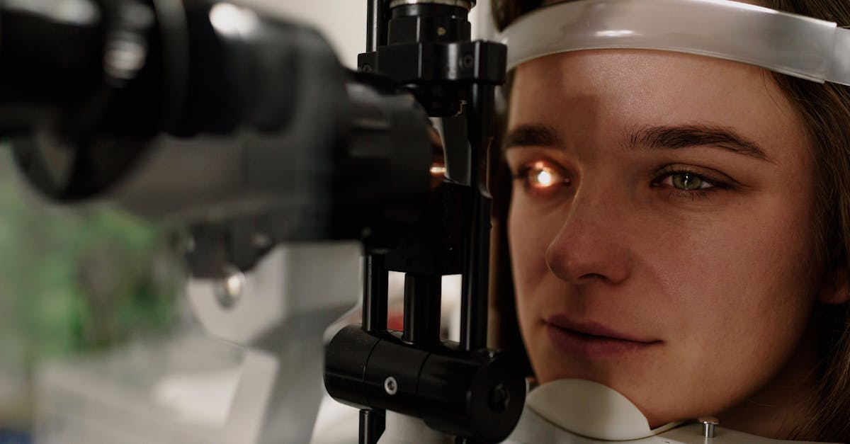
When a patient presents with persistently bloodshot eyes, a comprehensive ophthalmological examination is required to investigate the internal and external ocular architecture. The cornerstone of this exam is the slit-lamp biomicroscope. This device provides high-magnification, 3D views of the front of the eye.
Using the slit lamp, the ophthalmologist can evaluate the quality of the tear film, check the cornea for microscopic scratches or ulcers, and look deep into the anterior chamber of the eye to spot the floating white blood cells that definitively diagnose internal uveitis.
Tonometry is also performed to precisely measure the intraocular pressure, instantly ruling out glaucoma. To check for corneal damage, the physician may apply a specialized yellow dye called fluorescein. When illuminated with a cobalt blue light, the dye brilliantly highlights any microscopic abrasions or viral ulcers on the corneal surface.
13. Pharmacological Treatments
The treatment of bloodshot eyes relies entirely on reversing the specific underlying pathology. For severe dry eye syndrome, physicians frequently prescribe topical anti-inflammatory medications, such as cyclosporine or lifitegrast drops, which actively suppress the localized immune response and encourage natural tear production.
For severe allergic conjunctivitis, prescription-strength dual-action antihistamine and mast-cell stabilizer drops are utilized. These medications provide rapid itch relief while preventing further histamine release, shutting down the allergic cascade without causing rebound redness.
Infectious causes require targeted antimicrobial therapy. Bacterial ulcers or severe conjunctivitis are treated with broad-spectrum antibiotic drops. Viral herpetic eye infections require specific antiviral ointments to prevent the virus from permanently scarring the visual axis of the cornea.
14. Lifestyle and Environmental Adjustments
Mitigating chronic redness often requires sustained environmental modifications. For individuals who work extensively on computers, implementing the twenty-twenty-twenty rule is crucial: every twenty minutes, look at an object twenty feet away for twenty seconds. This practice breaks the prolonged stare, encouraging a full, restorative blink that redistributes the tear film over the dry cornea.
Utilizing a high-quality indoor humidifier adds vital moisture to the ambient air, preventing the rapid evaporation of tears in dry, climate-controlled office buildings. Shielding the eyes from harsh wind, ultraviolet light, and airborne debris with wrap-around sunglasses provides a necessary physical barrier against external irritants.
For contact lens wearers, strictly adhering to replacement schedules, switching to daily disposable lenses, and never sleeping in lenses drastically reduces the mechanical and hypoxic stress placed on the ocular surface, allowing the angry, engorged blood vessels to permanently recede.
15. When to Seek Urgent Ophthalmic Care
While mild, gritty redness from fatigue is easily managed, specific red flag symptoms indicate an immediate threat to vision and require urgent evaluation by an ophthalmologist or an emergency department.
Immediate medical care is strictly required if the red eye is accompanied by severe, deep, or throbbing pain, rather than simple surface scratching. Furthermore, any sudden decrease in visual acuity, blurry vision that does not clear with blinking, or extreme sensitivity to normal indoor lighting points strongly toward internal inflammation or pressure spikes.
If the redness occurs immediately following a blunt trauma to the eye or exposure to a chemical splash, emergency flushing and clinical assessment are mandatory to prevent catastrophic, irreversible damage to the sensitive optical structures.
16. Frequently Asked Questions FAQ
1. Is it safe to use redness-relief eye drops every day?
No. Daily use of cosmetic redness-relief drops causes rebound hyperemia, making your eyes permanently dependent on the drops and significantly redder over time. Use preservative-free artificial tears instead.
2. Can high blood pressure make my eyes perpetually red?
Systemic hypertension does not usually cause a chronic red eye. However, a sudden, massive spike in blood pressure can cause a tiny vessel in the eye to burst, resulting in a bright red subconjunctival hemorrhage.
3. Why do my eyes get so red and itchy during the spring?
This is classic allergic conjunctivitis. Airborne pollens land on the surface of the eye, triggering immune cells to release massive amounts of histamine, which violently dilates the blood vessels and causes severe itching.
4. Are red eyes a sign of lacking a specific vitamin?
A severe deficiency in Vitamin A can cause profound dry eyes and redness, but this is exceedingly rare in developed countries. Chronic redness is far more likely due to environmental dryness or screen fatigue.
5. How do I know if my red eye is contagious pink eye?
Contagious viral pink eye typically starts in one eye and spreads to the other within days, accompanied by a watery discharge and cold symptoms. Bacterial pink eye features thick, yellow, sticky pus that glues the eyelids shut overnight.
17. Bibliography
Disclaimer: The content is for informational purposes only and does not replace medical advice. Always consult your doctor for personalized treatment.

