
1. Introduction
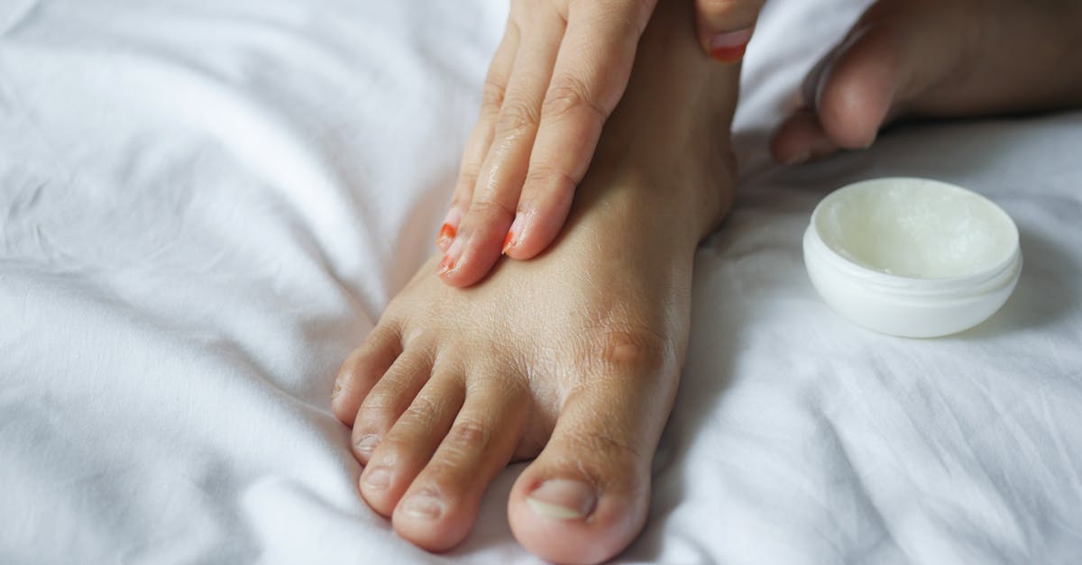
The fastest way to cure severely dry and cracked heels is to soak the feet in lukewarm water for fifteen minutes, gently debride the dead skin with a pumice stone, and immediately apply a keratolytic cream containing urea or salicylic acid. Following the keratolytic agent, a heavy layer of an occlusive ointment, such as pure petroleum jelly, must be applied and sealed under a pair of thick cotton socks overnight. This aggressive, layered approach halts trans-epidermal water loss, dissolves thickened calluses, and forces profound hydration into the deep epidermal fissures.
Severely cracked heels, medically known as heel fissures, represent a structural failure of the epidermis located on the plantar aspect of the foot. The skin on the soles is uniquely adapted to bear the entire weight of the human body. To withstand this immense mechanical pressure, the outermost layer of the skin, the stratum corneum, becomes exceptionally thick and tough. However, this specialized skin is inherently vulnerable because it completely lacks sebaceous glands, meaning it cannot produce natural moisturizing oils.
When this thick, dry skin is subjected to constant friction and repetitive pressure, especially in open-backed shoes, the rigid tissue cannot stretch. Instead of expanding, the hardened callus snaps, creating deep vertical fissures. These cracks expose the sensitive underlying dermal tissue and raw nerve endings, resulting in acute, sharp pain during ambulation.
Resolving this painful condition requires a methodical dermatological intervention. Simple lotions are entirely ineffective against thick calluses. Healing necessitates chemical exfoliation to break down the hardened protein bonds, mechanical debridement to remove the necrotic tissue, and profound occlusive hydration to restore the elasticity of the epidermal barrier.
2. Anatomy and Pathophysiology of the Heel
The skin on the heel is physiologically distinct from the skin on the rest of the body. The epidermis here features an additional layer called the stratum lucidum, which provides extra cushioning and friction resistance. The stratum corneum on the heel is also vastly thicker, composed of tightly packed, dead keratinocytes.
Because the plantar skin contains no sebaceous glands, it relies entirely on the sweat glands and internal systemic hydration to maintain its moisture balance. When the environment is dry, or when footwear allows moisture to evaporate rapidly, the keratinocytes lose their water content, shrinking and becoming brittle.
In response to repetitive mechanical stress, such as standing for prolonged periods on hard surfaces, the body defends the heel by accelerating the production of skin cells, forming a thick, yellowish callus known as hyperkeratosis. While this callus provides physical protection, it possesses zero elasticity.
During walking, the heel strikes the ground, and the fat pad beneath the heel bone expands outward. If the thick callus surrounding this fat pad is dry and inelastic, the outward expansion physically tears the skin apart. The depth of the fissure dictates the severity of the pain; superficial cracks are merely rough, while deep fissures penetrate the dermis, causing bleeding and providing a direct portal for bacterial infection.
3. The Role of Soaking and Mechanical Debridement
Attempting to apply moisturizing creams to a rigid, impenetrable callus yields poor results. The thick layer of dead keratin acts as a physical shield, preventing any active ingredients from reaching the living tissue below. The first step in rapid repair is softening and reducing this necrotic barrier.
Soaking the feet in lukewarm, soapy water for ten to fifteen minutes hydrates the dead cells, causing them to swell and soften. It is vital to use lukewarm water; hot water strips the minimal natural lipids from the surrounding healthy skin, ultimately worsening the dryness.
Once the callus is thoroughly softened, gentle mechanical debridement must be performed. Utilizing a clean pumice stone or a specialized foot file, the patient gently rubs the thickened edges of the fissures in a single direction. The objective is to slough off the top layers of the dead skin, not to aggressively file the heel down to raw tissue.
Aggressive, painful filing or the use of sharp razor tools, like callus shavers, should be strictly avoided in a home setting. Removing too much tissue at once triggers a defensive inflammatory response, causing the body to rapidly generate an even thicker, harder callus in retaliation.
4. Keratolytic Agents: Breaking the Bonds
To achieve profound, lasting smoothness, mechanical filing must be paired with chemical exfoliation. Keratolytics are specific dermatological agents designed to dissolve the desmosomes, which are the biological glue holding the dead keratinocytes together. By breaking these bonds, the thick callus naturally sheds its outer layers.
Urea is the gold standard keratolytic for severe foot calluses. It is a dual-action molecule; at concentrations between twenty and forty percent, urea effectively dissolves the tough keratin bonds, while simultaneously acting as a powerful humectant, drawing water deep into the cellular matrix.
Salicylic acid and lactic acid are alternative potent keratolytics. Lactic acid, an alpha-hydroxy acid, gently exfoliates the surface while improving the overall lipid barrier of the skin. Salicylic acid, a beta-hydroxy acid, penetrates deeply into the fissures, breaking down the hardened edges and smoothing the rough topography of the heel.
These active creams should be applied immediately after the feet are soaked and gently towel-dried. By applying the keratolytic directly to the freshly filed, damp skin, the active ingredients penetrate maximally into the newly exposed cellular layers.
5. Occlusive Ointments: Halting Water Loss
While keratolytics dissolve the callus and draw in water, they cannot hold the water inside the tissue on their own. The damaged epidermal barrier allows moisture to evaporate rapidly into the air, a process known as trans-epidermal water loss. Halting this evaporation is the critical final step in the nightly repair protocol.
Occlusives are heavy, large-molecule lipids that form an impermeable, waterproof shield over the skin. Pure white petroleum jelly is the most effective occlusive agent available in dermatology, blocking up to ninety-nine percent of water evaporation.
Immediately after applying the keratolytic cream, a thick, generous layer of petroleum jelly must be slathered over the entire heel, concentrating heavily on the deep fissures. This traps the urea, the absorbed water, and the natural body heat beneath an airtight seal.
In this artificially humid, occluded microenvironment, the migrating epidermal cells can multiply and repair the deep cracks exponentially faster than they could if exposed to the dry ambient air. This is the physiological secret to achieving overnight results.
6. The Moisture-Locking Sock Protocol
Applying heavy petroleum jelly to the feet creates a significant practical challenge, as the ointment is highly greasy and will immediately transfer to bedsheets or create a severe slipping hazard if the patient attempts to walk.
To secure the occlusive barrier, the patient must immediately don a pair of clean, one-hundred-percent cotton socks. Cotton is breathable, allowing heat to dissipate slightly while keeping the heavy ointment pressed firmly against the skin. Synthetic socks, such as polyester or nylon, can trap too much heat and sweat, leading to tissue maceration and increasing the risk of fungal infections like tinea pedis.
The socks act as a physical dressing, ensuring the active ingredients remain in continuous contact with the heel fissures throughout the entire sleep cycle. By morning, the majority of the ointment will have been absorbed or structurally integrated into the stratum corneum.
For exceptionally deep, painful fissures, specialized silicone-lined heel socks provide an even superior barrier. The internal silicone pad mimics the occlusive properties of the ointment, pressing the edges of the crack together and providing a shock-absorbing cushion for the morning walk.
7. Biomechanical Stress and Footwear
The physical forces acting upon the heel during ambulation directly dictate the formation of calluses and fissures. The type of footwear a patient chooses is often the primary etiological factor in severe heel cracking.
Open-backed shoes, such as sandals, flip-flops, or slingbacks, provide zero lateral support to the heel pad. When the heel strikes the ground, the fat pad splay outward unhindered. If the skin is dry, this uncontained lateral expansion rips the stratum corneum apart.
Furthermore, thin-soled shoes provide inadequate shock absorption, increasing the mechanical trauma to the epidermal layers and stimulating hyperkeratosis. To cure the condition and prevent recurrence, patients must transition to enclosed, supportive footwear.
Wearing a closed-heel shoe, particularly an athletic shoe with robust cushioning, physically cups the heel. This rigid cup contains the fat pad, preventing the lateral splay that causes the skin to split. This structural support is mandatory during the acute healing phase.
8. Liquid Bandages for Deep, Painful Fissures
When a heel fissure penetrates through the epidermis and into the highly vascularized and innervated dermis, every step causes the crack to pull apart, triggering agonizing, sharp pain. In these severe cases, simple ointments are insufficient to close the physical gap.
Medical-grade liquid bandages, composed of cyanoacrylate, offer an excellent clinical solution. When painted directly over a clean, dry fissure, the liquid polymerizes instantly, forming a rigid, waterproof, transparent seal that holds the edges of the cracked skin tightly together.
This physical splint prevents the crack from pulling apart during walking, eliminating the acute pain immediately. It also seals the raw nerve endings from the air and provides a sterile barrier against bacterial entry.
The liquid bandage should remain in place for several days until it naturally flakes off. During this time, the patient can continue to apply moisturizers to the surrounding intact skin, allowing the deep dermal tear to heal undisturbed beneath the chemical splint.
9. Preventing Bacterial and Fungal Infections
Deep heel fissures are not merely a cosmetic or comfort issue; they represent a significant breach in the primary immune defense of the body. The deep cracks provide a direct, open portal for bacteria and fungi present on the floor or inside shoes to enter the systemic circulation.
Infections such as cellulitis, a rapidly spreading bacterial infection of the deep dermal tissues, can easily originate from a neglected heel crack. Patients must monitor the fissures daily for signs of localized infection, including spreading redness, severe throbbing pain when not walking, or the presence of purulent discharge.
If the cracks appear red and inflamed, applying a thin layer of an over-the-counter topical antibiotic ointment, such as bacitracin, inside the fissure before applying the occlusive petroleum jelly provides targeted antimicrobial protection.
Fungal infections, such as athlete foot, can also severely dry out the skin, causing a powdery, white scaling on the heel that mimics simple dry skin. If the dryness is accompanied by intense itching or small blisters, a topical antifungal cream must be integrated into the treatment protocol to eradicate the underlying pathogen.
10. Systemic Hydration and Nutritional Factors
Topical treatments address the surface symptoms, but the fundamental health of the skin relies on systemic hydration and nutrition. If the body is chronically dehydrated, blood volume takes priority over peripheral tissue moisture, leaving the extremities, particularly the feet, completely desiccated.
Consuming adequate daily water is a non-negotiable baseline for healthy skin function. Furthermore, the synthesis of a strong, flexible epidermal barrier requires specific nutritional co-factors, primarily essential fatty acids.
Omega-three fatty acids, found in cold-water fish, walnuts, and flaxseeds, are critical components of the cellular membrane. A diet deficient in healthy fats results in brittle, rigid skin cells that are highly prone to cracking under mechanical pressure.
Vitamin deficiencies, particularly a lack of Vitamin E and Zinc, impair the skin ability to repair micro-trauma and synthesize new collagen. A balanced diet or targeted supplementation ensures the body has the biochemical tools required to build a resilient, elastic stratum corneum on the plantar surfaces.
11. Considerations for Diabetic Patients
For patients diagnosed with diabetes mellitus, a cracked heel is a potential medical emergency rather than a simple dermatological annoyance. Diabetes causes peripheral neuropathy, a gradual loss of protective sensation in the feet. A diabetic patient may not feel the pain of a deep, bleeding fissure.
Simultaneously, diabetes causes peripheral arterial disease, severely reducing the blood flow required to heal the wound. Furthermore, the elevated blood glucose levels provide an ideal feeding ground for invading bacteria, compromising the local immune response.
This combination of neuropathy, ischemia, and immune suppression means a simple heel crack can rapidly deteriorate into a severe diabetic foot ulcer, potentially leading to osteomyelitis (bone infection) or requiring amputation.
Diabetic patients must never perform aggressive home mechanical debridement with pumice stones or sharp files, nor should they use strong chemical keratolytics like salicylic acid without strict medical supervision. Any deep fissure in a diabetic patient mandates an immediate consultation with a podiatrist for professional, sterile wound care.
12. Addressing Obesity and Biomechanics
Body weight is directly proportional to the mechanical force exerted on the plantar heel pad. Obesity increases the sheer compressive force on the heels with every step, maximizing the lateral splay of the fat pad and drastically increasing the likelihood of the skin splitting.
While rapid weight loss is a long-term systemic goal, addressing the immediate biomechanical stress requires specialized orthotic support. Custom or high-quality over-the-counter orthotic insoles with a deep, rigid heel cup provide substantial mechanical relief.
These rigid cups physically contain the tissue, preventing the heel from flattening and spreading outward. Additionally, utilizing gel heel cups inserts provides superior shock absorption, dampening the impact force before it is transmitted to the fragile, calloused epidermis.
Correcting abnormal gait patterns, such as severe overpronation, where the foot rolls inward, ensures that the forces of walking are distributed evenly across the entire foot, preventing excessive, localized pressure buildup on the lateral edges of the heel.
13. Summary of the Dermatological Protocol
To ensure a structured and effective approach to healing severe heel fissures, the following table summarizes the sequential steps of the rapid overnight protocol and the physiological rationale for each action.
| Treatment Step | Physiological Action |
|---|---|
| Lukewarm Water Soak (15 mins) | Hydrates and softens the dense, dead keratinocytes. |
| Pumice Stone Debridement | Mechanically removes the top layers of the necrotic tissue barrier. |
| Urea-Based Cream (Keratolytic) | Chemically breaks the bonds between dead cells and acts as a deep humectant. |
| Petroleum Jelly (Occlusive) | Forms a waterproof seal to completely halt trans-epidermal water loss. |
| 100% Cotton Socks | Secures the ointment in place and provides a breathable, occluded environment. |
14. Differentiating from Psoriasis and Eczema
Not all thick, scaly skin on the heels is simple mechanical hyperkeratosis. Several systemic dermatological conditions present with thick, cracking plaques on the plantar surfaces and will not respond to standard moisturizing protocols.
Plantar psoriasis is a chronic autoimmune condition characterized by hyperproliferation of skin cells. It presents as highly defined, thick, red plaques covered with silvery scales, often accompanied by deep, agonizing fissures. Unlike simple dry skin, psoriasis is primarily an inflammatory disease driven by overactive T-cells.
Dyshidrotic eczema is another condition that can affect the soles of the feet, typically presenting initially as intensely itchy, deep-seated blisters that eventually burst and dry into thick, peeling, and cracked skin.
If a patient utilizes aggressive keratolytic and occlusive therapies for two weeks with zero clinical improvement, or if the cracking is accompanied by severe, deep itching or distinct red plaques, a dermatologist must be consulted. These immune-driven conditions require prescription topical corticosteroids or calcineurin inhibitors to halt the underlying inflammatory cascade, similar to treating a stubborn patch of itchy eczema.
15. Maintaining Long-Term Smoothness
Once the deep fissures are fully healed and the thick calluses reduced, preventing recurrence requires transitioning from an aggressive repair protocol to a sustainable maintenance routine. The skin on the heel remains permanently devoid of sebaceous glands; therefore, external moisture must be supplied continuously.
Applying a mild, ten percent urea cream or a ceramide-based lotion every night immediately after showering locks in the ambient moisture from the bathing process. The mechanical debridement with a pumice stone should be reduced to once a week to prevent excessive buildup without triggering a defensive callus response.
Choosing appropriate footwear is an ongoing requirement. Limiting the use of flip-flops and sandals to brief periods, and relying primarily on supportive, enclosed shoes with a proper heel counter, protects the fragile epidermal structure from mechanical splay.
By respecting the unique physiological limitations of the plantar skin and supplying consistent external hydration, patients can maintain smooth, intact heels and entirely prevent the painful recurrence of epidermal fissures.
16. Frequently Asked Questions (FAQ)
1. Is it safe to use a razor or callus shaver on my heels?
No, using sharp blades to shave calluses is highly dangerous. It removes too much protective tissue, triggering the body to grow a thicker callus, and carries a severe risk of cutting into the dermis and causing a bacterial infection.
2. Why do my heels crack more in the summer than the winter?
During the summer, people frequently wear open-backed sandals and flip-flops, which provide no lateral support to the heel, allowing the fat pad to smash outward and physically tear the dry skin apart.
3. Will soaking my feet in Epsom salts cure the cracks faster?
Epsom salts are excellent for sore muscles, but they pull moisture out of the skin. For severely dry heels, a plain, lukewarm water soak with a mild, moisturizing soap is superior for softening the tissue.
4. Can I use superglue to close a deep heel crack?
While medical-grade liquid bandages (cyanoacrylate) are safe and designed for the skin, household superglue contains toxic industrial chemicals that can cause severe irritation and should never be applied to an open wound.
5. Why won’t regular body lotion fix my dry heels?
Regular body lotions lack keratolytics (like urea) and heavy occlusives. They simply sit on top of the dead, hard callus without penetrating, whereas specialized foot creams dissolve the dead skin and trap moisture inside.
17. Bibliography
Disclaimer: The content is for informational purposes only and does not replace medical advice. Always consult your doctor for personalized treatment.
