1. Introduction
A digital nerve injury represents a profound disruption of the sensory architecture within the hand. The digital nerves are critical peripheral structures responsible for transmitting tactile, thermal, and nociceptive data from the fingers to the central nervous system. When a causative force severs or crushes these microscopic conduits, the immediate consequence is a profound loss of sensation in the affected digit, severely impairing the intricate motor control and protective feedback required for complex manual dexterity.
The human hand interacts continuously with the environment, rendering the superficial digital nerves highly vulnerable to sharp lacerations, industrial crush accidents, and severe abrasive trauma. Unlike localized soft tissue damage that heals through basic scar formation, an injured peripheral nerve undergoes a complex biological process of degeneration and highly unpredictable regeneration.
Effective clinical management relies on prompt identification of the neurological deficit prior to any surgical exploration. Achieving functional sensory recovery mandates meticulous microsurgical repair techniques. Understanding the precise cellular mechanisms of nerve regeneration allows hand surgeons to align the severed neural pathways accurately, minimizing painful neuroma formation and maximizing the patient’s long-term tactile recovery.
2. Anatomy of the Digital Nerves
The sensory innervation of the fingers is primarily supplied by the terminal branches of the median and ulnar nerves. As these major nerves enter the palm, they divide into common palmar digital nerves, which subsequently bifurcate at the web spaces to form the proper palmar digital nerves. Two proper palmar digital nerves run along the palmar-lateral and palmar-medial aspects of each digit.
These nerves are situated in close anatomical proximity to the digital arteries, collectively forming the neurovascular bundle. The nerve generally lies slightly anterior (palmar) to the artery. This intimate structural relationship dictates that a deep laceration causing significant digital bleeding highly correlates with a simultaneous disruption of the accompanying digital nerve.
Microscopically, a digital nerve is composed of several fascicles—bundles of individual axons—surrounded by a protective connective tissue sheath known as the perineurium. The entire nerve is enclosed by an outer layer called the epineurium. Successful surgical repair relies on aligning these microscopic fascicular structures to ensure regenerating axons reach their correct sensory targets in the fingertip.
3. Mechanisms of Nerve Trauma
Digital nerves are subjected to varying mechanisms of injury, each producing a distinct pattern of structural damage that dictates the potential for biological recovery. The most common etiology is a sharp laceration, typically caused by a knife, shattered glass, or a sharp industrial tool. A sharp laceration cleanly severs the epineurium, perineurium, and axons, creating a discrete anatomical gap that is highly amenable to primary surgical repair.
Crush injuries occur when a significant compressive force is applied to the digit, such as a finger caught in heavy machinery or crushed by a heavy object. The blunt force severely contuses the nerve, causing localized ischemia, intraneural edema, and widespread axonal destruction over a significant length of the nerve. These injuries have a poorer prognosis because the extensive zone of trauma complicates the surgical identification of healthy nerve ends.
Avulsion injuries represent the most severe mechanism. The nerve is subjected to a violent traction force, physically tearing it apart. This results in ragged, structurally destroyed nerve ends and profound damage to the surrounding vascular supply, frequently necessitating complex nerve grafting to bridge the resulting defect.
4. Pathophysiology of Axonal Disruption
When a digital nerve is completely severed, a highly orchestrated biological sequence known as Wallerian degeneration occurs in the distal portion of the nerve (the segment separated from the spinal cord). Within twenty-four to forty-eight hours of the injury, the axons and their protective myelin sheaths in the distal stump break down completely.
Tissue macrophages infiltrate the distal nerve sheath, clearing away the cellular debris. However, the empty connective tissue tubes (the endoneurium) remain structurally intact. These empty tubes serve as the crucial biological pathways that guide regenerating axons back toward the sensory receptors in the fingertip.
In the proximal stump (the segment still attached to the spinal cord), the severed axons sprout growth cones. These growth cones actively seek the empty endoneurial tubes of the distal stump. If the nerve ends are surgically aligned, the growth cones enter the tubes and regenerate at a rate of approximately one millimeter per day. If they cannot find the tubes, they form a disorganized, painful knot of nerve tissue.
5. Classification of Nerve Injuries
To standardize diagnosis and treatment planning, nerve injuries are classified based on the severity of the internal structural disruption, utilizing the Seddon classification system.
| Injury Classification | Structural Integrity | Clinical Prognosis |
|---|---|---|
| Neurapraxia | Axons intact; focal demyelination or conduction block. | Full spontaneous recovery within days to weeks. |
| Axonotmesis | Axons severed; connective tissue sheaths (epineurium/perineurium) intact. | Good potential for regeneration along intact pathways. |
| Neurotmesis | Complete transection of axons and all connective tissue sheaths. | No spontaneous recovery; requires surgical repair. |
A sharp laceration to a digital nerve almost invariably results in neurotmesis, necessitating microsurgical intervention. Crush injuries often result in severe axonotmesis, requiring close clinical observation to determine if spontaneous regeneration will occur before committing to surgical exploration.
6. Clinical Examination and Sensory Mapping
The clinical evaluation of a suspected digital nerve injury must be performed systematically before any local anesthetic is administered, as anesthesia abolishes the patient’s ability to report sensory deficits. The clinician assesses the resting posture of the hand and observes for obvious signs of arterial bleeding indicating a neurovascular bundle injury.
Sensory evaluation is conducted using two-point discrimination testing. Utilizing a specialized caliper, the clinician gently presses two points against the skin of the uninjured hand to establish a baseline, typically between two to five millimeters on the fingertips. The test is then repeated on the injured digit. An inability to distinguish two distinct points at distances greater than the baseline indicates significant sensory disruption.
The clinician maps the specific anatomical zone of numbness. Because each proper digital nerve supplies one-half of the palmar aspect of the finger, assessing the radial (thumb side) and ulnar (pinky side) aspects of the digit independently allows the clinician to determine exactly which digital nerve has been lacerated.
7. Differentiating Partial from Complete Transection
Distinguishing between a partially severed and a completely transected digital nerve presents a significant clinical challenge. A partial laceration implies that some fascicles remain anatomically continuous, preserving a degree of sensory function distal to the wound.
In a partial laceration, the patient may report diminished, altered, or hypersensitive sensation rather than complete numbness. Two-point discrimination may be delayed or widened but not entirely absent. However, a partial laceration still disrupts the epineurium and allows for the disorganized sprouting of injured axons, creating a high risk for painful neuroma formation.
If the clinical examination is ambiguous, or if the laceration is located in a high-risk zone with persistent sensory abnormalities, formal surgical exploration under magnification is indicated. The surgeon physically inspects the nerve to definitively ascertain the degree of structural disruption and determine if a primary repair is required.
8. Pre-Surgical Management and Wound Care
Immediate management of a lacerated digit prioritizes hemorrhage control and the prevention of secondary infection. Direct, sustained pressure is applied to the wound using sterile gauze. The use of blind clamping to stop bleeding is strictly contraindicated; indiscriminately placing a hemostat into a bleeding wound can crush the adjacent, highly delicate digital nerve, converting a clean laceration into an irreparable crush injury.
The wound is vigorously irrigated with sterile saline to remove particulate debris and minimize bacterial load. A sterile dressing is applied, and the hand is immobilized in a bulky splint to prevent further mechanical trauma to the severed nerve ends during transport or while awaiting surgical intervention.
Tetanus prophylaxis is administered if the patient’s vaccination status is outdated, and empirical prophylactic antibiotics are considered for heavily contaminated wounds or injuries involving crush mechanisms and devitalized tissue.
9. Principles of Microsurgical Repair
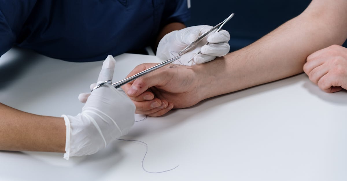
The definitive treatment for a completely transected digital nerve is microscopic surgical repair. This highly specialized procedure is performed in an operating room utilizing an operating microscope or high-powered surgical loupes, ensuring the surgeon can clearly visualize the microscopic fascicular architecture.
The primary objective is an epineurial repair. The surgeon carefully dissects the proximal and distal nerve stumps, cleanly excising any contused or ragged tissue to expose healthy, sprouting nerve fascicles. The goal is to coapt (join) the two nerve ends together without any tension.
Utilizing specialized micro-instruments and suture thread finer than a human hair, the surgeon places precise stitches solely through the outer epineurial sheath. This aligns the internal fascicles perfectly, providing the optimal biological conduit for the proximal growth cones to enter the distal endoneurial tubes.
10. Managing Nerve Gaps: Grafts and Conduits
If the trauma destroyed a significant length of the nerve, pulling the two ends together creates excessive tension. Tension causes localized ischemia at the repair site, leading to dense scar formation that physically blocks axonal regeneration. In these scenarios, the surgeon must bridge the anatomical gap.
For small gaps, typically less than two centimeters, a synthetic nerve conduit may be utilized. This absorbable, tube-like structure is sewn between the two nerve ends. It creates a protected microenvironment filled with growth factors, guiding the sprouting axons across the gap into the distal stump.
For larger defects, an autologous nerve graft is the gold standard. The surgeon harvests a segment of a non-critical sensory nerve, frequently the sural nerve from the leg or the medial antebrachial cutaneous nerve from the forearm. This biological graft provides the necessary structural scaffolding and living Schwann cells to support the regeneration of the digital nerve over the extended defect.
11. Postoperative Neuroma Formation
A traumatic neuroma is the most significant, painful complication following a digital nerve injury. When a nerve is completely severed and not surgically repaired, or if the surgical repair fails due to excessive scarring, the regenerating axons from the proximal stump have nowhere to go.
These growth cones continue to multiply, intertwining with localized scar tissue to form a dense, disorganized, spherical mass of highly sensitive nerve fibers. This is a terminal neuroma. A neuroma in continuity occurs when the nerve is partially damaged or repaired, but a subset of axons escapes the epineurial sheath and forms a painful knot along the otherwise intact nerve trunk.
Clinically, a neuroma presents as a distinct, palpable mass at the injury site that is exquisitely tender to even light touch. Tapping on the neuroma elicits a sharp, electrical shock radiating distally, known as a positive Tinel’s sign. Managing a painful neuroma often requires surgical excision of the mass and burying the proximal nerve stump deep into adjacent muscle or bone to silence the electrical firing.
12. Concomitant Tendon and Vascular Injuries
Because the digital nerves travel in immediate proximity to the flexor tendons and the digital arteries, isolated nerve injuries are relatively rare. A deep laceration on the palmar surface of the finger frequently disrupts the entire neurovascular bundle and the underlying flexor tendon complex.
Surgical management in these complex cases follows a strict hierarchy of repair. The structural integrity of the skeleton must be stabilized first if fractures are present. Following skeletal fixation, the flexor tendons are meticulously repaired to restore motor function.
Vascular repair takes precedence over nerve repair if the digit is severely ischemic; the surgeon must perform microvascular anastomosis of the digital arteries to restore blood flow and save the finger. Finally, the digital nerves are repaired. This comprehensive sequence ensures the repaired nerve is placed in a stable, well-vascularized tissue bed, optimizing the environment for regeneration.
13. Rehabilitation and Sensory Re-education
The surgical repair marks the beginning of a prolonged recovery process. Following a period of postoperative immobilization to protect the delicate microscopic sutures, the patient begins an intensive hand therapy protocol. Early therapy focuses on maintaining joint flexibility and preventing the repaired tendons from scarring down to the surrounding tissue.
As the nerve slowly regenerates down the finger over several months, the patient will begin to experience altered sensations, such as hypersensitivity, tingling, or an inability to accurately interpret tactile information. At this stage, a formal sensory re-education program is initiated.
Sensory re-education involves structured exercises designed to retrain the brain to correctly interpret the new, often distorted signals arriving from the regenerating nerve. The patient practices identifying different textures, shapes, and temperatures with the affected digit, utilizing visual feedback to help the central nervous system recalibrate the sensory map of the hand.
14. Timelines and Functional Outcomes
Patients must be thoroughly counseled regarding the slow biological timeline of nerve regeneration. The axons grow approximately one millimeter per day. Therefore, a laceration at the base of the finger may require several months for the sensation to reach the fingertip.
The ultimate functional outcome is dependent on several critical variables. Age is a profound determinant; children possess immense neuroplasticity and typically achieve near-normal sensory recovery, while older adults experience slower, less complete regeneration.
The mechanism of injury also plays a vital role. Clean, sharp lacerations repaired primarily have a highly favorable prognosis. Crush injuries or severe avulsions requiring extensive nerve grafting carry a significantly lower probability of returning protective two-point discrimination. Despite perfect surgical execution, achieving absolute normal sensation following a complete transection is exceedingly rare in the adult population.
15. Frequently Asked Questions (FAQ)
1. What are the immediate signs that I have cut a digital nerve?
If a cut on your finger results in a sudden, complete loss of feeling or a persistent numb, tingling sensation in the fingertip above the cut, it is highly likely that a digital nerve has been severed.
2. Does a cut digital nerve require surgery?
Yes. If the nerve is completely cut in half, it will not grow back together on its own. A specialized hand surgeon must use a microscope and tiny stitches to reconnect the two ends of the nerve so it can heal correctly.
3. How long will it take to get the feeling back in my finger after surgery?
Nerves grow very slowly, about one millimeter each day. Depending on where the cut is located, it can take anywhere from three to six months for the growing nerve to reach your fingertip, and even longer for the brain to relearn how to process the signals.
4. What is a neuroma and why does it hurt?
A neuroma is a hard, painful knot of nerve tissue that forms when a cut nerve cannot find its correct path to grow back. The nerve fibers become tangled in scar tissue and misfire, causing severe, electrical shock-like pain when touched.
5. Will my finger ever feel 100% normal again?
While surgery is highly successful in restoring protective sensation (the ability to feel hot, cold, and pain), it is very rare for an adult to regain absolute, 100% perfect feeling after a nerve has been completely cut.
16. Bibliography
Disclaimer: The content is for informational purposes only and does not replace medical advice. Always consult your doctor for personalized treatment.