1. Introduction
A hand laceration is a traumatic disruption of the complex soft tissues of the palm, dorsum, or digits. The human hand is an incredibly intricate biomechanical marvel, densely packed with tendons, nerves, and blood vessels operating within millimeters of the skin surface. Consequently, even a laceration that appears superficial visually can cause profound mechanical and neurological disability if a vital underlying structure is severed.
The medical management of a hand laceration is fundamentally different from a simple skin wound on the torso or thigh. The primary goal is not merely cosmetic closure, but the absolute preservation of mechanical function and tactile sensation. A missed tendon injury or an unaddressed nerve transection can permanently rob a patient of their ability to perform fine motor tasks, secure a strong grip, or perceive temperature and texture.
Achieving a successful outcome requires a methodical clinical examination to map the integrity of every anatomical layer. Through rigorous decontamination protocols, specialized regional anesthesia, and meticulous surgical repair techniques, physicians can effectively manage these complex injuries, setting the stage for aggressive occupational therapy and functional recovery.
2. Complex Anatomy of the Hand
To accurately diagnose the severity of a hand laceration, one must understand its highly compact anatomical architecture. The skin on the palmar surface is thick, specialized for gripping, and heavily tethered to the underlying palmar aponeurosis, a dense, fibrous connective tissue shield.
Beneath this shield lie the flexor tendons, which travel from the forearm muscles, through the tight carpal tunnel, and out to the tips of the fingers. These tendons glide inside lubricated synovial sheaths. On the dorsal surface, the skin is thin and loose, providing little protection for the extensor tendons, which sit immediately beneath the subcutaneous layer and are responsible for opening the hand and straightening the fingers.
Interwoven among these tendons is the neurovascular network. The median, ulnar, and radial nerves branch extensively to provide motor commands to the intrinsic hand muscles and exquisite sensory feedback from the skin. The superficial and deep palmar arterial arches ensure a robust, redundant blood supply to all tissues.
3. Mechanisms of Traumatic Laceration
The physics behind the injury dictates the characteristics of the wound and the likelihood of deep structural damage. Sharp trauma, frequently caused by kitchen knives, broken glass, or box cutters, creates clean, linear incisions. While these wounds cause less peripheral tissue damage, the sharp edge penetrates deeply with minimal resistance, carrying a remarkably high risk of completely severing taut tendons and delicate nerves.
Crush-avulsion injuries occur when the hand is caught in moving machinery, car doors, or gears. This violent mechanism crushes the tissue, bursting the skin and tearing tendons and nerves away from their attachments. The resulting wound is ragged, heavily contused, and features devitalized tissue edges that are highly prone to necrosis and virulent infection.
High-pressure injection injuries are a specialized, highly deceptive mechanism. When a fluid like paint or hydraulic fluid is injected into the hand under massive pressure from industrial equipment, it causes a tiny entry puncture but forces toxic chemicals deep along the fascial planes. This constitutes an absolute surgical emergency, frequently resulting in rapid tissue death and amputation if not aggressively decompressed.
4. Hemostasis and Tourniquet Application
Because of the redundant arterial arches in the hand, lacerations typically bleed profusely. The immediate first aid priority is achieving hemostasis through continuous, firm, direct pressure applied over the wound with sterile gauze for ten to fifteen minutes.
Elevating the injured hand above the level of the heart significantly reduces the arterial hydrostatic pressure, assisting the natural coagulation cascade in forming a stable platelet plug.
If direct pressure fails to control severe, pulsatile arterial bleeding, a pneumatic tourniquet may be applied to the upper arm in a controlled clinical setting. The tourniquet creates a bloodless surgical field, allowing the physician to safely explore the wound and identify retracted blood vessels without operating blindly in a pool of blood, which risks inadvertently clamping and destroying adjacent nerves.
5. Assessing Motor Function and Tendon Integrity
The most critical step in evaluating a hand laceration is a meticulous assessment of tendon integrity. Because tendons are under constant tension, a severed tendon will instantly retract like a snapped rubber band, hiding far proximal to the actual skin wound. The physician must independently test the function of every muscle group against resistance.
To assess the flexor tendons, the physician stabilizes specific joints and asks the patient to bend the isolated segments of their fingers. For the dorsal surface, the physician tests the extensor mechanism by asking the patient to straighten their fingers against the physician’s opposing pressure.
Even if the patient can perform the movement, a partial tendon laceration may be present. If the patient experiences sharp pain during the specific movement, or if the strength is markedly diminished compared to the uninjured hand, a partial rupture of the tendon fibers is highly suspected and requires specialized surgical exploration.
6. Sensory Evaluation and Nerve Damage
The hand relies on dense sensory innervation for protection and fine motor dexterity. The physician must systematically map the sensation of the entire hand, specifically testing the territories supplied by the median, ulnar, and radial nerves.
This evaluation relies on two-point discrimination testing using a specialized calibrated instrument. A normal fingertip can distinguish two distinct points applied simultaneously at a distance of three to five millimeters. An inability to distinguish the points, or a complete lack of light touch perception, strongly suggests a nerve transection.
If you have previously experienced neurological deficits related to muscle control, reviewing clinical information on muscle weakness can provide context regarding peripheral nerve motor functions. Any sensory deficit mandates microscopic surgical repair to realign the nerve fascicles, providing a structural tunnel for the slow regeneration of the nerve axons.
7. Animal Bites and High-Risk Contamination
Hand lacerations resulting from animal or human bites are treated with profound clinical caution. The complex, enclosed fascial spaces of the hand provide an ideal, oxygen-deprived environment for virulent oral bacteria to multiply rapidly.
Human bites, particularly those occurring over the knuckles during an altercation (a “fight bite”), are extraordinarily dangerous. The tooth frequently penetrates the joint capsule. When the hand is subsequently opened, the contaminated tendon glides back into the hand, dragging aggressive polymicrobial flora deep into the tissue planes, frequently resulting in devastating septic arthritis.
Bite wounds are generally not sutured closed immediately. Instead, they undergo aggressive high-pressure irrigation, surgical debridement, and are left open to heal by secondary intention while the patient is placed on broad-spectrum prophylactic antibiotics, typically amoxicillin-clavulanate.
8. Diagnostic Imaging for Hidden Injuries
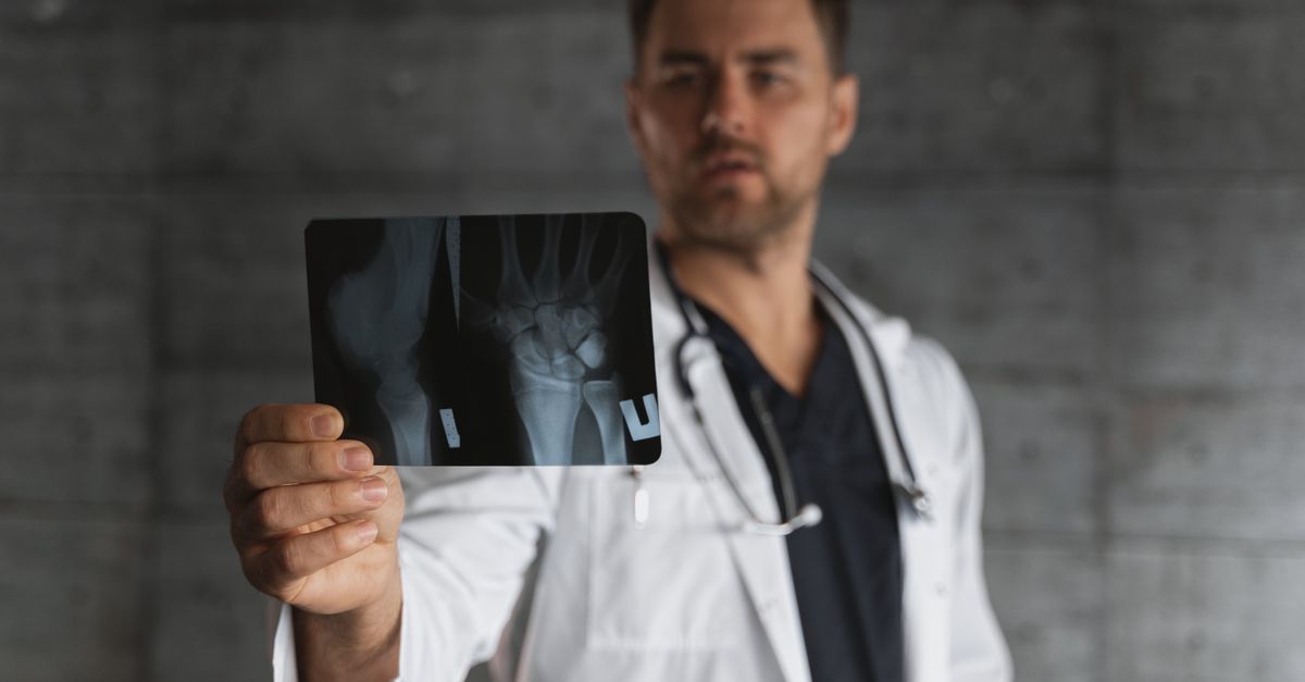
While the clinical physical examination drives most treatment decisions, radiological imaging is a mandatory component of assessing severe hand trauma. Standard multi-view radiographs (X-rays) of the hand and wrist are obtained to rule out underlying fractures, especially in crush injuries or deep lacerations over the knuckles.
Radiographs are also indispensable for locating radiopaque foreign bodies embedded within the wound bed. Shattered glass, metal fragments, or gravel hidden deep beneath the fascia must be identified and meticulously extracted.
Leaving a foreign body in the hand virtually guarantees chronic infection, severe inflammatory reactions, and the formation of a painful granuloma, which will continually interfere with the mechanical function of the hand.
9. Data Summary: Traumatic Hand Zones
Hand surgeons classify the palmar surface into specific anatomical zones to predict injury severity and dictate surgical approaches, particularly regarding flexor tendon injuries.
| Anatomical Zone | Location Description | Clinical Significance |
|---|---|---|
| Zone I | Distal to the middle finger joint | Involves only the flexor digitorum profundus tendon |
| Zone II | Base of finger to middle crease (No Man’s Land) | High risk of severe scarring; both flexor tendons tightly encased |
| Zone III | Palm of the hand (Lumbrical origin) | High risk of combined nerve, artery, and tendon injuries |
| Zone IV | Carpal Tunnel (Wrist crease) | Dense crowding of all tendons and the median nerve |
10. Wound Irrigation and Debridement
The foundation of preventing infection and promoting optimal healing is rigorous wound decontamination. Following the administration of regional anesthesia, the physician performs high-pressure, copious irrigation using sterile normal saline.
This mechanical flushing force separates and removes microscopic soil, coagulated blood, and environmental bacteria from the delicate internal structures of the hand. Antiseptic solutions like hydrogen peroxide are strictly avoided within the wound bed, as their cytotoxic properties heavily damage exposed nerves and tendons.
Surgical debridement involves using sharp instruments to meticulously trim away ragged, crushed, or devitalized tissue edges. Creating clean, viable margins ensures that the tissue will heal efficiently and minimizes the development of thick, restrictive scar tissue.
11. Regional Anesthesia and Nerve Blocks
Because the hand is densely innervated and highly sensitive, achieving profound anesthesia is necessary for thorough exploration and repair. Injecting large volumes of local anesthetic directly into a hand laceration can severely distort the anatomical landmarks and increase pressure within the tight tissue compartments.
Therefore, clinicians rely heavily on regional nerve blocks. By injecting a long-acting anesthetic, such as bupivacaine, around the median, ulnar, or radial nerves at the level of the wrist, the physician can completely numb the entire hand.
This technique provides a painless, undistorted surgical field for the physician and ensures the patient remains entirely comfortable during a potentially lengthy and complex reconstructive procedure.
12. Surgical Repair of Tendons and Nerves
Lacerations involving severed tendons or nerves require the expertise of a specialized hand surgeon or plastic surgeon. These repairs are typically performed in an operating room under magnification using specialized microsurgical instruments.
Severed tendons must be retrieved and intricately sutured together using a core locking stitch designed to withstand the immense mechanical tension of muscle contraction, combined with a fine running suture to smooth the outer surface so it glides easily through the tendon sheath.
Severed nerves require meticulous realignment of the microscopic inner fascicles. Using hair-thin sutures, the surgeon connects the outer protective sheath of the nerve ends. This creates a biological tunnel, allowing the proximal nerve fibers to slowly regenerate and grow down toward the fingertips at a rate of roughly one millimeter per day.
13. Principles of Skin Closure
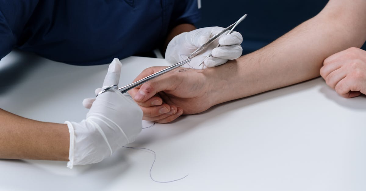
If the deep structures are intact and the wound is clean and recent, the physician proceeds with primary skin closure. The palmar skin is thick and under constant mechanical stress, requiring robust, non-absorbable synthetic sutures, such as nylon, placed with a technique that slightly everts the wound edges.
Conversely, the dorsal skin is thin and delicate, requiring finer sutures to prevent tearing through the tissue. Care is taken to avoid placing excessive tension on the skin edges, which can compromise the microvascular blood supply and lead to skin necrosis.
For superficial, linear lacerations not subjected to tension across a joint, medical-grade tissue adhesives or reinforced adhesive strips may occasionally be utilized to provide a painless, effective closure.
14. Splinting and Postoperative Immobilization
Following surgical repair, particularly if tendons or nerves were involved, the hand must be rigorously protected. The physician applies a custom-molded plaster or fiberglass splint to immobilize the hand in a highly specific, protective posture.
For example, if a flexor tendon was repaired on the palm, the hand is splinted with the wrist and fingers slightly bent forward. This position completely removes the mechanical tension from the healing tendon, preventing the fragile surgical sutures from snapping if the patient accidentally attempts to straighten their hand.
The splint is worn continuously for several weeks. The patient is instructed to keep the hand elevated above the heart to minimize inflammatory swelling and throbbing pain.
15. Occupational Therapy and Rehabilitation
The surgical repair of a complex hand laceration is only the first phase of treatment; rigorous rehabilitation is the ultimate determinant of final functional success. Because the hand structures operate in such close proximity, the natural inflammatory healing process aggressively forms dense scar tissue.
If this scar tissue matures unchecked, it will bind the tendons, nerves, and skin into a solid, immovable block. Occupational hand therapists implement specialized, early motion protocols. They guide the patient through highly controlled, specific movements designed to glide the healing tendons smoothly, preventing them from adhering to the surrounding scar tissue.
This rehabilitation process is frequently painful, arduous, and requires months of dedicated adherence to restore strength, range of motion, and fine motor dexterity.
16. When to Seek Immediate Hand Surgery
While minor, superficial cuts can be washed and bandaged at home, specific clinical signs mandate an immediate visit to an emergency department for evaluation by a hand specialist.
If a laceration results in an inability to bend or straighten a finger, a complete loss of sensation, or uncontrolled, pulsatile arterial bleeding that defeats ten minutes of direct pressure, it is a surgical emergency. Similarly, any laceration caused by a high-pressure injection device must be evaluated immediately, even if the entry wound looks like a harmless pinprick.
Delaying treatment for a severed tendon or nerve leads to massive retraction of the structures and severe scarring, making eventual surgical repair exponentially more difficult and significantly decreasing the likelihood of a successful functional recovery.
17. Frequently Asked Questions (FAQ)
1. Why does my whole hand ache when I only cut one finger?
The tendons and nerves in your fingers originate in your forearm and travel through your hand. Inflammation and swelling from a severe cut on a finger can easily travel down the tendon sheaths into the palm, causing widespread aching and stiffness.
2. How long do I have to wait before going to the doctor for a cut?
To minimize the risk of infection and ensure proper tissue alignment, hand lacerations should be evaluated and sutured within six to twelve hours. Delays beyond twenty-four hours heavily increase the risk of infection and complicate surgical repair.
3. Will the feeling in my hand come back?
If a sensory nerve was severed and surgically repaired, sensation can slowly return as the nerve regenerates (about one millimeter a day). However, the process takes many months, and the final sensation is rarely exactly as it was before the injury.
4. Can I remove the splint to shower?
No. If your hand was splinted after a complex repair, removing the splint to shower could allow your hand to move into a position that immediately tears the fragile tendon repairs. You must cover the splint with a waterproof bag while bathing.
5. Why did the doctor leave my dog bite wound open?
Animal mouths are filled with aggressive bacteria. Closing a bite wound with stitches seals those bacteria deep inside the hand, creating an ideal environment for a severe, limb-threatening infection. Leaving it open to drain is the safest protocol.
18. Bibliography
Disclaimer: The content is for informational purposes only and does not replace medical advice. Always consult your doctor for personalized treatment.