1. Introduction
A cyst on the earlobe indicates a benign encapsulation of epidermal cells or glandular secretions beneath the skin surface. The most common presentation is an epidermoid cyst, which forms when dead skin cells become trapped and multiply within a localized dermal sac, producing a firm, slow-growing nodule. While these lumps frequently induce anxiety due to their sudden appearance, they are overwhelmingly non-cancerous and primarily represent a structural anomaly in the normal cellular turnover process.
The earlobe is structurally unique, consisting of areolar connective tissue and a high density of sebaceous glands without any underlying cartilaginous support. This specialized dermal environment is prone to microscopic traumas from piercings, friction, and localized inflammation. When the narrow exit ducts of the skin glands or hair follicles become occluded, the continuous production of keratin or sebum has nowhere to drain, leading to the gradual expansion of a cystic cavity.
Proper clinical diagnosis is essential to differentiate a simple benign cyst from more concerning dermal lesions or reactive lymphadenopathy. Management focuses on preventing infection and, when necessary, performing a complete surgical excision of the cyst wall to ensure the nodule does not recur. Attempting to manage these structures through manual pressure is contraindicated and frequently exacerbates the underlying inflammation.
2. Dermal Anatomy of the Earlobe
To understand the pathophysiology of cyst formation, one must examine the histological anatomy of the earlobe. Unlike the upper portions of the auricle, which are supported by a rigid framework of elastic cartilage, the lobule is composed entirely of tough areolar connective tissue mixed with adipose cells.
This fleshy structure is covered by a thin layer of epidermis containing a high concentration of apopilosebaceous units. These units consist of a tiny hair follicle and an attached sebaceous gland. The sebaceous glands secrete a lipid-rich substance called sebum, which travels up the follicular duct to lubricate and protect the skin surface.
Because the earlobe lacks a rigid cartilaginous backing, the dermal layers are highly mobile and pliable. This pliability allows for the rapid, visible expansion of cysts as fluids and cellular debris accumulate within the connective tissue space without facing immediate structural resistance.
3. Epidermoid Cysts vs Sebaceous Cysts
The terms epidermoid cyst and sebaceous cyst are frequently used interchangeably by the general public, but they represent distinct clinical entities. The vast majority of cysts found on the earlobe are true epidermoid cysts.
An epidermoid cyst originates from the epidermal cells that make up the outermost layer of the skin. Normally, these cells continuously shed away. If a microscopic injury pushes these cells deeper into the dermis, they continue to multiply and form a closed sac. They secrete a thick, white, cheese-like substance composed of keratin protein.
True sebaceous cysts are considerably rarer and originate specifically from the sebaceous glands. They are filled with a clear, oily, liquid sebum rather than thick keratin. Clinically, they present similarly, but histological examination reveals their different cellular origins.
4. Pathophysiology of Cyst Formation
The formation of an epidermoid cyst is a slow, methodical biological process. It begins with the implantation of epidermal cells into the deeper dermal layers. Once trapped in this abnormal location, the epidermal cells survive and establish a spherical boundary, effectively creating an internal pocket lined with skin cells.
These lining cells behave exactly as they would on the skin surface; they mature, die, and shed keratin into the center of the newly formed sac. Because the sac is sealed, the keratin has no exit route. Over months or years, the continuous accumulation of this dense protein slowly stretches the cyst wall, causing the visible lump to expand.
The cyst wall is a crucial anatomical feature. It is a distinct fibrous capsule that physically separates the keratinous debris from the surrounding healthy connective tissue. If this wall remains intact, the cyst is generally painless and inert.
5. The Role of Apopilosebaceous Units
The dense concentration of apopilosebaceous units on the earlobe provides numerous potential sites for cyst development. The narrow ducts connecting the sebaceous glands and hair follicles to the skin surface are highly susceptible to physical obstruction.
Blockages can occur due to an overproduction of sebum, the accumulation of dried skin cells, or localized inflammation. When the ostium of the follicle becomes occluded, it creates a micro-comedone. As the gland continues to pump secretions into the blocked duct, the follicle dilates dramatically, eventually transforming into a cystic structure.
This follicular occlusion mechanism explains why earlobe cysts frequently feature a tiny, dark central punctum. This punctum represents the original, now massively dilated and oxidized, opening of the blocked hair follicle on the skin surface.
6. Earlobe Piercings and Microtrauma
Earlobe piercings are a leading mechanical catalyst for epidermoid cyst formation. The process of piercing the lobule creates a traumatic, penetrating wound through the epidermis and deep into the areolar tissue.
During the healing phase, epidermal cells aggressively proliferate to line the new piercing tract. Occasionally, a cluster of these rapidly multiplying cells becomes pinched off and buried within the dermal layers adjacent to the tract. These displaced cells form the foundation of a new epidermoid cyst.
Furthermore, the continuous insertion and removal of jewelry induce chronic microtrauma and friction. This mechanical irritation can trigger localized follicular inflammation or force surface keratin deeper into the piercing tract, initiating the encapsulation process characteristic of cyst development.
7. Keratin Accumulation and Inflammatory Response
While an intact cyst is a benign, painless lump, its internal contents are highly irritating to the surrounding tissues. The dense keratin paste contained within an epidermoid cyst is biologically recognized as foreign material by the body’s immune system.
If the cyst wall is subjected to mechanical trauma, such as aggressive squeezing or an accidental blow, it can rupture internally. This rupture spills the dense keratin directly into the delicate areolar tissue of the earlobe.
The immune system reacts instantly, launching a massive, localized foreign-body inflammatory response. Neutrophils and macrophages flood the area, causing the earlobe to become intensely red, swollen, hot, and exquisitely painful. This acute inflammatory phase is frequently misdiagnosed as a primary bacterial infection.
8. Clinical Characteristics of Benign Cysts
Identifying the physical characteristics of an earlobe cyst provides reassurance regarding its benign nature. An uncomplicated epidermoid cyst typically presents as a smooth, perfectly round, dome-shaped nodule located within the fleshy tissue of the lobule.
The lump feels firm but slightly rubbery to the touch and can usually be moved slightly beneath the overlying skin. It is completely painless unless actively inflamed. The skin over the cyst generally retains its normal color, though a prominent central blackhead or punctum may be visible.
These cysts grow at an exceedingly slow rate. A patient may notice a small bump that remains unchanged in size for several years before a period of mild inflammation causes a sudden, noticeable expansion.
9. Differentiating Cysts from Reactive Lymph Nodes
Clinical diagnosis requires differentiating a benign dermal cyst from other localized swellings, most importantly reactive lymph nodes.
| Characteristic | Epidermoid Cyst | Reactive Lymph Node |
|---|---|---|
| Anatomical Location | Within the fleshy earlobe tissue. | Directly beneath or behind the earlobe in the neck. |
| Attachment | Tethered to the overlying skin. | Mobile beneath the skin, deeper in tissue. |
| Central Punctum | Often present (small dark pore). | Never present. |
| Onset Speed | Slow, chronic growth. | Rapid swelling following a cold or infection. |
Recognizing that lymph nodes reside deeper in the fascial planes of the neck rather than within the dermal layers of the lobule is a key anatomical distinction during physical palpation.
10. Complications Involving Infection and Rupture
An inflamed, ruptured cyst is highly susceptible to secondary bacterial infection. When the cyst wall breaks and the overlying skin stretches, normal skin flora, primarily Staphylococcus aureus, can invade the necrotic keratin debris.
An infected cyst transforms into a localized abscess. The earlobe becomes dramatically engorged, turning deep red or purple, and throbs with severe, relentless pain. The center of the mass becomes fluctuant, yielding to pressure as liquid pus accumulates.
This clinical state requires prompt medical intervention. If left untreated, the abscess can spread, causing a generalized cellulitis of the auricle or leading to permanent tissue necrosis and cosmetic deformity of the earlobe.
11. Diagnostic Assessment and Palpation
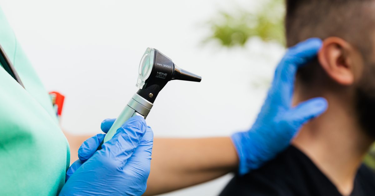
The diagnosis of an earlobe cyst is predominantly clinical, relying on visual inspection and tactile palpation by a healthcare provider. The physician assesses the mobility of the mass, its consistency, and its relationship to the overlying epidermis.
In cases where the diagnosis is ambiguous, or if the mass is unusually hard, fixed to deep structures, or rapidly growing without signs of inflammation, diagnostic imaging such as high-resolution ultrasound may be utilized. Ultrasound easily differentiates a fluid-filled or keratin-filled cyst from a solid soft tissue neoplasm.
A definitive diagnosis is ultimately confirmed through histological examination of the cyst wall by a pathologist following complete surgical excision.
12. The Danger of Self-Extraction
A critical clinical directive is that patients must never attempt to squeeze, pop, or extract an earlobe cyst at home. The mechanical force required to pop a cyst through the tiny skin pore almost inevitably causes the fragile cyst wall to rupture internally.
Squeezing forces the highly irritating keratin and trapped bacteria deep into the surrounding healthy tissues of the earlobe rather than outward. This transforms a small, painless lump into a massive, agonizing inflammatory abscess within hours.
Furthermore, manual squeezing cannot remove the fibrous cyst wall. As long as the lining cells of the sac remain in the dermal tissue, the cyst will continuously refill with keratin, guaranteeing a persistent recurrence.
13. Medical Management of Inflamed Cysts
When a patient presents with an acutely inflamed or infected cyst, definitive surgical removal is temporarily postponed. Operating on severely inflamed, friable tissue increases the risk of scarring and makes identifying the cyst capsule impossible.
Initial medical management focuses on calming the severe immune response. A physician may inject a small amount of a potent corticosteroid, such as triamcinolone, directly into the inflamed mass. This rapidly suppresses the inflammation, reducing the swelling and pain within forty-eight hours.
If a true bacterial infection is present, marked by fluctuance and systemic signs, the clinician will perform a simple incision and drainage under local anesthesia to evacuate the pus, followed by a course of targeted oral antibiotics. Understanding systemic inflammatory control is further explored in our tension headache clinical literature.
14. Surgical Excision Procedures
The only definitive cure for an epidermoid cyst is the complete surgical excision of the entire intact cyst wall. This is a minor, outpatient surgical procedure performed under local anesthesia once all acute inflammation has resolved.
The surgeon makes a small elliptical incision over the cyst, typically incorporating the central punctum. Using meticulous blunt dissection techniques, the surgeon separates the delicate fibrous capsule of the cyst from the surrounding areolar tissue.
It is imperative that the cyst is removed intact. If any fragment of the capsule is left behind, the epidermal cells will regenerate a new cyst. Following successful extraction, the surgical defect is closed with fine sutures, ensuring an optimal cosmetic outcome for the earlobe.
15. Prevention and Dermatological Care
While genetic predispositions and normal anatomical variations make some cyst formation inevitable, certain dermatological practices can reduce the risk. Maintaining excellent hygiene around ear piercings is paramount.
Cleaning piercing tracts regularly and avoiding heavy, abrasive jewelry prevents the chronic microtrauma that drives epidermal cell implantation. Patients prone to severe acne or follicular occlusion may benefit from regular use of mild exfoliating washes containing salicylic acid to keep the glandular ducts clear.
If a patient notices a new, small cyst developing, applying warm compresses can promote local vasodilation and sometimes encourage a minor blockage to resolve naturally before a permanent fibrous capsule forms.
16. When to Seek Medical Attention
Patients should consult a healthcare provider for any new, persistent lump on the earlobe to confirm its benign nature. Immediate medical evaluation is required if the cyst suddenly becomes red, severely swollen, hot to the touch, or begins to drain foul-smelling pus.
Urgent assessment is also necessary if the lump feels rock-hard, is immovably fixed to the deeper tissues, or causes numbness or paralysis in the surrounding facial areas, as these are clinical red flags for aggressive neoplastic growths requiring comprehensive oncological investigation.
Routine, elective surgical removal of a benign cyst prevents the unpredictable and highly painful complications associated with spontaneous rupture and infection.
17. Frequently Asked Questions (FAQ)
1. Will an earlobe cyst eventually go away on its own?
No, a true epidermoid cyst has a permanent fibrous wall that continuously produces keratin; it will not disappear without complete surgical removal.
2. Can I use acne cream to shrink a cyst on my earlobe?
Topical acne creams cannot penetrate deep enough to reach the fibrous capsule of the cyst and are entirely ineffective for shrinking structural dermal cysts.
3. Is it normal for an earlobe cyst to smell bad if it drains?
Yes, the accumulated keratin inside the cyst often has a strong, foul, cheese-like odor due to the breakdown of skin proteins, especially if bacteria have colonized the debris.
4. How long does it take to recover from cyst removal surgery?
The surgical excision is minor; the earlobe may be slightly bruised and tender for a few days, and the fine surface sutures are typically removed within one week.
5. Do ear piercings cause cysts?
Yes, the trauma of a piercing can push surface skin cells deep into the lobe tissue, and the chronic friction of jewelry can trigger the formation of an epidermoid cyst adjacent to the tract.
18. Bibliography
Disclaimer: The content is for informational purposes only and does not replace medical advice. Always consult your doctor for personalized treatment.