1. Introduction
A swollen and hard earlobe usually indicates the presence of an epidermoid cyst, a localized bacterial infection resulting from a piercing, or acute contact dermatitis. While often physically alarming and tender to the touch, these indurated lesions are overwhelmingly benign dermatological or infectious responses. The earlobe is composed entirely of soft areolar connective tissue and fat, lacking the rigid cartilage found in the upper portion of the ear. This unique anatomical structure makes it highly pliable but also highly susceptible to rapid, localized fluid accumulation and inflammatory encapsulation.
When skin cells, sebum, or foreign pathogens become trapped within this soft tissue, the body mounts an immediate defense. It isolates the offending material by forming a dense, fibrous wall around it, creating a distinct, hard nodule. If the area becomes actively infected, intense vascular dilation occurs, flooding the tissue with white blood cells and causing the entire earlobe to balloon outward, becoming hot, red, and exquisitely painful.
Understanding the specific characteristics of the swelling is essential for targeted resolution. A chronic, painless hard lump requires a vastly different clinical approach than a sudden, fiery red, and rapidly expanding swelling. Differentiating between a sterile cyst, an allergic reaction to jewelry, and an aggressive soft-tissue infection ensures that patients receive the appropriate topical or systemic therapy to prevent permanent tissue scarring or cosmetic deformity.
2. Anatomy of the Auricle and Earlobe
The external ear, or auricle, is a complex appendage designed to capture sound waves. The upper two-thirds of the ear derive their rigid, intricate shape from a continuous framework of elastic cartilage. In stark contrast, the lower third—the lobule or earlobe—contains absolutely no cartilage. It is a pendulous sac composed solely of adipose (fat) tissue, loose areolar connective tissue, and a rich network of microscopic blood vessels.
Because it lacks a rigid cartilaginous skeleton, the earlobe is extremely distensible. It can stretch significantly to accommodate heavy jewelry. However, this lack of dense structural boundaries also means that when inflammation occurs, fluid spreads rapidly and uniformly throughout the lobule.
The epidermis covering the earlobe contains numerous sebaceous glands. These glands secrete sebum to lubricate the skin. When the exit pathway of a sebaceous gland becomes physically blocked, the normal cellular debris and oils have nowhere to go, setting the stage for the most common cause of a hard nodule in the earlobe.
3. Epidermoid and Sebaceous Cysts
The most frequent cause of a distinct, hard, pea-sized lump inside the earlobe is an epidermoid cyst. These benign cysts form when epidermal cells, which should normally shed from the surface of the skin, multiply and move inward, creating a small sac deep within the tissue. The sac fills with keratin, a thick, white, cheese-like protein.
Epidermoid cysts often develop very slowly over months or years. They are typically painless, smooth, and firm to the touch. The patient can often roll the hard nodule slightly between their fingers. Occasionally, a tiny central black dot, called a punctum, is visible on the surface of the skin over the cyst.
While inherently harmless, these cysts can suddenly rupture internally if squeezed or bumped. When the keratin contents spill into the surrounding fat tissue, the immune system reacts violently, causing the entire earlobe to suddenly swell, become severely red, and throb with pain, mimicking an acute bacterial infection.
4. Localized Bacterial Infections and Piercings
Earlobe piercings act as deliberate, unhealed wounds through the soft tissue, providing a direct entry portal for environmental pathogens. Localized bacterial infections are highly common, particularly in new piercings or when utilizing non-sterile jewelry.
Staphylococcus aureus and Pseudomonas aeruginosa are the primary pathogens responsible for these infections. When bacteria colonize the piercing tract, they trigger a massive localized immune response. The earlobe quickly becomes swollen, firm, and hot. The patient will experience sharp, throbbing pain, and thick, yellow, or greenish purulent discharge (pus) will ooze from the piercing hole.
In severe cases, the localized infection can form an abscess—a confined, tense pocket of pus. An abscess feels like a hard, extremely tender marble inside the earlobe and requires physical drainage by a physician, as oral antibiotics alone often cannot penetrate the dense abscess wall effectively.
5. Contact Dermatitis and Nickel Allergies
A sudden, hard swelling of the earlobe accompanied by intense, unbearable itching strongly suggests allergic contact dermatitis. This is a delayed-type hypersensitivity reaction mounted by the immune system against a specific foreign substance, most notably metal alloys used in inexpensive jewelry.
Nickel is the most pervasive culprit. When a nickel-containing earring is inserted, the metal oxidizes slightly, releasing ions that penetrate the thin skin of the earlobe. The immune system identifies these ions as dangerous invaders and releases a flood of histamine and inflammatory cytokines into the tissue.
The resulting allergic reaction causes the earlobe to swell dramatically, turning red and feeling firm due to the massive influx of plasma fluid. The skin surface frequently develops dry, scaly patches, microscopic blisters, and weeping clear fluid. The only cure is the immediate and permanent removal of the offending jewelry.
6. Keloid Scar Formation
A uniquely hard, painless swelling that slowly expands beyond the original site of an injury or piercing is characteristic of a keloid scar. Keloids represent a pathological over-healing response. Following minor trauma, such as an ear piercing, the body’s fibroblasts produce a massive, unregulated excess of thick collagen tissue.
Unlike a normal scar that remains flat, a keloid grows into a prominent, dense, rubbery nodule. The earlobe is one of the most common anatomical sites for keloid formation. They are highly prevalent in individuals with darker skin phenotypes and possess a strong genetic component.
Keloids are strictly cosmetic and structural issues; they do not indicate infection or malignancy. However, they are notoriously difficult to treat. Simple surgical removal frequently results in the keloid growing back even larger. Successful management requires a combination of precise excision, localized corticosteroid injections, and pressure garments.
7. Cellulitis of the Auricle
If the red, hard swelling spreads beyond the earlobe and begins to engulf the upper cartilaginous portion of the ear or the surrounding facial skin, the diagnosis escalates to cellulitis. Cellulitis is a fast-spreading, deep bacterial infection of the dermis and subcutaneous tissues.
This condition is a significant medical concern. The ear becomes fiery red, immensely swollen, tight, and highly painful. The patient frequently develops systemic symptoms, including a high fever, chills, and swollen lymph nodes in the neck.
Cellulitis requires immediate medical evaluation and systemic antibiotic therapy to halt the rapid spread of the bacteria. If the infection reaches the cartilage in the upper ear, it can cause devastating, permanent deformities by destroying the structural framework of the auricle.
8. Pseudocyst of the Auricle
While primarily affecting the upper, cartilaginous portion of the ear, a pseudocyst of the auricle can occasionally involve the lower transitional zones. This is a rare, benign, asymptomatic swelling caused by the accumulation of sterile, olive-oil-like fluid within the layers of the ear cartilage.
It typically presents as a smooth, firm, non-inflammatory swelling. The exact cause is unknown, but it is heavily linked to chronic, minor mechanical trauma, such as sleeping continuously on a hard surface or wearing tight helmets that rub against the ear.
Unlike a true cyst, a pseudocyst lacks an epithelial lining. It is completely painless and lacks the redness or warmth associated with an infection. Proper diagnosis often requires a physician to aspirate the fluid with a needle to confirm its sterile, non-purulent nature.
9. Distinguishing Earlobe Nodules
Clinicians rely on visual cues and palpation to differentiate the underlying causes of earlobe swelling.
| Lesion Characteristics | Most Likely Diagnosis | Primary Intervention |
|---|---|---|
| Firm, painless, slow-growing internal lump | Epidermoid Cyst | Observation; surgical excision if bothersome |
| Red, hot, very painful, oozing yellow pus | Bacterial Piercing Infection | Remove jewelry, warm compress, antibiotics |
| Intensely itchy, red, scaly, weeping fluid | Contact Dermatitis (Nickel) | Remove allergen, apply topical corticosteroids |
| Dense, rubbery overgrowth of scar tissue | Keloid | Corticosteroid injections, specialized surgery |
10. The Danger of Auricular Perichondritis
While the earlobe itself contains no cartilage, infections that begin in a low earlobe piercing can easily track upward into the rigid upper ear. When an infection reaches the perichondrium—the thin layer of tissue that supplies blood and nutrients to the ear cartilage—it causes perichondritis.
This is a disastrous complication. Cartilage has no internal blood supply; it relies entirely on the surrounding perichondrium. If the perichondrium is lifted away by a layer of inflammatory pus, the underlying cartilage starves, dies, and literally melts away.
This necrosis results in a severe, permanent cosmetic deformity commonly known as “cauliflower ear.” Any earlobe infection that causes the upper, rigid part of the ear to become red, swollen, and agonizingly painful is a medical emergency requiring intravenous antibiotics and immediate surgical drainage to save the cartilage.
11. Clinical Diagnostic Assessment
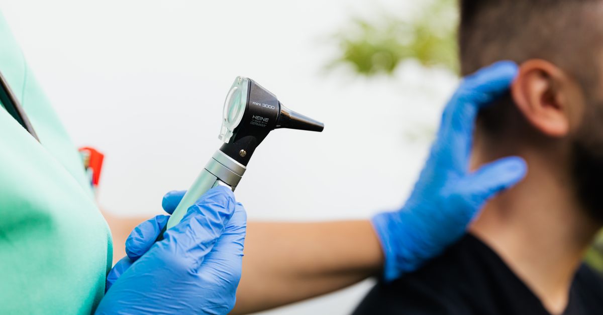
Evaluating a swollen, hard earlobe requires a meticulous physical examination. The physician will gently palpate the lesion to determine if it is solid (like a keloid), fluctuant and fluid-filled (like an abscess), or a discrete mobile sphere (like a cyst).
If active infection is suspected, the physician will check for tracking redness extending onto the face or neck and palpate the regional lymph nodes. If pus is present, a sterile swab is used to collect a sample for laboratory culturing, ensuring the prescribed antibiotics will be effective against the specific bacterial strain.
In rare cases where a firm, painless nodule does not fit the typical profile of a cyst or scar, a minor surgical biopsy may be performed to rule out rare soft tissue tumors or dermatological malignancies.
12. Conservative Home Management
For a mildly swollen, painful earlobe suspected to be an inflamed cyst or a minor piercing infection, targeted thermal therapy is the first line of defense. Applying a warm, moist compress to the earlobe for fifteen minutes, four times a day, increases localized blood flow.
The heat accelerates the body’s natural immune response and helps draw the trapped infection or pus to the surface of the skin, encouraging spontaneous, natural drainage.
If contact dermatitis is the cause, immediately removing all jewelry and applying a thin layer of over-the-counter hydrocortisone cream effectively suppresses the localized allergic immune response, allowing the swelling to subside rapidly.
13. Medical and Surgical Interventions
When home care fails, medical intervention is necessary. For an infected cyst or a localized abscess, a physician will perform an incision and drainage under local anesthesia. Using a sterile scalpel, a small cut is made to evacuate the trapped pus and keratin, providing immediate relief from the throbbing pressure.
For recurrent cysts, the physician must surgically excise the entire lesion. It is critical to remove the entire sac wall intact; if any part of the cystic lining is left behind in the tissue, the cyst will inevitably regrow and refill with keratin.
Bacterial infections accompanied by spreading redness or systemic symptoms mandate a course of oral antibiotics, typically targeting Staphylococcus and Streptococcus species, to eradicate the pathogen from the deep tissue networks.
14. Prevention and Hygiene Protocols
Preventing the recurrence of hard earlobe swellings relies heavily on hygienic practices. New ear piercings must be cleaned meticulously twice daily with sterile saline solution, avoiding harsh chemicals like rubbing alcohol or hydrogen peroxide, which damage healing tissue.
Individuals prone to epidermoid cysts should avoid aggressively squeezing or manipulating their earlobes, as this mechanical trauma forces microscopic skin cells deeper into the tissue, initiating cyst formation.
To prevent contact dermatitis, patients with known metal sensitivities must exclusively wear hypoallergenic jewelry. Medical-grade titanium, surgical stainless steel, niobium, and solid 14-karat or higher gold are the safest options, completely eliminating the risk of a massive histamine response and subsequent localized swelling.
15. Frequently Asked Questions FAQ
1. Should I squeeze the hard lump in my earlobe to pop it?
Absolutely not. Squeezing an epidermoid cyst frequently causes it to rupture internally, spilling irritating keratin into the surrounding tissue, which instantly turns a painless lump into a massive, painful, fiery red swelling.
2. Does a hard lump in the earlobe mean I have cancer?
It is very unlikely. The vast majority of hard nodules in the earlobe are benign cysts, keloid scars, or localized infections. However, any lump that bleeds, changes color, or grows rapidly should be evaluated by a dermatologist.
3. Will taking out my piercing make the infection trapped inside?
If there is a severe, active infection with a lot of pus, taking the jewelry out allows the hole to close, potentially sealing the infection inside and forming an abscess; you should see a doctor before removing jewelry from a severely infected piercing.
4. How do I know if my swollen earlobe is an allergy or an infection?
An allergic reaction to jewelry is typically intensely itchy, scaly, and involves both ears if both have the same metal. An infection usually affects only one ear, is hot, throbbingly painful, and oozes thick yellow or green pus.
5. Will a keloid scar on my earlobe go away on its own?
No, a keloid is an aggressive overgrowth of dense scar tissue that will not shrink or disappear naturally over time. It requires specialized medical treatments like corticosteroid injections or precise surgical removal to flatten it.
16. Bibliography
Disclaimer: The content is for informational purposes only and does not replace medical advice. Always consult your doctor for personalized treatment.