
1. Introduction
An infection of the external ear, clinically diagnosed as acute otitis externa, is a rapidly developing inflammatory condition involving the skin and subdermal tissues of the outer auditory canal. The bottom line up front is that this condition, frequently precipitated by retained moisture and minor mechanical trauma, allows opportunistic environmental bacteria or fungi to invade the delicate canal skin, requiring meticulous aural cleaning and the targeted application of topical antimicrobial and corticosteroid drops to resolve the intense pain and swelling without risking systemic antibiotic resistance.
Colloquially known as “swimmer’s ear,” this condition represents a distinct clinical entity entirely separate from middle ear infections located behind the eardrum. The external auditory canal is a unique anatomical structure—a dark, narrow, skin-lined tunnel terminating in a blind pouch. Understanding the physiological defenses of this specific environment is essential for diagnosing the breach in the epidermal barrier that allows pathogenic organisms to thrive.
2. Anatomy and Defense Mechanisms
The external auditory canal is approximately two and a half centimeters long in adults. The outer third is composed of elastic cartilage, containing numerous hair follicles and specialized ceruminous and sebaceous glands. The inner two-thirds consist of very thin, delicate skin resting directly on the temporal bone, devoid of glands or hair.
The primary defense mechanism of the canal is its slightly acidic environment and continuous epithelial migration. The skin cells inside the canal uniquely migrate outward, from the eardrum toward the external opening, acting like a slow conveyor belt to continuously sweep away cellular debris, dust, and microscopic organisms, keeping the deep canal remarkably clean.
3. The Protective Role of Cerumen
Cerumen, commonly known as earwax, is a critical component of the ear’s defense system. It is a complex mixture of desquamated epithelial cells and the lipid-rich secretions of the ceruminous glands located in the outer cartilaginous portion of the canal.
Cerumen coats the skin of the canal, creating a hydrophobic (water-repelling) barrier that prevents tissue maceration. Furthermore, cerumen is slightly acidic (pH 4 to 5) and contains specific antimicrobial peptides, lysozymes, and immunoglobulins. This acidic, lipid-rich coating creates a hostile environment that severely inhibits the survival and replication of pathogenic bacteria and fungi.
4. Pathophysiology of Epithelial Breakdown
An infection initiates when the protective cerumen barrier is depleted or the canal skin is mechanically compromised. The most frequent catalyst is the retention of moisture. Prolonged exposure to water from swimming, showering, or living in highly humid environments washes away the protective cerumen and alters the pH of the canal, shifting it toward an alkaline state.
This retained moisture causes the skin to become macerated (waterlogged), soft, and highly susceptible to microscopic tearing. The mechanical trauma of patients attempting to dry or scratch their ears with cotton swabs, bobby pins, or fingernails inflicts minute abrasions on the macerated skin, creating a direct physical portal of entry for colonizing pathogens.
5. Primary Bacterial Pathogens
Once the epithelial barrier is breached, the warm, moist, and now alkaline environment of the damaged canal provides an ideal culture medium. Acute otitis externa is overwhelmingly a bacterial disease.
The most frequently isolated pathogen is *Pseudomonas aeruginosa*, a ubiquitous environmental bacterium that thrives in aqueous environments. *Staphylococcus aureus* is the second most common causative organism. Both pathogens induce a rapid and intense localized inflammatory response, characterized by the dilation of blood vessels and the massive influx of white blood cells into the tightly bound tissues of the ear canal.
6. Fungal Otitis Externa (Otomycosis)
While bacteria are the primary culprits, approximately ten percent of external ear infections are caused by fungal pathogens, a condition termed otomycosis. *Aspergillus* species and *Candida* species are the most common fungal isolates.
Fungal infections frequently develop following prolonged use of broad-spectrum topical antibiotic drops, which eradicate the competing bacterial flora and allow the opportunistic fungi to flourish. Otomycosis presents with severe, unrelenting pruritus (itching) and the accumulation of thick, characteristic debris within the canal—often appearing as fine white, black, or grey fungal spores resembling wet newspaper or cotton.
7. Clinical Presentation and Symptoms
The hallmark clinical symptom of acute otitis externa is rapid, progressive, and severe otalgia (ear pain). Because the skin of the inner ear canal is tightly adhered directly to the underlying bone with almost no accommodating subcutaneous fat, even minor tissue swelling generates intense localized pressure and excruciating pain.
Patients frequently report that the pain is severely exacerbated by moving the jaw during chewing or speaking. A distinguishing physical sign is severe tenderness elicited by pulling upward and backward on the pinna (the outer ear) or by pushing firmly on the tragus (the small cartilage flap in front of the canal opening).
8. Diagnostic Examination and Otoscopy
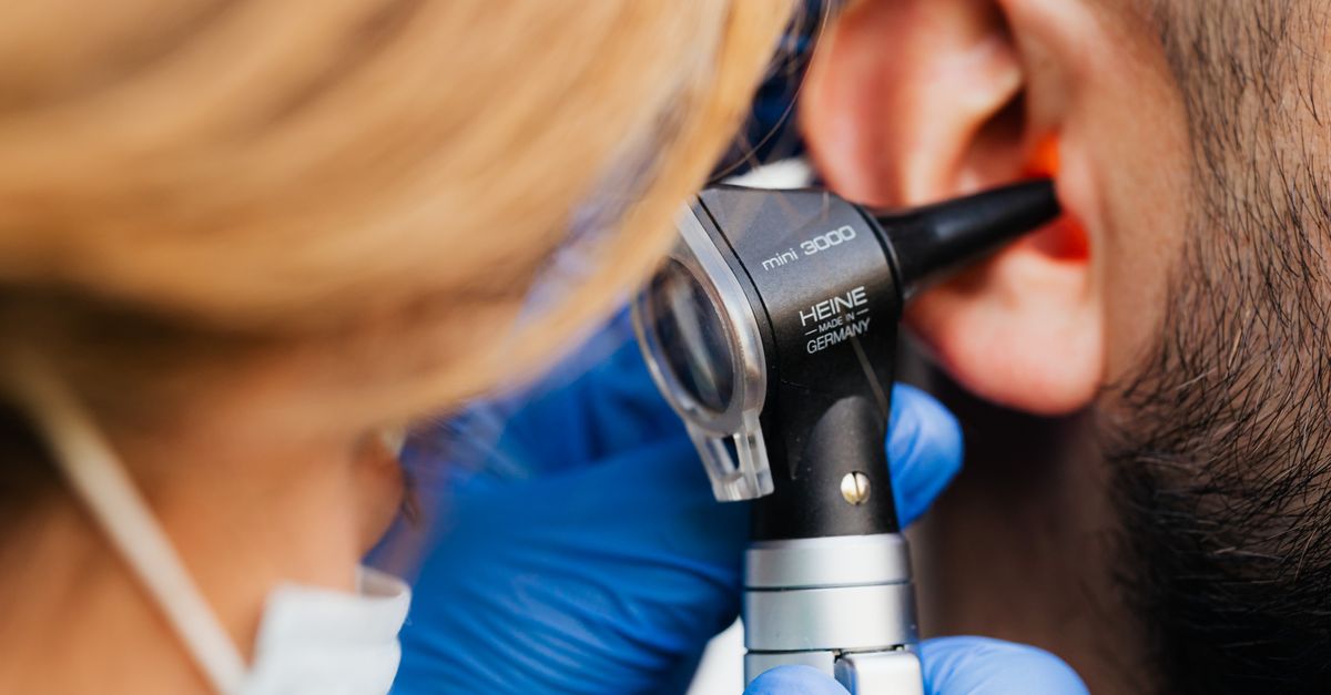
The diagnosis is established through a meticulous clinical history and direct visual inspection using an otoscope. The clinician carefully assesses the condition of the external canal and confirms the structural integrity of the tympanic membrane.
During the active infection, the canal skin appears highly erythematous (red) and markedly edematous (swollen). The swelling can become so severe that the canal is completely occluded, obscuring the view of the eardrum. The clinician will note the presence of purulent, foul-smelling exudate pooling in the canal. If fungal spores are visualized, the diagnosis shifts toward otomycosis.
| Clinical Finding | Acute Otitis Externa (Outer Ear) | Acute Otitis Media (Middle Ear) |
|---|---|---|
| Tragal Tenderness | Severe pain when pushing the tragus or pulling the earlobe | No pain upon manipulation of the outer ear structures |
| Canal Appearance | Red, swollen, narrowed, often filled with debris | Canal skin is typically normal and open |
| Eardrum Appearance | Normal or obscured by canal swelling | Bulging, red, opaque with fluid behind it |
9. Cleaning and Aural Toilet
The most critical and frequently overlooked initial step in medical management is the thorough cleaning of the ear canal, clinically referred to as aural toilet. The canal is often packed with thick, purulent exudate, sloughed skin cells, and cerumen.
If this dense debris is not removed, topical medicated drops cannot reach the infected tissue, resulting in treatment failure. The clinician uses specialized microscopic suction, curettes, or gentle dry mopping under direct visualization to carefully clear the canal. Irrigation with water is strictly avoided if the integrity of the eardrum cannot be verified.
10. Topical Pharmacological Therapies
Once the canal is cleansed, the primary treatment modality is the application of targeted topical ear drops. Topical therapy delivers extremely high concentrations of antimicrobial agents directly to the infected tissue—concentrations much higher than can be achieved safely with oral medications.
The standard formulation includes an antibiotic, such as ciprofloxacin or neomycin, to eradicate the *Pseudomonas* or *Staphylococcus* bacteria. This is universally combined with a topical corticosteroid, such as dexamethasone or hydrocortisone, to rapidly reduce the severe tissue inflammation, opening the canal and providing significant pain relief within twenty-four hours. For detailed discussions on managing skin barrier disruptions, protocols similar to topical dermatological therapies are often applied.
11. Managing Canal Edema with Ear Wicks
In severe cases where the inflammatory edema has completely swelled the canal shut, simply placing drops into the ear is ineffective, as the fluid will immediately run back out.
To bypass this obstruction, the clinician inserts an ear wick—a small, compressed sponge made of expanding polyvinyl alcohol or a strip of specialized gauze. The wick is carefully maneuvered past the swelling into the deep canal. When the patient applies the medicated drops, the wick absorbs the fluid, expands, and holds the antibiotic-steroid mixture firmly against the infected skin, continuously drawing the medication deep into the occluded space.
12. Systemic Therapy and Complications
Systemic (oral or intravenous) antibiotics are rarely necessary for uncomplicated otitis externa and should be avoided to prevent antibiotic resistance. However, their use is mandated if the infection spreads beyond the ear canal.
If the patient develops spreading cellulitis of the pinna or the skin of the face and neck, or exhibits systemic signs such as fever and swollen regional lymph nodes, oral antibiotics (frequently fluoroquinolones to cover *Pseudomonas*) must be initiated. Patients with poorly controlled diabetes or those who are immunocompromised require particularly aggressive monitoring for deep tissue spread.
13. Malignant Otitis Externa
The most feared complication of an external ear infection is malignant otitis externa, also known as necrotizing otitis externa. This is not a cancer, but an aggressive, life-threatening bacterial osteomyelitis (bone infection) of the temporal bone and skull base.
It occurs almost exclusively in elderly patients with diabetes or severe immunosuppression. The *Pseudomonas* bacteria invade through the skin of the canal and begin destroying the surrounding cartilage and bone. Patients present with deep, unrelenting, boring pain that is worse at night, and granulation tissue visible on the floor of the ear canal. This condition requires prolonged intravenous antibiotic therapy and urgent imaging to assess the extent of bone destruction.
14. Pain Management and Patient Education
Aggressive pain management is essential during the first forty-eight hours of treatment. The localized pressure causes substantial suffering. Systemic non-steroidal anti-inflammatory drugs (NSAIDs), such as ibuprofen, are highly effective at reducing both the pain and the underlying tissue inflammation.
Patients are instructed to keep the ear completely dry during the treatment course. This involves avoiding swimming entirely and placing a petroleum jelly-coated cotton ball in the outer ear while showering to prevent water entry. The patient must complete the entire prescribed course of drops, usually seven to ten days, even if symptoms resolve earlier.
15. Prevention Strategies
Preventing recurrent infections relies on maintaining the delicate acidic and hydrophobic balance of the ear canal. Patients must strictly avoid inserting anything into the ear canal, especially cotton swabs, which strip away protective cerumen and cause micro-abrasions.
For individuals prone to moisture retention, such as frequent swimmers, prophylactic measures are highly effective. Applying a few drops of a mixture containing equal parts rubbing alcohol and white vinegar immediately after swimming helps to evaporate trapped water rapidly and restores the naturally protective acidic pH of the canal, deterring bacterial colonization.
16. Frequently Asked Questions (FAQ)
1. Is swimmer’s ear the same as a regular ear infection?
No. Swimmer’s ear is an infection of the skin in the outer canal that leads to the eardrum. A “regular” ear infection, most common in young children, happens behind the eardrum in the middle ear space, usually due to a cold.
2. Why does my doctor want me to use ear drops instead of taking an antibiotic pill?
Ear drops put the medication directly onto the infected skin in a much higher concentration than a pill ever could. Drops also avoid the systemic side effects of oral antibiotics, like stomach upset or building dangerous antibiotic resistance in your gut.
3. Why did the doctor put a small sponge in my ear?
If your ear canal is swollen completely shut, drops will just roll down your cheek. The doctor inserts a tiny, safe sponge called a “wick.” When you put the drops on the wick, it expands and draws the medicine deep inside the swollen canal so it can work properly.
4. Should I clean the pus out with a cotton swab before putting my drops in?
Never put a cotton swab into your ear canal. It will physically push the pus and debris deeper, potentially damage your eardrum, and scratch the infected skin, making the infection much worse. Only a doctor should clean the deep canal.
5. How long will the intense pain last?
Once the appropriate antibiotic and steroid drops are started, the severe pain typically begins to improve within twenty-four to forty-eight hours. If the pain gets worse or you develop a fever after starting treatment, contact your physician immediately.
17. Bibliography
Disclaimer: The content is for informational purposes only and does not replace medical advice. Always consult your doctor for personalized treatment.


