1. Introduction to Infective Otitis Externa
Infective otitis externa, commonly known in the community as “swimmer’s ear,” is an acute inflammatory condition characterized by a bacterial or fungal infection of the skin lining the external auditory canal. The immediate clinical consequence is localized edema, intense pruritus, and severe ear pain. Prompt clinical evaluation is essential to clear the obstructive debris, administer targeted topical antimicrobials, and prevent the infection from spreading to surrounding osseous structures.
The external auditory canal is a unique anatomical cul-de-sac. It is a dark, warm, and narrow channel lined entirely by skin. While it possesses excellent self-cleaning and antimicrobial defense mechanisms, specific environmental triggers—most notably retained moisture and mechanical trauma—can swiftly disrupt this delicate equilibrium, allowing opportunistic environmental pathogens to flourish.
Clinical management generally involves localized therapy. Unlike infections of the middle ear, otitis externa is primarily an infection of the superficial epidermal layers. Successful treatment relies fundamentally on meticulously cleaning the canal to allow topical medications direct access to the inflamed epithelial surface, rather than relying solely on systemic antibiotics.
2. Anatomy of the External Auditory Canal
To understand the pathogenesis of this infection, one must examine the specific anatomy of the outer ear. The external auditory canal is a tubular structure extending from the auricle to the tympanic membrane. The outer third of the canal is cartilaginous and contains hair follicles, sebaceous glands, and specialized apocrine glands.
These apocrine glands produce cerumen, or earwax. The inner two-thirds of the canal is osseous, consisting of a very thin layer of skin tightly adherent directly to the underlying temporal bone, without a cushioning layer of subcutaneous fat. This tight adherence is the reason why even minor inflammation in this region causes such intense, disproportionate pain, as the swelling tissue immediately compresses the local sensory nerves against the rigid bone.
The canal also possesses a unique natural migratory mechanism. The epithelial cells grow outward from the center of the tympanic membrane toward the opening of the ear, acting as a slow conveyor belt that naturally carries debris and old cerumen out of the canal, negating the need for manual cleaning.
3. The Protective Role of Cerumen
Cerumen is not merely dirt; it is a vital, biologically active substance that provides the primary defense against infective otitis externa. It forms a hydrophobic layer over the delicate skin of the canal, physically repelling water and preventing the maceration of the underlying epidermal cells.
Furthermore, cerumen is distinctly acidic, maintaining a low pH environment within the canal. This acidity is inherently hostile to the growth of pathogenic bacteria and fungi. Cerumen also contains specific antimicrobial peptides and lysozymes that actively neutralize invading microorganisms.
When individuals aggressively clean their ears using cotton swabs or other implements, they strip away this essential protective layer. This action not only removes the chemical and physical barrier but also creates microscopic abrasions in the skin, providing a direct portal of entry for ambient pathogens.
4. Pathophysiology of Epithelial Breakdown
The cascade leading to infective otitis externa almost invariably begins with a breakdown of the epidermal barrier. The most frequent trigger is prolonged exposure to moisture, typically from frequent swimming, bathing, or residing in humid environments. Retained water within the canal causes the stratum corneum to macerate and soften.
This maceration shifts the naturally acidic pH of the canal to a more alkaline state, destroying the hostile environment that normally suppresses bacterial growth. Concurrently, the softened skin becomes exceptionally vulnerable to physical trauma from hearing aids, earplugs, or the ubiquitous use of cotton swabs.
Once the epidermal integrity is compromised, opportunistic bacteria rapidly colonize the superficial layers of the skin. The host immune system mounts an inflammatory response, leading to profound vasodilation and increased capillary permeability. The resulting massive accumulation of plasma fluid within the confined space of the canal causes the characteristic severe swelling and agonizing pain.
5. Primary Bacterial and Fungal Pathogens
Pseudomonas aeruginosa is the overwhelmingly predominant bacterial pathogen isolated in acute infective otitis externa. This Gram-negative bacterium is ubiquitous in the environment, particularly thriving in soil and aquatic environments. It is remarkably resilient and rapidly exploits the moist, alkaline conditions of a compromised ear canal.
Staphylococcus aureus is the second most common causative agent. Unlike the diffuse inflammation caused by Pseudomonas, Staphylococcus aureus infections often localize within the hair follicles of the outer cartilaginous canal, occasionally forming a distinct, painful furuncle or localized abscess.
In approximately ten percent of cases, the infection is driven by fungal organisms, a condition termed otomycosis. Aspergillus species and Candida species are the most frequent culprits. Fungal infections are particularly common in tropical climates, in patients using long-term topical antibacterial drops, or in individuals with underlying immunosuppressive conditions.
6. Environmental and Behavioral Risk Factors
Frequent exposure to recreational water, particularly in lakes or poorly maintained pools, significantly increases the risk of acquiring pathogenic strains of Pseudomonas. The condition earns the moniker “swimmer’s ear” precisely because the combination of retained moisture and exposure to aquatic bacteria provides the perfect storm for infection.
Aggressive aural hygiene is a massive behavioral risk factor. The insertion of any object into the ear canal pushes cerumen deeper, impacts debris against the tympanic membrane, and invariably causes micro-trauma to the thin epithelium covering the osseous canal.
Individuals with narrow, tortuous ear canals, or those suffering from chronic dermatological conditions such as eczema, psoriasis, or seborrheic dermatitis, face an elevated risk. The chronic inflammation and excessive scaling associated with these skin conditions disrupt the normal self-cleaning mechanism, causing debris to accumulate and trap moisture.
7. Clinical Manifestations and Pain
The hallmark symptom of acute infective otitis externa is severe, unrelenting otalgia. The pain is frequently disproportionate to the visual signs of inflammation. Crucially, the pain is drastically exacerbated by any manipulation of the auricle, particularly pulling upward and backward on the pinna, or pressing inward on the tragus.
Patients frequently report intense pruritus preceding the onset of pain, particularly if the infection is fungal in origin. As the inflammation progresses, a feeling of fullness or pressure develops within the ear. A distinct, purulent discharge, termed otorrhoea, often begins to weep from the canal.
This discharge may be clear, purulent, or foul-smelling. Depending on the severity of the edema and the volume of accumulated debris, the patient may experience conductive hearing loss, as the physical obstruction prevents sound waves from efficiently reaching the tympanic membrane.
8. Progression to Malignant Otitis Externa
A profoundly dangerous complication, termed malignant or necrotizing otitis externa, can occur when the infection spreads beyond the superficial skin and invades the underlying temporal bone. This aggressive osteomyelitis is almost exclusively caused by Pseudomonas aeruginosa.
Malignant otitis externa is a life-threatening condition observed primarily in elderly patients with poorly controlled diabetes mellitus or individuals with profound immunosuppression. The reduced microvascular circulation and impaired leukocyte function in these patients allow the bacteria to destroy the bone and spread toward the base of the skull.
The clinical presentation involves deep, boring, unremitting otalgia that is characteristically worse at night. Granulation tissue is frequently visible on the floor of the osseous canal. If left untreated, the infection rapidly involves the cranial nerves, leading to facial paralysis and central nervous system complications.
9. Otoscopic Examination and Diagnosis
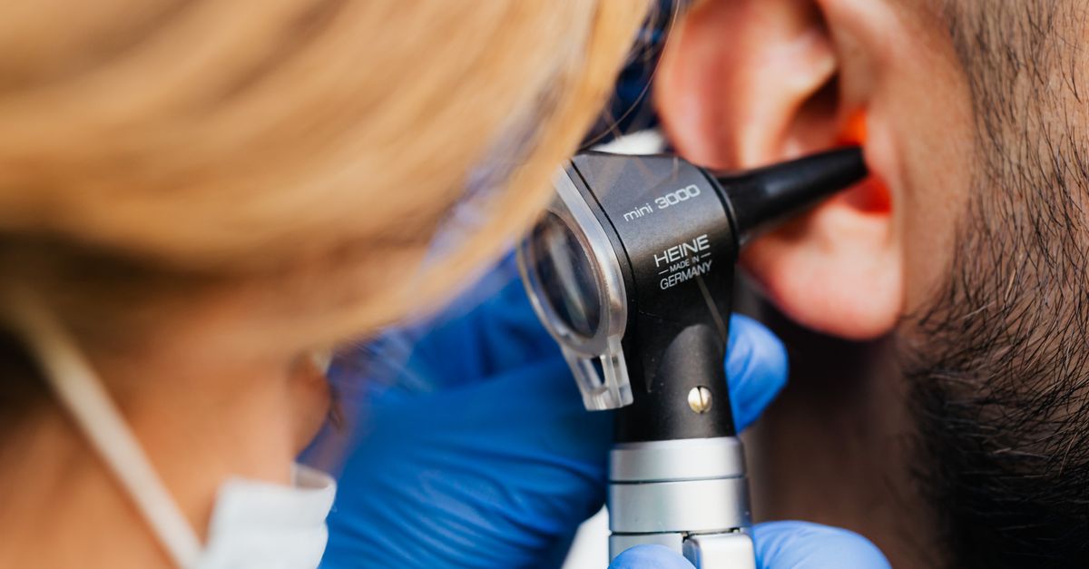
The diagnosis of infective otitis externa is primarily clinical, relying on a focused history and a meticulous otoscopic examination. The clinician attempts to visualize the entire canal and the tympanic membrane to assess the extent of the inflammation and rule out a concurrent middle ear infection.
In acute cases, the canal appears intensely erythematous and swollen, often to the point where the tympanic membrane is completely obscured by the edematous tissue. The canal is typically filled with a mixture of purulent exudate, sloughed epithelial cells, and cerumen.
If a fungal etiology is present, the clinician may observe distinctive fungal elements. Aspergillus infections often present with a thick, mold-like debris displaying distinct black or white spores (conidiophores) sprouting from the canal walls. Fungal infections generally present with more intense pruritus and less severe pain compared to bacterial etiologies.
10. Differentiating External and Middle Ear Infections
Accurately distinguishing between an infection of the ear canal and an infection behind the eardrum is a critical clinical skill, as the treatments differ substantially. The table below outlines the primary differentiating features.
| Diagnostic Feature | Infective Otitis Externa (Outer Ear) | Acute Otitis Media (Middle Ear) |
|---|---|---|
| Pain upon pulling the pinna or tragus | Severe pain exacerbated by movement. | No pain upon external manipulation. |
| Otoscopic Appearance | Canal is red, swollen, and filled with debris. | Canal is clear; eardrum is bulging and red. |
| Nature of Discharge | Common, often thick or purulent. | Absent, unless the eardrum has ruptured. |
| Systemic Symptoms | Usually absent. | Common, including fever and upper respiratory symptoms. |
11. Aural Toilet and Canal Cleaning
The foundational step in treating any severe case of infective otitis externa is a thorough aural toilet. Topical antimicrobial drops cannot penetrate thick layers of purulent exudate and sloughed skin to reach the infected epithelial surface. The canal must be physically cleared.
This procedure is typically performed by a physician using a specialized operating microscope and delicate micro-suction instruments. The clinician gently vacuums the debris from the canal, providing immediate relief from the feeling of fullness and significantly improving the efficacy of subsequent topical medications.
Manual irrigation with water is generally avoided in acute cases. The introduction of water can exacerbate the maceration of the tissue, push infected debris deeper against the tympanic membrane, or cause severe vertigo if the water temperature is not precisely matched to the patient’s body temperature.
12. Topical Antimicrobial and Steroid Therapies
Following aural toilet, the primary pharmacological treatment involves the direct application of topical otic drops. These medications provide a very high concentration of antimicrobial agents directly to the site of infection while completely avoiding the systemic side effects associated with oral antibiotics.
Combination drops containing an antibiotic and a topical corticosteroid are the standard of care. Medications like ciprofloxacin-dexamethasone or neomycin-polymyxin-hydrocortisone effectively eradicate the common bacterial pathogens while the steroid component rapidly suppresses the intense localized inflammation and pain.
If the tympanic membrane is known to be ruptured, or if its integrity cannot be verified due to severe swelling, medications containing aminoglycosides (like neomycin) must be strictly avoided due to their potential ototoxicity. In these scenarios, fluoroquinolone-based drops are utilized, as they are proven safe for the middle ear space.
13. Use of Ear Wicks for Severe Edema
In severe cases, the inflammatory edema may cause the walls of the ear canal to swell completely shut. When the canal is occluded, it is physically impossible for the patient to instill topical drops deep enough to reach the active site of infection.
To overcome this, the physician will carefully insert a small, expandable sponge, known as an ear wick, directly into the swollen canal. The patient then applies the prescribed antibiotic drops directly onto the exposed end of the wick.
The wick rapidly absorbs the medication and expands, maintaining constant, direct contact with the inflamed epithelial walls. The wick also provides mild, continuous mechanical pressure that helps reduce the swelling. As the inflammation subsides over a few days, the canal opens, and the wick falls out spontaneously or is easily removed by the physician.
14. Systemic Therapy and High-Risk Patients
Oral systemic antibiotics are rarely indicated for uncomplicated cases of infective otitis externa, as topical drops deliver a significantly higher concentration of the drug directly to the infected tissue. However, specific clinical scenarios mandate systemic intervention.
Oral antibiotics, specifically fluoroquinolones with excellent anti-pseudomonal coverage, are prescribed if the infection has spread beyond the ear canal, causing cellulitis of the auricle, face, or neck. Systemic therapy is also required if the patient exhibits systemic signs of illness, such as a high fever or regional lymphadenopathy.
Furthermore, any patient presenting with diabetes mellitus, severe immunosuppression, or a history of radiation therapy to the head and neck must be evaluated aggressively. Due to the high risk of progressing to malignant otitis externa, these vulnerable patients are frequently treated concurrently with both topical and systemic antimicrobial regimens.
15. Frequently Asked Questions FAQ
1. Why does my ear hurt so much when I chew food?
The jawbone connects to the skull right next to the ear canal. When you chew, the movement of the jaw joint presses directly against the severely inflamed and swollen tissues of the infected ear canal, causing sharp, excruciating pain.
2. Should I use a cotton swab to clean the pus out of my ear?
Absolutely not. Inserting a cotton swab will pack the infected debris deeper into the canal, scrape away the remaining protective wax, and create tiny cuts in the skin, driving the bacteria deeper into the tissue and worsening the infection.
3. Are oral antibiotics necessary for swimmer’s ear?
In most cases, no. Specially formulated antibiotic ear drops are actually much more effective because they deliver the medicine directly onto the infected skin in very high concentrations, without the side effects of taking pills.
4. How do doctors treat the infection if my ear canal is completely swollen shut?
A doctor will insert a tiny, compressed sponge called an ear wick into the tight space. When you put your prescribed ear drops onto the wick, it swells up and holds the medicine directly against the inflamed skin, slowly opening the canal.
5. How can I prevent this infection if I swim frequently?
Prevention involves keeping the ears dry. Shake your head to remove trapped water after swimming, or use a hair dryer on the lowest, cool setting held several inches away from the ear to gently evaporate the moisture. Avoid removing the natural protective earwax.
16. Bibliography
Disclaimer: The content is for informational purposes only and does not replace medical advice. Always consult your doctor for personalized treatment.