1. Introduction
A foot injury involves structural damage to the complex arrangement of bones, ligaments, tendons, and fascial connective tissues that form the anatomical foundation of human locomotion. Because the foot is required to bear the entire weight of the body while absorbing significant kinetic forces during ambulation, any mechanical disruption frequently results in profound functional impairment. The clinical presentation is typically characterized by localized pain, immediate edema, and an inability to bear weight.
The human foot consists of twenty-six distinct bones articulated through thirty-three joints, supported by a dense network of over one hundred muscles, tendons, and ligaments. This intricate architecture is broadly divided into the hindfoot, midfoot, and forefoot. Understanding the specific biomechanical demands placed on each of these regions is essential for identifying occult fractures and subtle ligamentous instabilities that might otherwise be overlooked during an initial clinical assessment.
Effective management of pedal trauma relies on a rigorous diagnostic approach. Clinicians must meticulously palpate the bony landmarks, assess neurovascular integrity, and utilize targeted radiographic imaging. By accurately identifying the specific anatomical deficit, healthcare providers can implement appropriate immobilization strategies or surgical interventions, ensuring proper tissue repair and preventing chronic mechanical dysfunction.
2. Biomechanics of the Foot
The primary function of the foot is to act as both a flexible shock absorber upon striking the ground and a rigid lever for forward propulsion. The medial longitudinal arch, supported heavily by the plantar fascia and the posterior tibial tendon, provides the necessary spring-like elasticity to dissipate kinetic energy.
During a normal walking cycle, the hindfoot absorbs the initial impact. The kinetic energy then transfers through the midfoot complex, which locks into a rigid structure, allowing the forefoot to push off against the ground. An injury to any component of this kinetic chain disrupts the entire sequence, placing abnormal stress on adjacent joints and frequently leading to compensatory ankle pain and abnormal gait patterns.
Tendons crossing the ankle joint insert into various pedal bones, facilitating complex movements such as inversion and eversion. A sudden, forceful stretch of these tendons during a misstep can cause severe structural tearing or forcefully detach a fragment of bone at the insertion site, creating an avulsion fracture.
3. Metatarsal Fractures
The five metatarsal bones form the structural framework of the forefoot. Fractures in this region are exceptionally common and typically result from a direct blunt impact, such as a heavy object dropping onto the dorsum of the foot.
A specific and clinically significant injury is the Jones fracture, which involves the proximal diaphysis of the fifth metatarsal. This particular anatomical zone receives a notably poor blood supply. Consequently, fractures in this region demonstrate a pronounced tendency toward delayed healing or complete nonunion, frequently necessitating prolonged immobilization or surgical fixation with an intramedullary screw.
Stress fractures of the metatarsals, colloquially known as march fractures, develop from repetitive mechanical overload rather than a single acute impact. These injuries are prevalent among military recruits and endurance athletes. Patients report a deep, aching pain that progressively worsens with weight-bearing activity and strictly resolves with rest.
4. Lisfranc Joint Disruption
The Lisfranc joint complex represents the critical articulation between the midfoot tarsal bones and the forefoot metatarsals. A Lisfranc injury occurs when the ligaments stabilizing this junction are severely sprained or when the corresponding bones are fractured and dislocated.
This injury typically arises from an indirect force applied to a plantarflexed foot, such as stumbling into a hole or being tackled while the foot is firmly planted. The patient experiences severe midfoot pain, profound swelling on the top of the foot, and an absolute inability to rise onto their toes. Plantar ecchymosis, a distinct bruising pattern on the sole of the foot, is a hallmark clinical sign.
Lisfranc injuries are notoriously difficult to identify on standard non-weight-bearing radiographs. Because failure to stabilize this joint leads to severe, debilitating midfoot arthritis, clinicians must maintain a low threshold for ordering weight-bearing films or advanced tomographic imaging to detect subtle structural displacement.
5. Plantar Fascia Trauma
The plantar fascia is a thick, unyielding band of connective tissue originating from the calcaneus and extending to the base of the toes. While chronic inflammation of this structure is common, acute traumatic tearing can occur during sudden, explosive acceleration, such as sprinting or jumping.
An acute plantar fascia rupture presents with a sudden, sharp popping sensation in the arch of the foot. The patient may subsequently develop a palpable defect or lump in the plantar tissue accompanied by significant localized bruising.
Management of an acute fascial rupture is predominantly conservative. The patient is placed in a rigid walking boot to relieve tension on the tissue, allowing the dense collagen matrix to heal. Structured physical therapy is eventually initiated to restore flexibility to the arch and strengthen the intrinsic foot musculature.
6. Calcaneal and Talar Fractures
The calcaneus and the talus form the hindfoot, bearing the most intense compressive loads in the lower extremity. Fractures in this region are almost exclusively the result of profound vertical impact, such as falling from a significant height or enduring a severe motor vehicle collision.
A calcaneal fracture severely flattens the heel, creating a widened, shortened appearance of the hindfoot. Because the impact required to shatter the heel bone is so intense, clinicians must routinely evaluate these patients for concurrent compression fractures of the lumbar spine.
Talar fractures are particularly precarious. The talus is covered largely by articular cartilage and receives a fragile, retrograde blood supply. A displaced fracture of the talar neck frequently severs this vascular network, placing the bone at exceptionally high risk for avascular necrosis, a condition where the bone tissue starves of oxygen, dies, and structurally collapses.
7. Ligamentous Sprains and Instability
While ankle sprains are universally recognized, the small stabilizing ligaments throughout the midfoot and forefoot are equally susceptible to traumatic stretching. A midfoot sprain involves the dense dorsal and plantar ligaments binding the navicular, cuboid, and cuneiform bones.
These sprains typically result from severe twisting motions. The foot becomes diffusely swollen and exquisitely tender to palpation over the central dorsal aspect. Because the midfoot relies entirely on ligamentous integrity for its rigid arch structure, inadequate healing of a severe sprain leads to a gradual flattening of the arch and chronic mechanical fatigue.
Turf toe is a specific ligamentous sprain affecting the main joint of the great toe. The plantar capsule is forcefully hyperextended when the toe is driven upward against a rigid athletic surface. This injury cripples the push-off phase of the running cycle, demanding strict taping or rigid orthotic inserts to restrict movement during the prolonged healing phase.
8. Tendon Ruptures in the Foot
Tendons are responsible for transferring muscular power from the lower leg to the foot. The Achilles tendon is the most robust tendon in the body, inserting into the posterior calcaneus. A complete rupture typically occurs during sudden push-off movements, resulting in a palpable gap behind the heel and an inability to actively point the foot downward against resistance.
The posterior tibial tendon courses down the inside of the ankle and attaches under the midfoot, serving as the primary dynamic stabilizer of the medial arch. Acute traumatic tearing or chronic degenerative rupture of this tendon causes the arch to collapse completely, leading to a condition known as adult-acquired flatfoot deformity.
Surgical intervention is frequently recommended for complete tendon ruptures in active individuals. The torn collagen bundles are meticulously sutured together to restore appropriate resting tension, allowing the muscle to effectively govern the mechanical movement of the foot once rehabilitation is complete.
9. Crush Injuries and Compartment Syndrome
When a heavy object compresses the foot, the resulting crush injury involves multiple tissue layers simultaneously. The dense bones may shatter, but the primary clinical concern is the severe contusion to the surrounding muscle and vascular tissues.
The foot contains several distinct muscular compartments bounded by rigid, unyielding fascia. Following a severe crush injury, the damaged tissues swell rapidly, and bleeding occurs within these closed spaces. The internal pressure rises relentlessly, compressing the delicate microvascular networks and choking off arterial blood flow to the muscles and nerves.
This phenomenon is known as compartment syndrome. The patient experiences pain that is entirely out of proportion to the visible injury and is not relieved by standard analgesic medications. If the pressure is not surgically released through a fasciotomy, the muscle tissue undergoes irreversible ischemic necrosis, resulting in permanent, crippling deformity.
10. Clinical Examination and Gait Analysis
Evaluating pedal trauma begins with a systematic physical assessment. The clinician observes the resting posture of the foot, looking for gross deformity, significant swelling, or pronounced ecchymosis. A detailed neurovascular check is mandatory, assessing the dorsalis pedis and posterior tibial pulses, along with sensory perception across all dermatomes.
Palpation is conducted precisely along the bony contours, specifically testing the bases of the metatarsals, the navicular prominence, and the entire length of the calcaneus. The clinician gently manipulates each joint to evaluate mechanical stability and assess for hidden ligamentous laxity.
If the patient can tolerate weight-bearing, a brief gait analysis provides invaluable diagnostic clues. An inability to push off suggests Achilles or forefoot pathology, while a reluctance to strike the heel indicates severe hindfoot contusion or fracture.
11. Diagnostic Imaging Modalities
Standard radiographic imaging is the fundamental diagnostic tool for evaluating structural foot trauma. A comprehensive series includes anteroposterior, lateral, and oblique views. Whenever clinically possible, these radiographs should be taken while the patient bears weight. Weight-bearing films expose subtle joint space widening that remains hidden when the foot is relaxed.
Computed Tomography is essential for surgical planning in complex fractures, particularly those involving the Lisfranc joint, the calcaneus, or the talus. It provides detailed, cross-sectional maps of the shattered bone fragments and evaluates the precise alignment of the articular surfaces.
Magnetic Resonance Imaging is exceptionally sensitive for diagnosing soft tissue pathology. It is the preferred modality for confirming suspected plantar fascia ruptures, evaluating the integrity of the posterior tibial tendon, and identifying early bony stress reactions before they become visible fractures on plain radiographs.
12. Principles of Immobilization
The cornerstone of conservative management for stable pedal injuries involves strict immobilization. Restricting movement protects the injured tissues from mechanical sheer forces, allowing the inflammatory cascade to subside and the primary collagen matrix to organize appropriately.
A short-leg walking cast or a removable pneumatic walking boot is frequently utilized. These devices provide rigid external support, distributing the compressive forces of walking away from the specific injury site and up toward the sturdy lower leg.
For injuries requiring absolute offloading, such as severe calcaneal fractures or complex midfoot disruptions, patients are instructed to use crutches or knee scooters to strictly avoid placing any weight on the affected limb, preventing the displacement of bone fragments during the early healing phase.
13. Surgical Fixation Techniques
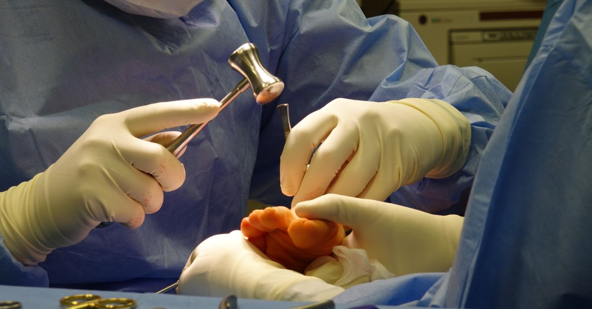
When fractures are significantly displaced or when critical ligaments are completely ruptured, surgical intervention is necessary to restore anatomical continuity. The objective of open reduction and internal fixation is to align the bones perfectly and secure them rigidly, preventing post-traumatic arthritis.
Surgeons utilize anatomically contoured titanium plates and low-profile screws to stabilize metatarsal and calcaneal fractures. The hardware acts as an internal splint, holding the bone fragments tightly together while natural osseous bridging occurs.
For severe Lisfranc injuries, the disrupted joint spaces may be temporarily stabilized with screws or permanently fused together to eliminate painful motion across the damaged cartilage. Postoperative care demands strict non-weight-bearing compliance, as the delicate metal hardware cannot support the full mechanical load of the human body without failing.
14. Rehabilitation and Orthotic Support
Following the acute healing phase, structured rehabilitation is critical for restoring functional mobility. Prolonged immobilization invariably leads to muscular atrophy and profound joint stiffness. Physical therapy initially focuses on aggressive range-of-motion exercises to prevent the formation of dense, restrictive scar tissue.
As flexibility returns, the regimen shifts toward progressive resistance training. Strengthening the intrinsic muscles of the foot and the powerful extrinsic muscles of the lower leg restores the dynamic stability required to support the arch and propel the body forward.
Custom orthotic inserts play a vital role in long-term recovery. By altering the biomechanical angles of the foot strike, orthotics can permanently offload stress from a recovering metatarsal or provide essential mechanical support to a weakened plantar fascia, minimizing the risk of recurrent chronic injury.
15. Frequently Asked Questions
1. How can I tell if my foot is broken or just severely sprained?
While sprains can be incredibly painful, an absolute inability to bear any weight on the foot, combined with severe pain when pressing directly on the bones, strongly suggests a fracture requiring formal radiographic evaluation.
2. What is a Jones fracture?
A Jones fracture is a specific break on the outer edge of the foot, specifically the fifth metatarsal bone. It occurs in an area with a remarkably poor blood supply, making it notoriously slow to heal and often requiring a specialized walking boot or surgery.
3. Why is my foot bruised on the bottom after twisting it?
Bruising on the sole of the foot is a hallmark sign of a Lisfranc injury, which involves torn ligaments or broken bones in the middle of the foot. This is a severe injury that requires immediate clinical assessment.
4. Can a torn tendon in the foot heal without surgery?
Some partial tendon tears can heal using a rigid walking boot and extensive physical therapy. However, complete ruptures in young or active individuals typically require surgical repair to restore the mechanical strength needed for running and jumping.
5. What should I do immediately after dropping something heavy on my foot?
You should immediately elevate the foot above heart level and apply a cold compress to minimize swelling. Do not attempt to walk on it if the pain is severe. Seek medical attention promptly to rule out a fracture or a dangerous buildup of pressure inside the foot.
16. Bibliography
Disclaimer: The content is for informational purposes only and does not replace medical advice. Always consult your doctor for personalized treatment.