1. Introduction
A sudden, sharp headache behind the ear during straining is typically a primary exertional headache caused by a transient spike in intrathoracic and intracranial pressure. The physical act of straining, known as the Valsalva maneuver, temporarily impedes venous blood return from the head, rapidly expanding the delicate blood vessels in the meninges. This rapid vascular expansion mechanically stretches the surrounding sensory nerves, particularly the occipital nerves, triggering a sharp, localized pain response.
The human circulatory system must carefully balance the blood flowing into the brain through the arteries and the blood draining out through the veins. When you lift a heavy object, cough forcefully, or bear down, you instinctively hold your breath and bear down, clamping the vocal cords shut. This action drastically increases the pressure inside your chest and abdomen, providing core stability. However, this high pressure acts as a physical roadblock for the jugular veins, preventing blood from effectively draining out of the skull.
As arterial blood continues to pump in, but venous blood cannot escape, the blood vessels surrounding the brain become rapidly engorged. The meninges, the protective layers encasing the brain, are highly sensitive to this mechanical stretching. The resulting pain is sudden, often explosive, and closely linked to the exact moment of physical strain.
While this phenomenon is largely a benign physiological reflex, the sudden onset of severe head pain always warrants careful analysis. Understanding the neural pathways, specifically the occipital nerve branches located just behind the ear, explains the precise location of the pain and helps differentiate a harmless exertion headache from a serious vascular event.
2. The Valsalva Maneuver and Intracranial Pressure
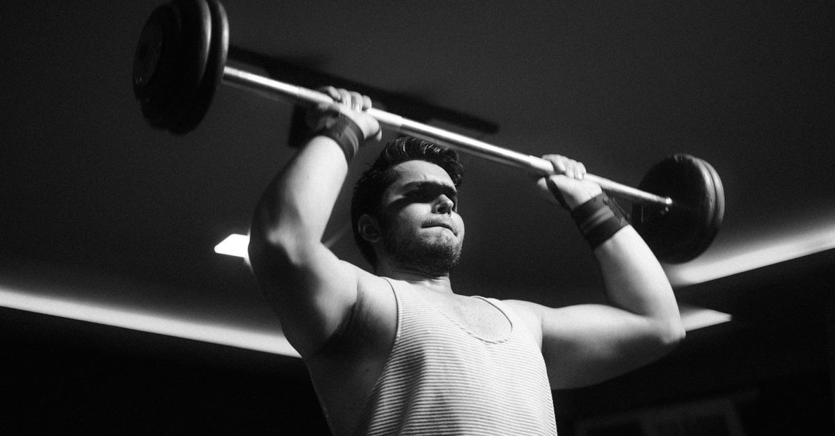
The Valsalva maneuver is a fundamental physiological action performed daily during activities like lifting, coughing, or straining during a bowel movement. It involves forced expiration against a closed glottis. This action elevates intrathoracic pressure, which stabilizes the spine and torso to facilitate heavy muscular exertion.
However, the pressure inside the chest directly dictates the pressure gradient for venous blood returning from the head. Because the jugular veins rely on a low-pressure environment in the chest to drain blood downward via gravity and pressure differentials, a sudden spike in chest pressure brings this drainage to an immediate halt.
The blood backs up into the cranial vault. Because the skull is a rigid, closed container of bone, any increase in fluid volume directly translates into an increase in intracranial pressure. The brain itself does not feel pain, but the intricate network of blood vessels and the meningeal coverings are richly supplied with pain-sensing nerve fibers.
The rapid, forceful distension of these intracranial and extracranial vessels during the Valsalva maneuver acts as a mechanical trigger, activating the nociceptors and generating the acute, throbbing pain characteristic of an exertion headache.
3. Anatomy of the Occipital Nerves
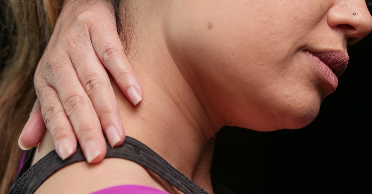
The specific location of the pain—directly behind the ear—points to the involvement of the occipital nerves. The lesser occipital nerve and the greater occipital nerve originate from the upper cervical spine, specifically the C2 and C3 nerve roots. They travel upward, piercing through the dense musculature of the upper neck to provide sensory innervation to the back of the head and the area surrounding the ears.
These nerves are situated in a highly vulnerable anatomical position. As they exit the spinal column and travel toward the scalp, they pass closely to the major blood vessels supplying the posterior skull, including the occipital artery.
When physical straining causes these arteries to suddenly dilate and engorge with blood, the expanding vessels can mechanically press against the adjacent occipital nerves. The nerve fibers are highly sensitive to sudden compression or chemical irritation from surrounding inflammatory processes.
The brain perceives this sudden nerve compression as a sharp, electrical, or stabbing pain localized precisely to the anatomical distribution of the nerve, which sits prominently behind the mastoid bone of the ear.
4. Occipital Neuralgia as a Contributing Factor
In some individuals, the sharp pain is not solely a vascular issue but involves pre-existing irritation of the nerve itself, a condition known as occipital neuralgia. Occipital neuralgia is characterized by chronic or episodic shooting, shocking pain radiating from the base of the skull up to the ears or eyes.
If the occipital nerves are already inflamed or tightly compressed by tense neck muscles, they exist in a state of hyper-excitability. The threshold required to trigger a pain signal is abnormally low. In this state, even a minor increase in vascular pressure from mild straining can push the nerve past its threshold, causing a sudden, explosive pain behind the ear.
Occipital neuralgia is often rooted in chronic poor posture, previous whiplash injuries, or cervical spine arthritis. When an individual with this underlying nerve irritation performs a heavy lift, the combination of tight neck muscles stabilizing the spine and the sudden vascular engorgement creates a perfect storm for a severe neuralgic pain spike.
The pain of occipital neuralgia is distinctly sharp and lancinating, closely resembling an electrical shock, which clearly differentiates it from the dull, heavy throb of a standard muscular tension headache.
5. Cervicogenic Headache Overlap
The structures of the upper neck are deeply integrated with the pain pathways of the head. A cervicogenic headache originates from a structural disorder in the cervical spine, such as an irritated facet joint or a bulging disc, but the pain is felt in the head.
During heavy straining, the muscles of the neck contract forcefully to brace the upper body. If there is an underlying joint dysfunction in the upper neck, this intense muscular contraction can temporarily jam the facet joints together or compress a cervical nerve root.
Because the upper cervical nerves converge with the trigeminal nerve system in the brainstem, the brain frequently misinterprets pain originating in the neck joints as pain occurring behind the ear or in the temple.
If the sharp headache behind the ear during exertion is accompanied by significant neck stiffness or restricted range of motion, a cervicogenic etiology is highly probable, highlighting the mechanical link between cervical stability and cranial pain.
6. Primary Exertional Headache
When no underlying structural, vascular, or neurological defect can be found, the condition is classified clinically as a primary exertional headache. This is a recognized headache disorder provoked explicitly by physical exercise, most commonly weightlifting, running, or sudden explosive movements.
Primary exertional headaches typically present as a pulsating, severe pain that can last anywhere from five minutes to forty-eight hours after the triggering event. The exact pathophysiology remains a subject of ongoing research, but it is largely attributed to an abnormal vascular response.
In healthy individuals, the blood vessels in the brain can rapidly constrict or dilate to maintain constant cerebral blood flow despite massive changes in systemic blood pressure during exercise. In individuals prone to primary exertional headaches, this autoregulatory mechanism is believed to be sluggish or flawed.
The vessels over-dilate during the strain and fail to recover promptly, leaving the meninges stretched and the pain receptors constantly firing until the vascular tone eventually normalizes.
7. The Role of Dehydration and Heat
Environmental and systemic factors can lower the threshold for developing an exertion headache. Dehydration is a potent trigger. When the body is dehydrated, the total blood volume decreases, causing the blood to become more viscous.
To maintain adequate oxygen delivery to the brain during physical strain, the cerebral blood vessels must dilate wider than they normally would if the individual were properly hydrated. This exaggerated dilation increases the mechanical stretch on the meninges.
Furthermore, exercising in a hot environment exacerbates the issue. Heat forces widespread peripheral vasodilation to cool the body. The cardiovascular system must work significantly harder to maintain blood pressure, creating chaotic shifts in vascular tone when a sudden Valsalva maneuver is introduced.
Ensuring optimal systemic hydration and managing environmental heat drastically reduces the likelihood of encountering sudden, sharp cranial pain during physical exertion.
8. Warning Signs of a Subarachnoid Hemorrhage
While most exertion headaches are benign, the sudden onset of severe head pain during physical strain is the classic presentation of a subarachnoid hemorrhage, a life-threatening medical emergency.
A subarachnoid hemorrhage occurs when a weakened blood vessel in the brain, typically an aneurysm, ruptures under the sudden spike of intracranial pressure caused by the Valsalva maneuver. The blood rapidly fills the space surrounding the brain, causing immediate, catastrophic irritation of the meninges.
| Characteristic | Benign Exertional Headache | Subarachnoid Hemorrhage |
|---|---|---|
| Onset | Sudden, during strain. | Instantaneous, “Thunderclap” onset. |
| Pain Severity | Moderate to severe, throbbing. | Worst headache of the person’s entire life. |
| Neurological Signs | None. Normal cognition. | Confusion, slurred speech, visual loss, weakness. |
| Associated Symptoms | Mild nausea, neck tension. | Projectile vomiting, stiff neck, loss of consciousness. |
Any headache described as a “thunderclap”—reaching maximum, unbearable intensity within seconds of the strain—mandates an immediate call to emergency services for an urgent cranial computed tomography scan.
9. Diagnostic Evaluation
Because benign and dangerous exertion headaches share an identical trigger, new onset exertional pain must always be evaluated by a physician. The primary goal of diagnosis is to rule out secondary causes, such as vascular malformations, tumors, or arterial dissections.
A neurologist will typically order a magnetic resonance imaging scan of the brain to visualize the soft tissues and rule out any structural abnormalities or masses that might be increasing baseline intracranial pressure.
Additionally, a magnetic resonance angiogram is crucial. This specialized imaging technique maps the blood vessels of the brain and neck in high detail, looking for unruptured aneurysms or arteriovenous malformations that could be expanding dangerously during the physical strain.
Only after imaging confirms normal anatomy can the diagnosis of a benign primary exertional headache or occipital neuralgia be confidently established, providing the patient with safety and peace of mind.
10. Immediate Management and Relief
If a sharp headache strikes behind the ear while straining, the physical activity must be terminated immediately. Pushing through the pain will only sustain the elevated blood pressure and prolong the vascular distension, making the headache significantly worse and longer-lasting.
Moving to a cool, quiet, and dimly lit room reduces sensory input, helping to calm the hypersensitive nervous system. Applying an ice pack to the base of the skull or directly over the painful area behind the ear is a highly effective intervention.
The cold temperature induces local vasoconstriction, shrinking the dilated blood vessels and reducing the mechanical pressure on the occipital nerves. The cold also acts as a topical analgesic, slowing the nerve conduction velocity and dulling the sharp, electrical pain.
Over-the-counter non-steroidal anti-inflammatory drugs, such as ibuprofen or naproxen, can be taken to reduce vascular inflammation and inhibit the pain pathways, provided they do not conflict with other personal medical conditions.
11. Preventative Pharmacological Therapies
For individuals who experience frequent, debilitating exertion headaches despite modifying their physical activities, a physician may prescribe preventative medications. Indomethacin is a potent prescription non-steroidal anti-inflammatory drug that is remarkably effective for this specific condition.
Indomethacin possesses unique vasoconstrictive properties in the cerebral circulation. Taking a dose thirty to sixty minutes prior to the physical activity prevents the excessive vascular dilation during the Valsalva maneuver, effectively blocking the mechanical trigger of the headache.
Beta-blockers, such as propranolol, are also frequently utilized. These medications blunt the cardiovascular response to exertion, preventing massive spikes in heart rate and blood pressure, thereby providing a more stable hemodynamic environment in the brain during exercise.
For pain rooted specifically in occipital neuralgia, a physician may offer an occipital nerve block—a localized injection of anesthetic and corticosteroid directly around the nerve behind the ear—to calm the hyper-excitable tissue for several months.
12. Correcting Breathing Mechanics During Exercise
The most fundamental behavioral modification to prevent these headaches is correcting the breathing technique during physical strain. The extreme pressure spike of the Valsalva maneuver is entirely preventable.
When lifting weights or performing heavy labor, individuals must train themselves to exhale continuously through the most strenuous portion of the movement. For example, during a squat, the individual should inhale smoothly on the descent and exhale forcefully through pursed lips on the ascent.
This continuous exhalation acts as a pressure release valve. It prevents the intrathoracic pressure from trapping the venous blood, allowing the circulatory system to constantly clear the cranial vault and avoiding sudden vascular engorgement in the head.
Working with a physical therapist or a certified strength coach to unlearn the instinctual habit of breath-holding is the most effective long-term strategy for individuals prone to exertional cranial pain.
13. Addressing Cervical Spine Health
Because tight neck muscles and cervical joint dysfunction play a massive role in occipital nerve compression, maintaining the health of the upper spine is critical. Postural stress, such as staring down at a smartphone for hours, creates chronic tension in the suboccipital muscles at the base of the skull.
Regular stretching of the cervical spine, particularly neck retractions (chin tucks), strengthens the deep neck flexors and relieves the posterior muscular tension that entraps the occipital nerves.
Heat therapy applied to the neck prior to physical exertion warms up the musculature, ensuring the tissues are pliable and less likely to clamp down aggressively on the underlying nerves when stabilizing the head during a lift.
Professional physical therapy or massage therapy can assist in breaking down chronic fascial adhesions in the upper neck, restoring normal glide to the soft tissues and preventing mechanical friction against the delicate sensory pathways behind the ears.
14. Gradual Warm-Up Protocols
A sudden transition from a resting state to maximum physical exertion is a primary trigger for vascular headaches. The cardiovascular system requires time to adjust the intricate balance of arterial supply and venous return.
Implementing a comprehensive, gradual fifteen-minute warm-up before any heavy lifting or strenuous activity is non-negotiable. Light aerobic exercise, such as brisk walking or cycling, gently elevates the heart rate and blood pressure, allowing the cerebral autoregulation system time to adapt.
Dynamic stretching and performing the planned physical movements with very light weights further prime the nervous system and the muscular stabilizing pathways. By the time heavy exertion is required, the vascular system is fully prepared to handle the load without erratic, painful fluctuations in vessel diameter.
Respecting the body’s physiological need for transition is a powerful tool in avoiding acute neural and vascular distress.
15. Frequently Asked Questions
1. Is it normal for the pain behind my ear to pulse with my heartbeat?
Yes. Because the pain is driven by dilated blood vessels pressing against a nerve, the throbbing sensation perfectly matches your pulse as the arterial pressure surges with each heartbeat.
2. Should I push through a minor headache when lifting weights?
No. Continuing to strain while experiencing an exertion headache will sustain the high intracranial pressure, worsening the pain and potentially increasing the risk of a more severe vascular event. Stop immediately and rest.
3. Why does coughing sometimes cause the exact same sharp pain?
A forceful cough is essentially a rapid, intense Valsalva maneuver. It instantly spikes chest pressure, blocks venous return from the head, and causes the exact same sudden vascular distension in the brain as lifting a heavy weight.
4. Can lack of sleep make exertional headaches more likely?
Yes. Sleep deprivation significantly lowers the central nervous system’s pain threshold and impairs vascular autoregulation, making you much more susceptible to neuralgic pain and headaches during physical stress.
5. How do I know if the pain behind my ear is a dangerous aneurysm?
A benign headache peaks over several seconds to minutes and feels throbbing. A ruptured aneurysm is an instantaneous “thunderclap” of maximum, agonizing pain, often accompanied by vomiting, visual changes, or a stiff neck. Seek immediate emergency care if these red flags occur.
16. Bibliography
Disclaimer: The content is for informational purposes only and does not replace medical advice. Always consult your doctor for personalized treatment.