1. Introduction to High Birth Weight
High birth weight, clinically designated as fetal macrosomia, refers to a newborn significantly larger than average. While the birth of a robust, sizable infant is often viewed anecdotally as a sign of vigorous health, in modern obstetric medicine, an excessively high birth weight presents a complex matrix of mechanical and metabolic challenges for both the mother and the newborn.
The clinical threshold for defining this condition is precise, recognizing that as fetal weight crosses specific biological percentiles, the statistical probability of complications during labor and the immediate postpartum period rises substantially. Managing a pregnancy complicated by macrosomia requires meticulous clinical surveillance, accurate prenatal imaging, and careful strategic planning regarding the mode and timing of delivery.
Understanding the physiological drivers of excessive fetal growth, particularly the profound influence of the maternal metabolic state, is essential for implementing preventative measures during gestation and ensuring a safe, atraumatic delivery process for both patients.
2. Defining Fetal Macrosomia
In obstetrics, fetal macrosomia is strictly defined by an absolute weight metric, regardless of the gestational age of the fetus. The universally accepted clinical threshold for diagnosing macrosomia is an estimated fetal weight or an actual birth weight exceeding 4,000 grams.
Some medical guidelines utilize a higher threshold of 4,500 grams to define severe macrosomia, as the risk of catastrophic birth trauma increases exponentially beyond this specific weight.
It is important to distinguish macrosomia from the term “large for gestational age.” A fetus is considered large for gestational age if its weight falls above the ninetieth percentile for its specific week of gestation. While a fetus can be both large for gestational age and macrosomic, the absolute weight definition of macrosomia is the primary metric utilized by obstetricians when calculating the mechanical risks of vaginal delivery.
3. The Physiology of Fetal Growth
Fetal growth is a complex, dynamic process driven by an intricate interplay of genetic potential, placental function, and the availability of maternal nutrients. The primary physiological driver of excessive fetal growth is hyperinsulinemia, an overproduction of insulin by the fetus itself.
Glucose from the mother’s bloodstream crosses the placenta freely to provide the fetus with the energy necessary for development. However, maternal insulin, a large protein molecule, cannot cross the placental barrier. Therefore, the fetus must produce its own insulin to manage the glucose it receives.
In a normal physiological state, this supply and demand are perfectly balanced. However, if the maternal blood contains excessive amounts of glucose, this massive glucose load crosses the placenta and floods the fetal circulation. In response, the fetal pancreas secretes vast amounts of insulin to process the sugar.
4. Insulin as a Fetal Growth Hormone
The critical pathophysiological factor in macrosomia is that, for a developing fetus, insulin acts as a powerful, primary growth hormone. The persistent fetal hyperinsulinemia triggered by high maternal glucose levels stimulates excessive, accelerated cellular growth across multiple fetal organ systems.
Insulin specifically promotes the rapid accumulation of adipose tissue, particularly around the fetal abdomen and shoulders. Furthermore, it stimulates the enlargement of vital internal organs, such as the liver and the heart, a condition known as visceromegaly.
This asymmetrical growth pattern—where the fetal abdomen and shoulders grow disproportionately larger than the fetal head—is the hallmark of macrosomia driven by metabolic dysfunction. This specific disproportion poses the greatest mechanical threat during a vaginal delivery, as the head may pass through the birth canal successfully while the oversized shoulders become physically trapped.
5. Maternal Diabetes and Hyperglycemia
The single most significant and modifiable risk factor for delivering a macrosomic infant is maternal diabetes. This encompasses both pre-existing type 1 or type 2 diabetes mellitus and gestational diabetes, which specifically develops during the course of the pregnancy.
When a mother has poorly controlled diabetes, her blood glucose levels remain chronically elevated. This persistent maternal hyperglycemia translates directly into constant fetal hyperglycemia, triggering the continuous, relentless overproduction of fetal insulin that drives the excessive anatomical growth.
Meticulous glycemic control throughout the entire duration of the pregnancy is the absolute cornerstone of preventing macrosomia in diabetic mothers. Obstetricians employ rigorous dietary interventions, frequent blood glucose monitoring, and precise pharmacological therapies, including insulin or oral hypoglycemic agents, to maintain the maternal blood sugar within strict physiological limits and normalize the fetal growth trajectory.
6. Maternal Obesity and Nutritional Factors
Maternal obesity is a profound, independent risk factor for high birth weight, entirely separate from the presence of clinical diabetes. A mother with a high pre-pregnancy body mass index possesses a substantially elevated baseline risk of delivering a macrosomic infant.
The pathophysiology behind this risk involves severe maternal insulin resistance, which is characteristic of obesity. Even if the mother does not meet the diagnostic criteria for frank gestational diabetes, her body’s resistance to insulin leads to subtle, constant elevations in blood glucose and circulating lipids. These excess nutrients cross the placenta, fueling accelerated fetal adiposity.
Furthermore, excessive gestational weight gain during the pregnancy significantly compounds this risk. Clinical guidelines mandate specific, tailored weight gain targets based on the mother’s initial body mass index to optimize fetal growth and prevent the unchecked nutrient transfer that drives macrosomia.
7. Genetic and Demographic Influences
While metabolic factors are the primary drivers, specific genetic and demographic variables also influence the ultimate birth weight of the infant. The genetic height and weight of the parents naturally dictate the baseline growth potential of the fetus.
| Demographic Risk Factor | Clinical Correlation |
|---|---|
| Previous Macrosomic Infant | Mothers who have delivered a large baby previously possess a markedly high risk of recurrence in subsequent pregnancies. |
| Maternal Age | Advanced maternal age (over thirty-five) is statistically correlated with an increased incidence of high birth weight. |
| Post-Term Pregnancy | Pregnancies extending beyond forty-one weeks allow for continued, unrestrained fetal growth in utero. |
Male fetuses also demonstrate a physiological tendency to weigh slightly more than female fetuses at equivalent gestational ages. Recognizing these static demographic factors helps the clinician accurately risk-stratify the pregnancy during the early prenatal visits.
8. Clinical and Ultrasonic Estimation of Weight
Accurately estimating the fetal weight prior to delivery is exceptionally difficult, yet it is essential for formulating a safe delivery plan. Clinical estimation involves the obstetrician manually palpating the maternal abdomen (Leopold’s maneuvers) and measuring the fundal height. While useful for tracking general growth, manual estimation lacks the precision required for critical surgical decision-making.
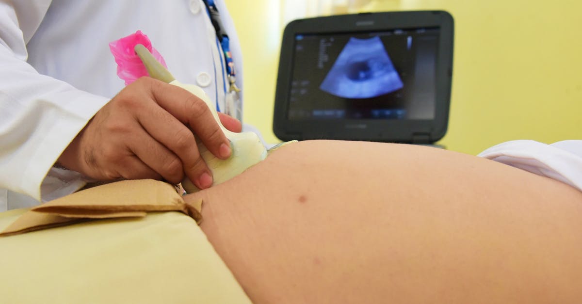
Obstetric ultrasound is the primary diagnostic tool utilized to calculate estimated fetal weight. The sonographer precisely measures specific fetal biometrics, including the biparietal diameter (head size), head circumference, abdominal circumference, and femur length. A complex mathematical algorithm computes these measurements to produce the estimated weight.
However, ultrasound estimations are notoriously subject to a margin of error, frequently overestimating or underestimating the true weight by ten to fifteen percent. This inherent inaccuracy, particularly pronounced in the third trimester and in obese mothers, creates a significant clinical dilemma when deciding between a trial of vaginal labor or a scheduled surgical delivery.
9. Maternal Risks During Delivery
Attempting a vaginal delivery of a macrosomic infant exposes the mother to substantial physical risks. The sheer mechanical force required to expel an oversized fetus through the pelvic canal frequently results in prolonged, exhausting labor.
The incidence of severe obstetric lacerations is markedly increased. The mother faces a high probability of sustaining third-degree or fourth-degree perineal tears, which involve extensive tearing through the vaginal mucosa and deep into the anal sphincter muscles, requiring complex surgical repair and carrying long-term risks of pelvic floor dysfunction or incontinence.
Furthermore, the excessive stretching of the uterine muscle by the massive fetus frequently leads to uterine atony postpartum. The exhausted, overdistended uterus fails to contract vigorously after the placenta is delivered, resulting in a postpartum hemorrhage, a critical obstetric emergency characterized by massive, rapid maternal blood loss.
10. The Catastrophe of Shoulder Dystocia
The most feared and devastating mechanical complication associated with a high birth weight is shoulder dystocia. This is a profound, life-threatening obstetric emergency that occurs during a vaginal delivery.
Shoulder dystocia occurs when the fetal head successfully passes through the birth canal and is delivered, but the anterior shoulder of the fetus becomes physically wedged tightly behind the mother’s pubic bone. The delivery process halts abruptly.
The fetus is trapped. Because the chest is tightly compressed within the birth canal, the infant cannot expand its lungs to breathe, and the umbilical cord is frequently compressed, cutting off the oxygen supply from the placenta. The obstetrician has only a matter of minutes to perform highly specific, forceful mechanical maneuvers to dislodge the shoulder and deliver the infant before severe, irreversible hypoxic brain damage or fetal death occurs.
11. Neonatal Birth Trauma
When shoulder dystocia occurs, the mechanical forces applied to expedite the delivery can result in significant structural trauma to the newborn. The most frequent injury is a brachial plexus palsy.
The brachial plexus is a complex network of delicate nerves traversing the neck and shoulder, controlling the movement of the arm and hand. During a severe dystocia, the forceful downward traction applied to the fetal head to dislodge the trapped shoulder can violently stretch or completely tear these nerves.
This results in a temporary or permanent paralysis of the infant’s arm, known as Erb’s palsy. Additionally, the extreme mechanical pressure of the birth process frequently causes fractures of the fetal clavicle (collarbone) or the humerus (upper arm bone). While these fractures generally heal flawlessly, they cause significant acute pain and distress for the newborn in the immediate postpartum period.
12. Neonatal Hypoglycemia and Metabolic Distress
The risks of macrosomia extend beyond the delivery room. In the immediate hours following birth, the macrosomic infant, particularly one born to a diabetic mother, faces a profound risk of severe metabolic instability, most notably neonatal hypoglycemia.
Throughout the pregnancy, the fetus was exposed to continuous high levels of maternal glucose, leading the fetal pancreas to pump out massive quantities of insulin. The moment the umbilical cord is clamped at birth, the abundant maternal glucose supply is severed instantly.
However, the newborn’s pancreas cannot adjust immediately. It continues to hyper-secrete insulin. This massive volume of circulating insulin rapidly depletes the newborn’s own limited glucose reserves, causing the blood sugar to plummet to dangerous levels. Unrecognized and untreated neonatal hypoglycemia can lead to acute seizures and irreversible neurological damage.
13. Delivery Planning and Cesarean Considerations
Given the profound mechanical and metabolic risks, the mode of delivery must be planned meticulously. When a fetus is suspected to be severely macrosomic, the obstetrician engages the mother in a detailed discussion regarding the risks of a trial of labor versus an elective cesarean section.
Clinical guidelines from major obstetric organizations generally recommend offering an elective cesarean delivery to diabetic mothers if the estimated fetal weight exceeds 4,500 grams, due to the high risk of asymmetrical growth and shoulder dystocia.
For non-diabetic mothers, the threshold for offering a scheduled cesarean section is typically higher, usually considered when the estimated weight exceeds 5,000 grams. The decision is highly individualized, taking into account the mother’s clinical history, pelvic anatomy, previous delivery experiences, and her personal informed preferences regarding surgical intervention.
14. Postpartum Care and Long-Term Projections
Immediately following the delivery of a macrosomic infant, the neonatal care team initiates strict metabolic protocols. The infant’s blood glucose levels are monitored rigorously for the first twenty-four to forty-eight hours. Early, frequent feedings with breast milk or formula are prioritized to stabilize the blood sugar. If the hypoglycemia is severe or persistent, the infant is transferred to the neonatal intensive care unit for intravenous dextrose therapy.
The clinical implications of a high birth weight extend far into the child’s future. Epidemiological studies demonstrate a profound correlation between fetal macrosomia and long-term metabolic consequences.
Infants born with a significantly high birth weight possess a markedly elevated risk of developing childhood obesity, severe insulin resistance, and early-onset type 2 diabetes later in life. Consequently, pediatricians emphasize the critical importance of implementing healthy nutritional habits, encouraging vigorous physical activity, and maintaining vigilant metabolic screening as the child grows to mitigate these programmed genetic and metabolic risks.
15. Frequently Asked Questions (FAQ)
1. Can I do anything to prevent my baby from getting too large?
Yes. Meticulously controlling your blood sugar if you have diabetes, managing your diet to prevent excessive weight gain during pregnancy, and engaging in regular, physician-approved exercise are the most effective preventative measures.
2. Will I definitely have to have a cesarean section if my baby is large?
No. Many women successfully deliver large babies vaginally. A cesarean section is offered as a safer alternative when the estimated weight crosses specific safety thresholds or if labor fails to progress normally.
3. Does ultrasound always accurately predict the baby’s weight?
No. Ultrasounds late in pregnancy have a significant margin of error and can overestimate or underestimate the true birth weight by up to a pound, which complicates delivery planning.
4. Why is a large baby at risk for low blood sugar after birth?
The baby’s body is used to producing huge amounts of insulin to handle the mother’s high blood sugar. When the cord is cut, the sugar supply stops, but the high insulin levels continue, rapidly dropping the baby’s own blood sugar.
5. Will a macrosomic baby automatically become an obese adult?
Not automatically, but the statistical risk is substantially higher. Establishing healthy dietary habits and an active lifestyle early in childhood is essential to combat this programmed metabolic risk.
16. Bibliography
Disclaimer: The content is for informational purposes only and does not replace medical advice. Always consult your doctor for personalized treatment.