
1. Introduction to Holoanencephaly
Holoanencephaly is a severe, profound congenital anomaly of the central nervous system. It represents the most extensive and clinically devastating form of anencephaly, a condition categorized broadly under neural tube defects. In this profound developmental failure, the embryonic neural tube fails to close completely at its cranial end, leading to the absolute absence of the major portions of the brain, the overlying meninges, the skull vault, and the scalp.
This condition is a catastrophic structural failure that occurs during the earliest stages of embryonic development. Because the vital higher cortical structures do not form, the condition is uniformly lethal. The fetus lacks the neuroanatomical architecture required for consciousness, cognition, and the sustenance of extrauterine life.
Clinical management in obstetrics focuses on early, definitive diagnosis through advanced prenatal screening and high-resolution ultrasound imaging. Meticulous diagnosis allows the medical team to provide immediate, compassionate, and transparent prognostic counseling to the expectant parents, enabling them to make informed decisions regarding the management of the pregnancy and prepare for the inevitable clinical outcome.
2. Embryonic Neural Tube Development
To comprehend the pathophysiology of this defect, it is necessary to examine the normal embryological sequence of the human nervous system. Development begins in the third week of gestation with the formation of the neural plate, a flat band of specialized cells on the dorsal surface of the embryo.
As development proceeds, the edges of this plate fold upward and fuse together in the midline to form a hollow, cylindrical structure known as the neural tube. This fusion begins in the cervical region and zips outward simultaneously toward the head (cranial neuropore) and the tail (caudal neuropore) of the embryo.
The entire process of neural tube closure must be flawlessly completed by the twenty-eighth day following conception. The fully closed neural tube serves as the foundational architecture for the entire central nervous system; the cranial portion will expand and differentiate to form the brain and skull, while the caudal portion will form the spinal cord and vertebral column.
3. Pathophysiology of Holoanencephaly
Holoanencephaly occurs when the fusion process fails entirely at the cranial neuropore. Because the neural tube remains open at the top, the rapidly developing embryonic brain tissue remains continuously exposed to the surrounding amniotic fluid.
The amniotic fluid contains enzymes and mechanical forces that are highly toxic to unprotected, delicate neural tissue. As the pregnancy progresses, the exposed forebrain and midbrain tissue is subjected to continuous chemical degradation and physical friction, causing it to necrose and degenerate completely.
Simultaneously, because the neural tube failed to close, the induction signals required for the surrounding mesoderm to form the bones of the cranial vault (the calvaria) and the overlying skin do not occur. The result is the complete absence of the frontal, parietal, and occipital bones, leaving a totally exposed base of the skull covered only by a hemorrhagic, fibrotic mass of degenerated tissue known as the area cerebrovasculosa.
4. Genetic and Environmental Risk Factors
The etiology of neural tube defects is multifactorial, involving a complex interplay between genetic susceptibilities and environmental influences. While the exact triggering mechanism is often unidentifiable in a specific pregnancy, robust epidemiological data have highlighted significant risk factors.
Maternal metabolic conditions pose a substantial risk. Pre-gestational diabetes mellitus, particularly if glycemic control is poor during the critical first month of embryonic development, significantly increases the likelihood of structural anomalies. Maternal obesity is also a recognized, independent risk factor for neural tube defects.
Pharmacological exposures during early pregnancy are heavily implicated. Certain antiepileptic medications, notably valproic acid and carbamazepine, actively interfere with folate metabolism and drastically elevate the risk of cranial closure defects. Environmental factors, including severe maternal hyperthermia (e.g., prolonged high fever or hot tub use) during the first trimester, also represent recognized teratogenic threats.
5. The Critical Role of Folic Acid
The single most significant, modifiable factor in the prevention of holoanencephaly and all other neural tube defects is maternal folic acid status. Folic acid, a synthetic form of vitamin B9, is a crucial coenzyme required for DNA synthesis, cellular division, and epigenetic regulation during rapid embryonic growth.
Clinical studies conclusively demonstrate that a deficiency in maternal folate during the periconceptional period—the weeks immediately before and after conception—is the primary driver for the failure of neural tube closure. Because the neural tube closes by day twenty-eight, the critical window for prevention occurs before many women even realize they are pregnant.
Consequently, global obstetric guidelines mandate that all women of reproductive age consume a daily supplement of four hundred micrograms of folic acid. For women with a previous pregnancy complicated by a neural tube defect, the recommended prophylactic dose is increased substantially to four thousand micrograms daily, beginning months prior to a planned conception.
6. Maternal Alpha-Fetoprotein Screening
The diagnosis of a neural tube defect is often initially suspected based on routine maternal serum screening performed during the second trimester. The primary biochemical marker utilized is maternal serum alpha-fetoprotein (MSAFP).
Alpha-fetoprotein is a major protein produced by the fetal liver. In a normal pregnancy, small amounts of this protein cross the placenta and enter the maternal bloodstream. However, when a fetus has an open neural tube defect like holoanencephaly, the exposed, vascular tissue allows massive quantities of alpha-fetoprotein to leak directly into the amniotic fluid and subsequently into the maternal circulation.
An MSAFP level that is significantly elevated (typically greater than 2.5 multiples of the median) serves as a critical red flag. While an elevated level is not a definitive diagnosis—as it can also indicate multiple gestation or incorrect pregnancy dating—it immediately prompts a detailed, high-resolution obstetric ultrasound to visually confirm the structural anatomy of the fetus.
7. Obstetric Ultrasound Diagnosis
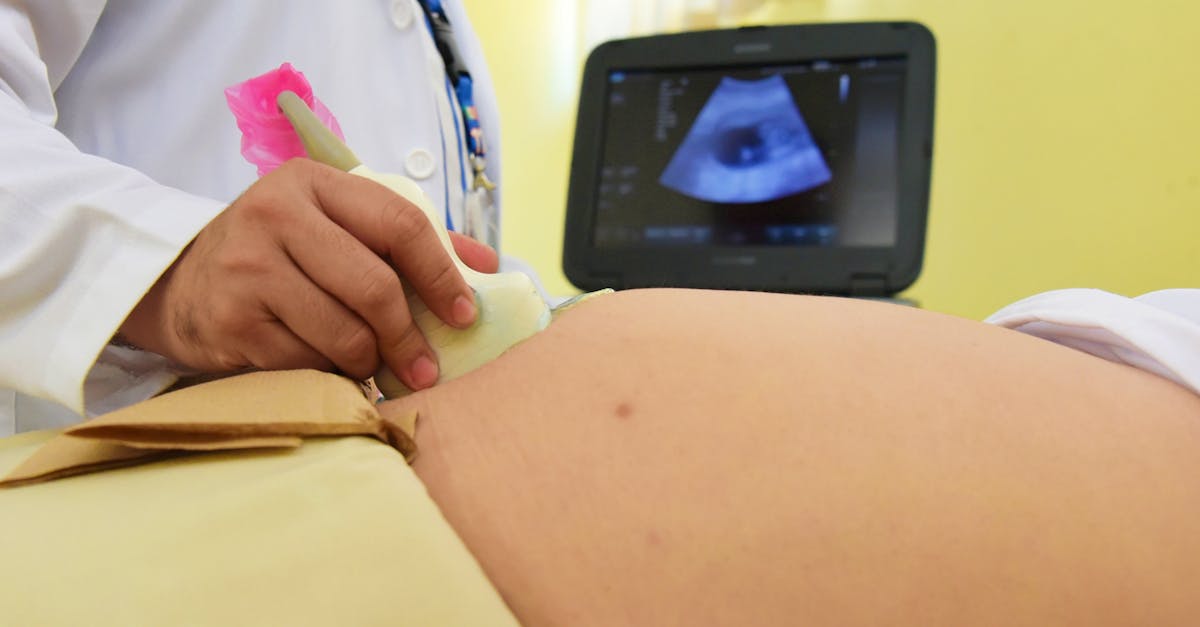
High-resolution obstetric ultrasonography is the definitive diagnostic modality for holoanencephaly. The profound structural absence of the cranial vault allows experienced sonographers to identify the anomaly with near absolute certainty, often as early as the late first trimester (eleven to fourteen weeks gestation).
The classic sonographic sign is the complete absence of the normally rounded, echogenic (bright) skull vault above the orbits. The normal brain hemispheres are entirely missing. The fetal face typically appears structurally intact up to the level of the brow, but the forehead slopes sharply backward due to the missing frontal bones.
In coronal ultrasound views, the prominent, bulging orbits combined with the absent cranial vault give the fetal face a characteristic “frog-like” appearance. The sonographer will also visualize the area cerebrovasculosa as an irregular, heterogeneous mass of tissue floating in the amniotic fluid directly above the fetal face.
8. Differentiating Neural Tube Defects
While the diagnosis of an open cranial defect is straightforward on ultrasound, clinicians categorize the exact type based on the extent of the missing tissue.
| Neural Tube Defect | Anatomical Description |
|---|---|
| Holoanencephaly | The most severe form; complete absence of the brain hemispheres, midbrain, and the entire skull vault down to the foramen magnum. |
| Meroanencephaly | Partial absence; some rudimentary brain tissue and partial skull bone formation may be present at the base of the skull. |
| Encephalocele | The neural tube closed, but a gap in the skull bones allows fully formed brain tissue to herniate outward in a sac. |
Regardless of whether the diagnosis is holoanencephaly or meroanencephaly, the clinical outcome remains universally fatal, as the critical higher cortical structures necessary for life are irrevocably destroyed.
9. Clinical Signs During Pregnancy
While the fetus suffers catastrophic structural failure, the mother may remain completely asymptomatic, save for the routine symptoms of pregnancy like fatigue. However, as the pregnancy progresses into the second and third trimesters, a specific clinical complication often arises: polyhydramnios.
Polyhydramnios is the excessive accumulation of amniotic fluid within the uterus. In a healthy pregnancy, the volume of amniotic fluid is regulated precisely because the fetus continuously swallows the fluid, processes it through the kidneys, and urinates it back out.
Fetuses with holoanencephaly lack the neurological reflexes required to swallow efficiently. Because the swallowing mechanism is impaired or absent, the amniotic fluid continues to build up unchecked. This massive fluid volume causes severe, uncomfortable stretching of the maternal uterus, significantly increasing the risk of premature rupture of membranes and preterm labor.
10. Associated Fetal Anomalies
While holoanencephaly is the primary and fatal diagnosis, it is frequently accompanied by a spectrum of other severe congenital malformations. The failure of the cranial neural tube to close often disrupts the embryological signaling pathways required for the proper formation of other organ systems.
Spina bifida, an open defect of the lower spinal cord, is concurrently present in a significant percentage of these fetuses. Cleft lip and cleft palate are frequent facial anomalies. Furthermore, severe structural defects of the heart, gastrointestinal tract malformations, and clubfoot (talipes equinovarus) are commonly identified during the detailed anatomical ultrasound survey.
The presence of multiple, widespread anomalies may indicate an underlying, overarching chromosomal disorder, such as Trisomy 13 or Trisomy 18. Therefore, the clinical team typically offers amniocentesis to evaluate the fetal karyotype, providing the parents with precise genetic information for future family planning.
11. Maternal Health Considerations and Delivery
When a diagnosis of holoanencephaly is firmly established, obstetric management requires a delicate balance of protecting the mother’s physical health while providing profound psychological support.
Because the condition is invariably fatal for the fetus, clinical protocols generally recommend against performing a cesarean section purely for fetal indications, as major abdominal surgery carries inherent, unnecessary risks for the mother. The preferred clinical approach is a planned vaginal delivery.
However, the onset of natural labor can be delayed. The fetal brain lacks a functional pituitary gland, which normally secretes hormones that help trigger the cascade of natural labor. Consequently, these pregnancies often extend post-term if left to progress naturally. The clinical team frequently utilizes medical induction of labor to initiate a safe vaginal delivery at an appropriate time chosen by the parents.
12. Prognosis and Clinical Outcomes
The prognosis for holoanencephaly is absolute. It is a uniformly lethal condition. The vast biological requirements for consciousness, autonomous respiration, and basic metabolic regulation cannot be met due to the profound absence of the cerebrum, midbrain, and hypothalamus.
Approximately half of fetuses with this diagnosis are stillborn, succumbing in utero prior to the onset of labor. Of those born alive, survival is measured in hours, or very rarely, a few days. The brainstem, which controls basic autonomic functions like heartbeat, may be partially intact, allowing the newborn to breathe briefly and exhibit basic reflex movements.
However, respiratory failure inevitably occurs shortly after birth. The neonatal care protocol shifts entirely to palliative, comfort care. The clinical team focuses exclusively on keeping the infant warm, comfortable, and pain-free, allowing the parents undisturbed time to bond and grieve with their child.
13. Psychological Support and Counseling
The diagnosis of a lethal fetal anomaly is one of the most profoundly devastating events an expectant parent can endure. The emotional trauma is immense, requiring immediate, structured, and compassionate psychological intervention.
The clinical team must deliver the diagnosis with absolute transparency, using clear, non-technical language to ensure the parents fully comprehend the fatal nature of the condition. False hope must be avoided, but empathy is paramount.
Parents are immediately connected with specialized grief counselors, perinatal bereavement teams, and social workers. These professionals guide the parents through the complex, agonizing decisions regarding the continuation or termination of the pregnancy, the formulation of a specialized birth plan, and the coordination of palliative care and memorial arrangements, providing a vital framework of support during an overwhelming crisis.
14. Preconception Planning for Future Pregnancies
Following the loss of a pregnancy to a neural tube defect, comprehensive preconception counseling is a mandatory clinical requirement before the parents attempt to conceive again.
The statistical risk of recurrence for any neural tube defect in a subsequent pregnancy rises to approximately two to three percent. To mitigate this elevated risk, aggressive nutritional prophylaxis is implemented.
The mother is prescribed a high-dose folic acid regimen—typically four milligrams daily—beginning at least three months prior to discontinuing contraception and continuing through the first trimester of the subsequent pregnancy. Furthermore, the clinical team will meticulously optimize any maternal metabolic conditions, such as diabetes or obesity, and review all maternal medications to eliminate any teratogenic exposures, ensuring the safest possible environment for the future embryo.
15. Frequently Asked Questions (FAQ)
1. Is there any surgical procedure that can fix this condition before birth?
No. Because the brain tissue completely fails to form during the first few weeks of pregnancy, there is no tissue to repair. The condition is absolutely irreversible and fatal.
2. Did I do something wrong during my pregnancy to cause this?
No. This defect occurs within the first 28 days after conception, often before a woman knows she is pregnant. It is a complex genetic and developmental event, not the result of an action taken by the mother.
3. Will an ultrasound catch this defect early in the pregnancy?
Yes. Modern, high-resolution ultrasounds performed by experienced sonographers can clearly identify the missing skull and brain tissue, usually between the 11th and 14th week of gestation.
4. Why did my doctor recommend taking such a high dose of folic acid for my next pregnancy?
Because you have had a pregnancy affected by a neural tube defect, your risk of it happening again is statistically higher. A massive dose of folic acid (4mg) is clinically proven to drastically reduce that recurrence risk.
5. Does the baby feel pain during the pregnancy or birth?
Pain perception requires a highly developed, functioning cerebral cortex. Because this portion of the brain is completely absent, the fetus lacks the neurological structures required to process or feel pain.
16. Bibliography
Disclaimer: The content is for informational purposes only and does not replace medical advice. Always consult your doctor for personalized treatment.
