1. Introduction
Hyperosmolar coma due to type 2 diabetes mellitus is a life-threatening complication defined by profoundly elevated blood sugar levels and severe systemic dehydration leading to unconsciousness. Because individuals with type 2 diabetes produce trace amounts of insulin, they avoid severe acid accumulation but suffer from intense fluid loss through the kidneys. Treatment centers on aggressive rehydration and meticulous electrolyte stabilization in an intensive care setting.
This condition, historically known as Hyperosmolar Hyperglycemic Nonketotic Coma, represents the most severe end of the metabolic spectrum for patients with type 2 diabetes. The mortality rate is notable, primarily because the condition frequently affects older adults who may possess concurrent cardiovascular or renal comorbidities. The onset is insidious, evolving over days or weeks, allowing blood glucose and osmolality to reach staggering levels before the profound neurological depression prompts emergency medical intervention.
2. The Dynamics of Type 2 Diabetes
Type 2 diabetes is fundamentally characterized by peripheral insulin resistance and a progressive decline in pancreatic insulin secretion. Unlike type 1 diabetes, where insulin production ceases entirely, the pancreas in type 2 diabetes continues to produce some insulin.
This residual insulin is a crucial protective factor. It is generally sufficient to suppress lipolysis, the breakdown of fat stores, which prevents the massive generation of acidic ketone bodies. However, this small amount of insulin is entirely inadequate to facilitate the entry of glucose into the resistant muscle and fat cells. Consequently, glucose accumulates relentlessly in the bloodstream, setting the stage for a hyperosmolar crisis without the immediate warning signs of severe acidosis.
3. Defining the Hyperosmolar State
Osmolality measures the concentration of chemical particles dissolved in the blood plasma. In normal physiology, sodium and urea dictate this concentration, maintaining a precise balance that keeps cells perfectly hydrated.
When blood glucose levels rise to severe levels, glucose becomes a dominant, pathological osmotic force. The blood becomes profoundly hyperosmolar. Because nature abhors a concentration gradient, water is forcefully drawn out of the surrounding cells and tissues into the hyper-concentrated bloodstream in a physiological attempt to dilute it. This extraction dehydrates the cells, severely impairing their structural integrity and biochemical function.
4. The Absence of Severe Ketoacidosis
The distinctive clinical feature of this condition is the general absence of severe ketoacidosis. In a diabetic crisis where insulin is completely absent, the body burns fat for fuel, creating acidic ketones that rapidly lower the blood pH, causing deep, rapid breathing and severe abdominal pain.
In a hyperosmolar coma due to type 2 diabetes, the trace amounts of circulating insulin are enough to tell the liver to refrain from breaking down fat. Because the blood pH remains relatively normal and the dramatic symptoms of acidosis are absent, the patient may endure the rising blood sugar for weeks, feeling generally unwell but delaying medical care until severe dehydration and cognitive decline occur.
5. Chronic Hyperglycemia and Insulin Resistance
The pathway to a hyperosmolar coma begins with chronic, unmanaged hyperglycemia. As insulin resistance worsens, the liver continues to inappropriately release stored glucose into the bloodstream, interpreting cellular starvation as a need for more fuel.
Simultaneously, the high blood sugar environment acts toxically on the remaining pancreatic beta cells, further suppressing their ability to release insulin. This creates a relentless upward trajectory of blood glucose. As the levels climb over four hundred to six hundred milligrams per deciliter, the physical properties of the blood change, becoming increasingly viscous and difficult to pump through the microscopic capillaries of the body.
6. Renal Filtration and Fluid Loss
The kidneys act as the primary filter for the blood, attempting to reclaim valuable nutrients while excreting waste. There is a physiological threshold for glucose reabsorption; when blood sugar exceeds this limit, the kidneys can no longer hold onto it, and glucose spills into the urine.
This glycosuria is the engine of dehydration. Glucose is an osmotic diuretic; as it flows through the renal tubules, it drags massive volumes of water out of the body. The patient begins to urinate frequently and in large volumes. Over several days or weeks, this uncompensated fluid loss severely depletes the total body water, shrinking the intravascular blood volume and compounding the concentration of the remaining blood.
7. The Onset of Severe Dehydration
As the osmotic diuresis continues, the patient experiences profound systemic volume depletion. The physical signs of dehydration become starkly evident. The skin loses its elasticity, and the mucous membranes become parched.
The reduction in blood volume directly compromises cardiac output and blood pressure. The patient may experience severe orthostatic hypotension, becoming dizzy or fainting upon standing. Furthermore, as blood flow to the kidneys diminishes due to the low volume, the kidneys begin to fail. This acute kidney injury stops the excretion of glucose, causing a sudden, exponential spike in blood sugar levels that accelerates the hyperosmolar crisis.
8. Neurological Deterioration to Coma
The central nervous system bears the brunt of the hyperosmolar insult. As the hyper-concentrated blood pulls water out of the cerebral tissues, the neurons in the brain physically shrink and pull away from their supporting structures.
This cellular dehydration disrupts neurotransmitter function and alters the electrical gradients necessary for nerve conduction. The clinical presentation evolves from mild confusion and severe lethargy to profound disorientation and visual hallucinations. The patient may exhibit focal neurological signs, such as localized muscle weakness, that closely mimic an acute stroke. Ultimately, the profound cerebral dehydration depresses the brainstem, resulting in a hyperosmolar coma.
9. Common Triggers in Older Adults
A hyperosmolar coma is typically precipitated by a significant physiological stressor. In the elderly population, the most frequent trigger is an occult infection, particularly pneumonia or a severe urinary tract infection.
Infections provoke a systemic release of stress hormones, including cortisol and adrenaline, which directly antagonize insulin action and command the liver to produce more glucose. Other common triggers include acute medical events like a myocardial infarction, or the initiation of medications that impair glucose tolerance, such as high-dose corticosteroids or thiazide diuretics. Limited access to water, common in elderly patients with mobility issues or dementia, rapidly exacerbates the dehydration.
10. Clinical Signs of Volume Depletion
Emergency medical personnel rely on physical examination to gauge the severity of the volume depletion upon initial contact with the patient.
The patient will frequently present with tachycardia, a rapid heart rate representing the heart’s attempt to maintain blood pressure with a severely diminished fluid volume. The eyes may appear sunken, and capillary refill time will be significantly delayed. The patient’s breathing is typically shallow and normal in rate, sharply contrasting with the deep, labored breathing seen in ketoacidosis. The profound dryness of the tissues is the hallmark physical finding.
11. Cardiovascular Complications
The severe dehydration and increased blood viscosity place an immense burden on the cardiovascular system. The thick, sluggish blood is inherently pro-thrombotic, substantially increasing the risk of dangerous blood clots forming in the deep veins of the legs or traveling to the lungs.
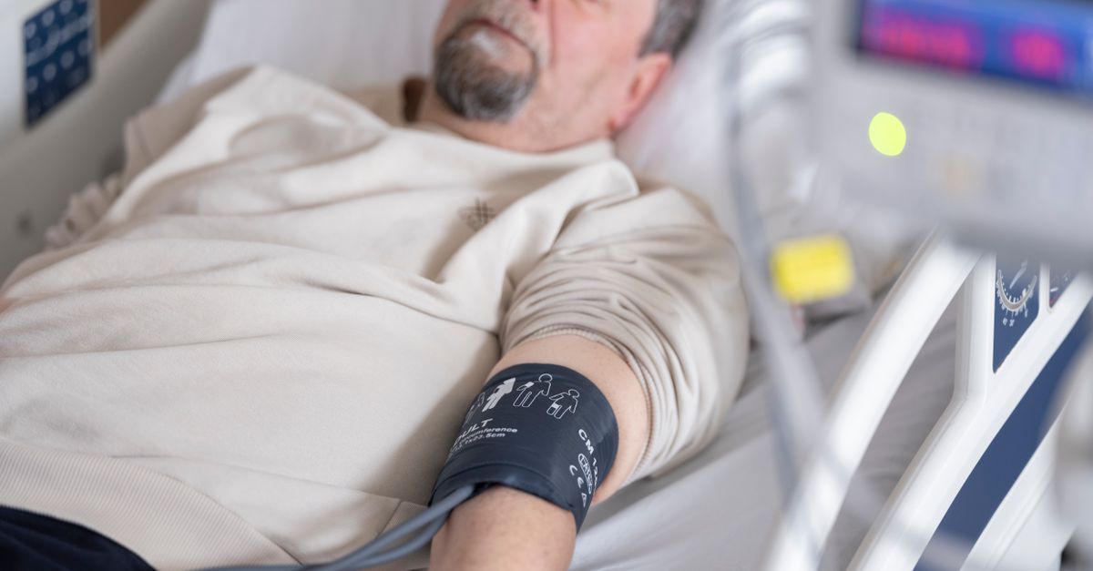
Furthermore, the lack of adequate blood volume impairs coronary perfusion. In older adults with preexisting coronary artery disease, this reduced blood flow can precipitate an acute myocardial infarction, even while the patient is comatose. Meticulous cardiovascular monitoring is essential during the initial resuscitation phase in the intensive care unit.
12. Differentiating Coma Types
Accurate differentiation dictates the urgency and specific nature of medical interventions.
| Clinical Marker | Hyperosmolar Coma (Type 2) | Ketoacidotic Coma (Type 1) |
|---|---|---|
| Onset Timeline | Insidious; develops over days to weeks. | Rapid; develops over 24 to 48 hours. |
| Blood Glucose | Profoundly high, frequently > 800 mg/dL. | Moderately high, usually 300 – 600 mg/dL. |
| Acid-Base Status | Normal blood pH, negligible ketones. | Severe acidosis (low pH), high ketones. |
| Primary Symptom | Severe dehydration and neurological signs. | Abdominal pain, deep breathing, fruity breath. |
13. Emergency Diagnostic Workup
Upon arrival at the emergency department, immediate laboratory analysis is required. A comprehensive metabolic panel will reveal the precise glucose level and the essential electrolyte concentrations.
The calculated serum osmolality confirms the diagnosis; values exceeding 320 milliosmoles per kilogram correlate strongly with profound neurological impairment. Renal function markers, blood urea nitrogen and creatinine, are evaluated to assess the extent of acute kidney injury. Blood and urine cultures are routinely obtained to identify underlying infectious triggers, and a twelve-lead electrocardiogram is performed to evaluate for silent ischemia or electrolyte-induced arrhythmias.
14. Principles of Fluid Replacement
The absolute priority in managing a hyperosmolar coma is aggressive and calculated intravenous fluid resuscitation. The primary goal is to safely restore the depleted intravascular blood volume and re-establish blood flow to the kidneys.
Therapy typically begins with the rapid infusion of isotonic saline. Once the blood pressure is stabilized, the clinician carefully adjusts the type of intravenous fluid to a hypotonic solution based on the patient’s corrected sodium levels. The fluids must be administered thoughtfully; rehydrating the brain too rapidly can cause cerebral edema, a fatal swelling of the brain tissue. The total fluid deficit, often reaching nine to ten liters, is replaced steadily over twenty-four to forty-eight hours.
15. Managing Blood Glucose Safely
While intravenous fluids address the severe dehydration, continuous intravenous regular insulin is administered to halt hepatic glucose production and drive glucose into the cells.
Insulin therapy is initiated only after aggressive fluid resuscitation has begun and the patient’s potassium levels have been verified to prevent a sudden cardiovascular collapse. Because patients with type 2 diabetes retain some insulin sensitivity once hydrated, the blood glucose frequently drops significantly with fluid administration alone. The insulin drip is meticulously titrated based on hourly glucose checks, aiming for a slow, steady decline in blood sugar rather than a rapid plunge.
16. When to Consult a Healthcare Provider
A hyperosmolar coma is a medical emergency requiring immediate hospitalization. Caregivers of individuals with type 2 diabetes must seek emergency medical care if the patient exhibits severe confusion, an inability to stay awake, or a complete lack of urine output.
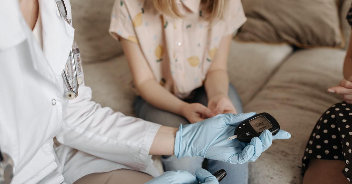
Routine home monitoring is the primary defense. If blood glucose readings remain consistently high, particularly during a concurrent illness, and the patient feels profoundly weak or dizzy, proactive medical intervention is required. Delaying care in the presence of severe dehydration invites critical neurological and renal complications.
17. Frequently Asked Questions (FAQ)
1. Can a person recover fully from a hyperosmolar coma?
Yes. With prompt intensive care treatment focusing on slow, careful fluid replacement and insulin therapy, the dehydration can be reversed, and the patient can regain full consciousness, provided no permanent stroke or cardiac event occurred during the crisis.
2. Why is there no acid buildup in this condition?
Individuals with type 2 diabetes still produce a tiny amount of insulin. This small amount is enough to stop the body from aggressively burning fat, which prevents the production of acidic ketones that cause acidosis.
3. Why do doctors focus on fluids before insulin in the hospital?
If insulin is given before your fluid volume is restored, the insulin will rapidly push the remaining sugar and water out of your bloodstream into your cells. This sudden loss of fluid from your blood vessels can cause your blood pressure to crash fatally.
4. How does an infection trigger this coma?
An infection causes the body to release stress hormones like cortisol. These hormones block the action of insulin and tell the liver to produce extra sugar to fight the illness, which rapidly drives up blood glucose levels in a diabetic patient.
5. How can this condition be prevented?
Prevention relies on consistent management of blood sugar, taking prescribed medications, and maintaining hydration, especially during illnesses. Regular blood glucose monitoring at home helps catch rising levels before severe dehydration sets in.
18. Bibliography
Disclaimer: The content is for informational purposes only and does not replace medical advice. Always consult your doctor for personalized treatment.